Stromal Cell-Derived Factor-1 Promotes Myeloma Cell Growth in Both Autocrine and Paracrine Manners
Seong-Woo Kim, Ha-Yon Kim, Hyo-Jin Lee, M.D., Hwan-Jung Yun, M.D., Samyong Kim, M.D. and Deog-Yeon Jo, M.D.
Division of Hematology/Oncology, Department of Internal Medicine, College of Medicine, Chungnam National University, Daejeon, Korea
Background: The question as to whether stromal cell-derived factor-1 (SDF-1) stimulated myeloma cell growth has been controversial. We explored the possibility that SDF-1 may function as an autocrine growth factor of myeloma cells.
Methods: CD138+ primary bone marrow myeloma cells and myeloma cell lines (RPMI8226, U266, and ARH77) were used. Chemotaxis in response to SDF-1 of the cells was analyzed using TranswellsTM. Cell proliferation was measured by a colorimetric assay. SDF-1 mRNA expression was analyzed by RT-PCR (reverse-transcription-polymerase chain reaction). SDF-1 and interleukin-6 (IL-6) receptor ex- pression as well as signaling molecule phosphorylation levels, were examined using Western blot analysis.
Concentrations of SDF-1 in the cell culture supernatants were measured by ELISA assay.
Results: SDF-1 alone had no discernible effect on the proliferation of CD138+ primary myeloma cells or myeloma cell lines. In contrast, SDF-1 significantly enhanced IL-6-induced proliferation of these cells.
SDF-1 up-regulated the expression of IL-6 receptor and enhanced phosphorylation of AKT in an additive manner with IL-6. Co-culture of the myeloma cells with umbilical vein endothelial cells over-expressing the SDF-1 gene revealed that SDF-1 played an important role in not only the migration of the cells underneath the stromal cells but also the proliferation of the cells in contact with stromal cells. All the myeloma cell lines expressed SDF-1 mRNA, and SDF-1 was detected in the culture supernatants of the cells. The G protein-coupled receptor inhibitor, pertussis toxin, inhibited the proliferation of these cells in suspension cultures.
Conclusion: SDF-1, most likely in concert with IL-6, enhanced the proliferation of myeloma cells in a both paracrine and autocrine manner. (Korean J Hematol 2008;43:127-137.)
Key Words: Multiple myeloma, SDF-1, CXCR4, Interleukin-6, Bone marrow stromal cells
127 접수:2008년 4월 15일, 수정:2008년 5월 7일
승인:2008년 7월 10일
교신저자:조덕연, 대전시 중구 대사동 640
301-721, 충남대학교병원 혈액종양내과 Tel: 042-280-7162, Fax: 042-257-5753 E-mail: [email protected]
이 연구는 한국학술진흥재단의 지역대학우수과학자 지원사업 의 지원으로 이루어진 것임(과제번호: KRF-2005-202-E00085).
Correspondence to:Deog-Yeon Jo, M.D.
Division of Hematology/Oncology, Department of Internal Medicine, Chungnam National University Hospital
640, Daesa-dong, Jung-gu, Daejeon 301-721, Korea Tel: +82-42-280-7162, Fax: +82-42-257-5753 E-mail: [email protected]
INTRODUCTION
Chemokine stromal cell-derived factor-1 (SDF-
1), which is constitutively expressed and pro- duced in bone marrow stromal cells (BMSCs), in- duces the migration and homing of hematopoietic stem/progenitor cells1) and lymphocytes,2) by sig-
naling via its G protein-coupled receptor, CXCR4.
Myeloma cells also express CXCR43) and respond to SDF-1, resulting in the trafficking and local- ization of these cells in the bone marrow (BM) microenvironment.4-6) Serum levels of SDF-1 are elevated in patients with multiple myeloma,7) suggesting that SDF-1 is involved in the develop- ment or progression of the disease. Bone marrow endothelial cells (BMECs) in multiple myeloma secrete CXC chemokines, including SDF-1, that mediate the interaction with myeloma cells.8) In addition, SDF-1 plays an important role in tumor neoangiogenesis, and the blockade of the SDF- 1/CXCR4 axis attenuates tumor growth.9) These observations raise the possibility that the modu- lation of CXCR4 on myeloma cells or SDF-1 pro- duction in BMSCs could influence the biology of myeloma cells and the disease course. However, direct effects of SDF-1 on the proliferation of myeloma cells are not well understood. It has been reported that SDF-1 promotes proliferation and protects against dexamethasone-induced apop- tosis in myeloma cells; however, these effects were only marginal.10) In this study, we explored the possibility that SDF-1 stimulates myeloma cell growth in an autocrine as well as a paracrine manner.
MATERIALS AND METHODS 1. Cells and reagents
BM samples were obtained, with informed con- sent, from patients with multiple myeloma and from healthy donors undergoing bone marrow harvest. CD138+ cells were purified from BM us- ing the MACS system (Miltenyi Biotec, Auburn, CA, USA) according to the manufacturer’s instructions. Only cell preparations that con- tained >95% CD138+ cells, as assessed by flow cytometric analysis, were used in the experi- ments. Human umbilical vein endothelial cells (HUVECs) were prepared as previously descri- bed11) and were cultured in medium-199 (Gibco BRL Life Technologies, Grand Island, NY, USA),
supplemented with 20% fetal bovine serum (FBS;
Gibco), L-glutamine (2mM), heparin (5U/mL), penicillin (100IE/mL), streptomycin (100μg/mL;
Sigma Chemical Co., St. Louis, MO, USA), and endothelial cell growth supplements (50μg/mL;
ECGS; Biomedical Technologies, Inc., Stoughton, MA, USA). Early-passage HUVEC cultures were used in the experiments. BMSCs were prepared by culturing CD34-negative cells in minimal es- sential medium-alpha (MEM-α, Gibco), supple- mented with 12.5% FBS, 12.5% horse serum (Gi- bco), hydrocortisone (10−6M, Sigma), 2-mercap- toethanol (5×10⁻⁵M, Sigma), streptomycin, and penicillin (referred to as LTC medium). After confluence was reached, the adherent cells were harvested and maintained in LTC medium with weekly passage. After three passages, neither CD45+ nor PECAM1+ cells were detected by flow cytometry. Murine BM stromal MS-5 cells were grown in MEM-α supplemented with 10%
FBS and were passaged weekly. These cells sup- port the proliferation of primitive human hema- topoietic cells in long-term cultures and secrete SDF-1.2) The human myeloma cell lines RPMI8226, U266, and ARH77 were cultured in RPMI1640 (Gibco) supplemented with 10∼15% FBS. Inter- leukin-6 (IL-6), SDF-1, and the 12G5 func- tion-blocking monoclonal antibody to CXCR4 were purchased from R&D Systems (Minneap- olis, MN, USA).
2. Flow cytometry
Cells were incubated with fluorescein iso- thiocyanate (FITC)- and/or phycoerythrin (PE)- conjugated monoclonal antibodies at 4oC for 30 min and were analyzed using a Coulter Elite flow cytometer (Coulter Electronics Ltd., Hialeah, FL, USA). The monoclonal antibodies used in the study were FITC- or PE-conjugated anti-CD138 (Mi15; BD PharMingen, San Diego, CA, USA) and PE-conjugated anti-CXCR4 (12G5; BD Phar- Mingen). To detect cytoplasmic CXCR4, the cells were permeabilized with saponin-based reagents (BD PharMingen) before labeling.
3. Cell proliferation assay
The effects of SDF-1 and IL-6 on the myeloma cell proliferation were measured using a colori- metric assay kit (CCK-8 assay kit, Dojindo Labo- ratories, Tokyo, Japan) according to the instruc- tions provided by manufacturer. Briefly, 5×10³ cells were incubated in 96-well plates in serum- free X-VIVO medium (BioWhittaker, Walkerwil- le, MA, USA) in the presence or absence of cyto- kines. After a 3-day incubation, 10μL of CCK-8 solution was added into each well. Two to four hours later, optical density (OD) was measured using a spectrophotometer (Molecular Devices Co., Sunnyvale, CA, USA) and the fold-increase in the OD compared with that of the control (proliferation index) was calculated. To analyze the effects of blocking the G protein-coupled re- ceptor signaling on the myeloma cell prolifera- tion, 10~15×104 cells were incubated in X-VIVO medium in 24-well plates in the presence or ab- sence of pertussis toxin (PTX, 200ng/mL, Sigma) for up to 4 days. After incubation, viable cells were counted using hemocytometer after trypan blue treatment.
4. Transmigration assay
For the transmigration experiments, cells (2.0×
105 to 2.5×10⁵ cells/well) were loaded into the upper chamber of a 24-well TranswellTM plate containing a 5-μm microporous membrane (Cor- ning-Costar, Cambridge, MA) and were allowed to migrate into the lower chamber for 4 hr. The migrated cells were counted using flow cytome- try, and the fold-increase in the number of mi- grated cells compared with that of the control (migration index) was calculated.
5. Short-term co-culture of myeloma cells on MS-5 feeder cells
The CD138+ or RPMI8226 cells were added onto MS-5 feeder layers in X-VIVO medium. The migration of the cells beneath the feeder layer was identified under an inverted microscope 24
hr later. To examine whether SDF-1 plays a cen- tral role in the cell migration, the added cells were pre-treated with 40μg/mL 12G5 or 200 ng/mL PTX for 30 min.
6. Preparation of recombinant adenoviruses car- rying the human SDF-1α and LacZ genes The Adeno-X expression system (Clontech, Bed- ford, MA, USA) was used to construct recombi- nant adenoviral vectors encoding the human SDF-1α gene and the LacZ gene. The mRNA was obtained from human BMSCs, and a human SDF-1α cDNA was constructed by reverse tran- scription-polymerase chain reaction (RT-PCR).
7. Transfection of HUVECs with adenoviral vectors HUVECs were seeded in T25 culture flasks (Corning-Costar, Cambridge, MA, USA). When the cells reached confluence, the layers were transfected at a multiplicity of infection (m.o.i.) of 20 in serum-free X-VIVO medium. After a 4 hr incubation, the adenoviral vector-containing supernatant was removed and replaced with IMDM supplemented with 20% FBS. Two to four parallel flasks were established for each con- dition. HUVECs transfected with E1(−)E4(+) replication-deficient adenovirus vectors remained quiescent and were viable for prolonged periods.12)
8. Co-culturing of myeloma cells and HUVECs After adenoviral infection, the CD138+ or RPMI8226 cells were placed onto previously es- tablished HUVEC layers in X-VIVO medium and incubated at 37°C in 5% CO2 in air. Cobblestone areas, which appeared as phase-dark clones of at least five cells beneath the HUVEC layer, were scored under a phase-contrast inverted micro- scope.
9. Measurement of the concentrations of SDF-1 in cell culture supernatants
Culture supernatants were prepared from MS-5 and myeloma cells grown in X-VIVO medium in T25 culture flasks. After a 72-hr incubation at
37oC, the supernatants were harvested, filtered through 2-μm membranes, and stored at 4oC un- til analyzed. The concentration of SDF-1α in the supernatants was determined using a commercial ELISA kit (R&D Systems), according to the man- ufacturer’s instructions. The optical density was measured using a spectrophotometer. Standard curves and calculated levels of cytokine were ob- tained using the program Softmax™ (Molecular Devices Co.).
10. RT-PCR
Total RNA was prepared from cells using Trizol reagent (Gibco) according to the manufac- turer’s instructions. After purification, RNA (1μg) was reverse-transcribed using SuperScript reverse transcriptase (Gibco) and the universal primer oligo, (dT)15 (Promega, Madison, WI, USA). For each PCR reaction, 1μL of the cDNA product was added to 24μL of PCR buffer (Gibco) sup- plemented with MgCl2 (2mM), specific primers (0.2μM), and 1U of Koma Taq polymerase (Koma International, Seoul, Korea). Each PCR cycle consisted of 1 min at 94°C, 45 sec at 55∼
65°C, and 1min at 72°C; 30 cycles were carried out using a GeneAmp PCR system (Perkin El- mer, Norwalk, CT, USA). The following primers were used: human CXCR4 (sense, AAT CTT CCT GCC CAC CAT CTA CTC C; antisense, GCG GTC ACA GAT ATA TCT GTC ATC TGC C); human SDF-1 (sense, AGA ATT CAT GAA CGC CAA GG; anti-sense, AGG ATC CTC ACA TCT TGA ACC); GAPDH (sense, CAT GTG GGC CAT GAG GTC CAC CAC; antisense, TGA AGG TCG GAG TCA ACG GAT TTG GTC).
11. Western blot analysis
Western blotting was used to detect the ex- pression of SDF-1, CXCR4, and IL-6 receptor as well as the phosphorylation of signaling mole- cules. The cells were collected by centrifugation, washed in phosphate-buffered saline, and lysed by the addition of SDS sample buffer [62.5mM Tris-HCl (pH 6.8), 6% (w/v) SDS, 30% glycerol,
125mM DTT, and 0.03% (w/v) bromophenol blue]. The whole-cell samples were sonicated, lysed, and denatured by boiling for 5 min. Equal amounts of protein from each sample were elec- trophoresed on 12∼18% SDS-polyacrylamide gels and transferred to nitrocellulose membranes (Amersham Life Science, Arlington Heights, IL, USA). The membranes were blocked for 1hr with Tris-buffered saline (TBS) containing 5% (w/v) milk and 0.1% Tween, and then incubated with the primary mouse or rabbit monoclonal antibody overnight at 4ºC. The blots were washed with TBS containing Tween, incubated with anti- mouse or anti-rabbit secondary antibody (Cell Signaling Technology Inc., Danvers, MA, USA) for 2 hr, and developed using West-Zol Plus (iNtRON Biotechnology, Seoul, Korea). For phos- phorylation studies, the cells were starved in se- rum-free medium for 16 hr, and then stimulated with cytokines. The following antibodies were used: anti-SDF-1 monoclonal antibody (R&D Sys- tems), anti-CXCR4 monoclonal antibody (Affini- ty Bio Reagents, Golden, CO, USA), anti-IL-6 re- ceptor polyclonal antibody (R&D Systems), an- ti-phospho-AKT polyclonal antibody (Ser473), anti-AKT polyclonal antibody, anti-phospho-ERK polyclonal antibody (Thr202, Tyr204), anti-ERK polyclonal antibody, anti-phospho-Stat3 poly- clonal antibody (Tyr705), and anti-Stat3 poly- clonal antibody (all purchased from Cell Signa- ling Technology).
12. Statistical analysis
The results are expressed as the mean±stand- ard deviation (SD) of at least three experiments.
Data were analyzed using Student’s t-test for paired samples; a value of P<0.05 was consid- ered statistically significant.
RESULTS
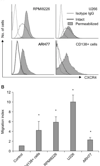
1. Myeloma cells express functional CXCR4 RPMI8226 and U266 cells strongly expressed CXCR4 on the cell surface (Fig. 1A) and re-
Fig. 1. Myeloma cells express functional CXCR4. (A) Flow cytometric analysis of the expression of CXCR4 on RPMI8226 cells, U266 cells, ARH77 cells, and CD138+ primary myeloma cells. ARH77 cells express CXCR4 minimally on the cell surface; however, analysis after permeabilization reveals CXCR4 in the cytoplasm of the majority of the cells. (B) Chemoattraction of myeloma cells induced by SDF-1 (100 ng/mL) over 4 hr measured using 24-well Transwells with 5-μm pores. Values represent the mean migration index (±SD) obtained from triplicate experiments for cell lines and those obtained from the experiments using five different specimens for CD138+ cells. *P<0.05 compared with control.
sponded to SDF-1, resulting in chemotaxis (Fig.
1B). A very small fraction of ARH77 cells ex- pressed CXCR4 on the cell surface; however, flow cytometric analysis after permeabilization re- vealed CXCR4 in the cytoplasm of most of the cells (Fig. 1A). BM CD138+ cells obtained from five patients with multiple myeloma showed vari-
able expression of CXCR4 (median, 43.3%; range, 17.3~56.6%) and exhibited chemotactic re- sponses to SDF-1 (Fig. 1).
2. SDF-1 enhances the proliferation of myeloma cells in concert with IL-6 via up-regulation of IL-6 receptor
SDF-1 alone (at concentrations of up to 200 ng/mL) had no discernible effect on the pro- liferation of myeloma cell lines or primary BM CD138+ myeloma cells in serum-free conditions.
IL-6 modestly stimulated the proliferation of RPMI8226 cells and primary BM CD138+ cells, and the combination of SDF-1 and IL-6 sig- nificantly enhanced cell proliferation (Fig. 2). IL- 6 did not influence CXCR4 expression on myelo- ma cells, whereas SDF-1 significantly up-regu- lated the expression of IL-6 receptor in RPMI8226 and U266 cells, but not in ARH77 cells, as de- termined by Western blot analysis (Fig. 3). IL-6 strongly increased phosphorylation of AKT, ERK, and Stat3 in RPMI8226 cells. SDF-1 modestly in- creased the phosphorylation of AKT and ERK, but not Stat3. Together, SDF-1 and IL-6 addi- tively enhanced the phosphorylation of AKT, but not ERK or Stat3 (Fig. 4).
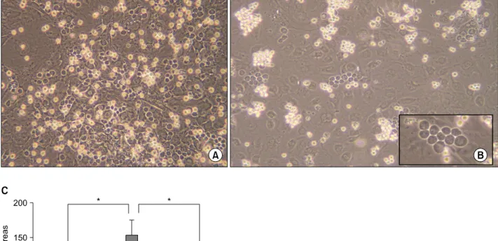
3. SDF-1 enhances the maintenance and growth of myeloma cells in contact with BMSCs To examine whether SDF-1 induces the migra- tion of myeloma cells beneath stromal cells, RPMI8226 cells were added onto MS-5 feeder layers in X-VIVO medium. After 12~24 hr, the migration of many cells beneath the feeder layer could be easily identified under an inverted mi- croscope (Fig. 5A). Pre-treatment of the RPMI8226 cells with PTX and CXCR4-blocking antibody 12G5 abolished the migration (data not shown), indicating that SDF-1 plays an important role in the migration of these cells. Next, we examined whether this migration leads to the growth of myeloma cells in contact with or under the super- vision of stromal cells. RPMI8226 cells and
Fig. 2. SDF-1 enhances the proliferation of myeloma cells in concert with IL-6. Cells were incubated in X-VIVO medium for 3 days in the absence or presence of SDF-1 (100ng/mL), IL-6 (20ng/mL), or both. (A) Proliferation of RPMI8226 cells. Data are the means and SD of the proliferation index of the cells from three independent experiments. (B) Proliferation of CD138+
primary myeloma cells. Data are the means and SD of the proliferation index of the cells from three independent experiments using three different specimens. *P<0.05.
Fig. 3. Effects of IL-6 on the expression of CXCR4 and effect of SDF-1 on the expression of IL-6 receptor in myeloma cells.
(A) IL-6 (20ng/mL) does not alter CXCR4 expression on the cell surface, as analyzed by flow cytometry. (B) In contrast, SDF-1 treatment (100ng/mL) for 24 hr up-regulates IL-6 receptor in RPMI8226 and U266 cells, as assessed by Western blot analysis. A representative result of two inde- pendent experiments is shown.
Fig. 4. Effects of SDF-1 and IL-6 on phosphorylation of AKT, ERK, and Stat3 in RPMI8226 cells. (A) Both SDF-1 (100ng/mL) and IL-6 (20ng/mL) increase the phsphorylation of AKT, and the combination enhances the phosphorylation of AKT com- pared to each cytokine. (B) SDF-1 modestly increases the phosphoryoation of ERK, and IL-6 strongly increases the phosphorylation of ERK. However, the combination does not enhance the phosphorylation. (C) SDF-1 does not cause the phosphorylation of Stat3 and does not enhance the pho- sphorylation induced by IL-6. A representative result of two independent experiments is shown.
CD138+ myeloma cells were cultured on estab- lished HUVEC layers, which had been trans- duced with the LacZ or SDF-1 gene using ad- enoviral vectors, in serum-free medium. The co-culture with HUVECs transduced with the SDF-1 gene gave rise to many cobblestone areas at week 2; this was inhibited by pretreating the
Fig. 5. SDF-1 induces both the migration of myeloma cells beneath the stromal cells and the formation of cobblestone areas.
(A) RPMI8226 cells were added onto MS-5 cell feeder layers in X-VIVO medium. The migration of the cells beneath the feeder layer was identified under an inverted microscope 24 hr later. (B) CD138+ primary myeloma cells were cultured in X-VIVO medium on established HUVEC layers, which had been transduced with the LacZ or SDF-1 gene using adenoviral vectors.
A representative result of the 2-week culture with HUVECs transduced with the SDF-1 gene is shown. Many cobblestone areas are observed. (C) CD138+ primary myeloma cells (1×10⁴ cells) were cultured on HUVEC layers transduced with the LacZ (AdeLacZ) or SDF-1 gene (AdeSDF-1). As indicated, the cells were pretreated with pertussis toxin (PTX; 200ng/mL) before co-culture. After 2 weeks, cobblestone areas were scored under a phase-contrast inverted microscope. Data are the means and SD of the numbers of cobblestone areas from three independent experiments using different specimens. *P<0.05.
myeloma cells with PTX. HUVECs transduced with the LacZ gene did not support the for- mation of cobblestone areas (Fig. 5B, C).
4. Myeloma cells produce and secrete SDF-1 RPMI8226, U266, and ARH77 cells all ex- pressed SDF-1 mRNA and produced SDF-1 pro- tein (Fig. 6A, B). SDF-1 was detected by ELISA in the supernatants of these cell cultures grown for 3 days in serum-free medium (Fig. 6C), dem- onstrating that myeloma cells can produce and
secrete SDF-1. PTX significantly inhibited the proliferation of RPMI8226, U266, and ARH77 cells in cultures grown for 4 days under serum- free conditions (Fig. 7). These results suggest that SDF-1 acts on myeloma cells in not only a para- crine but also an autocrine manner.
DISCUSSION
In the present study, we confirmed that SDF-1 induces the chemoattraction of myeloma cells in
Fig. 6. Myeloma cells express SDF-1 mRNA and produce SDF-1. (A) The expression of the mRNA for SDF-1 and CXCR4 in myeloma cells, as analyzed by RT-PCR. (B) Western blot analysis for SDF-1 and CXCR4 in the cells. (C) SDF-1 concentrations in 3-day culture supernatants of the cells, as measured by ELISA.
a Transwell system and the migration of cells be- neath BMSCs. SDF-1 alone had no discernable effect on the proliferation of myeloma cells; how- ever, it clearly stimulated cell proliferation in concert with IL-6. It has been reported that the growth of myeloma cells induced by BMSC cul- ture supernatants was only partially inhibited by IL-6 antagonists, suggesting the presence of solu- ble growth factor(s) for myeloma cells other than IL-6.13) Taken together, these findings suggest that SDF-1 is a candidate for a soluble growth factor. Other candidates, such as hepatocyte
growth factor14) and insulin-like growth factor-1 (IGF-1)15) have also been suggested.
Myeloma cells typically reside in the BM. The BM microenvironment produces cytokines and provides close contacts with myeloma cells, pro- ducing optimal conditions for survival and growth.16,17) The adhesion of myeloma cells to BMSCs stimulates the secretion of IL-618) and VEGF,10) which protect myeloma cells against apoptosis.19) In the presence of BMSCs, myeloma cells become independent of the IL-6/gp130/Stat pathway.20) Furthermore, cell-cell contact between BMSCs and myeloma cells via VCAM-1 and α4 β1-integrin enhances the production of osteo- clast-stimulating activity.21) Osteoclasts enhance myeloma cell growth and survival via cell-cell contacts, resulting in a vicious cycle, promoting the progression of the disease. In stroma-depend- ent, long-term bone marrow cultures (LTBMC), cobblestone areas, which are the phase-dark hem- atopoietic clones beneath the stromal layer, are observed throughout the cultures. Cobblestone area-forming cells, which initiate the formation of cobblestone areas, are required for the main- tenance of long-term cultures.22,23) In the present study, we demonstrated that co-culturing of CD138+ myeloma cells with HUVECs over-ex- pressing the SDF-1 gene leads to the formation of cobblestone areas similar to those in LTBMCs.
We believe that these cobblestone areas are equiv- alent to those seen in LTBMCs. To our knowl- edge, this is the first study to show that SDF-1 supports the formation of myeloma cell cobble- stone areas. These observations suggest that SDF- 1 plays a central role not only in the migration of myeloma cells beneath the endothelium or stroma but also in the maintenance and growth of these cells in contact with BMSCs.
The possibility that SDF-1 functions as an au- tocrine growth factor has been described in hem- atopoietic progenitor cells. That is, CD34+ cells purified from normal adult peripheral blood ex- pressed and produced SDF-1,24) and SDF-1 has been shown to stimulate the growth of hema-
topoietic progenitor cells in synergy with hema- topoietic growth factors.24,25) In the present study, we showed that myeloma cells are able to produce and secrete SDF-1 and that blocking the G-pro- tein-coupled receptor signaling by PTX inhibits myeloma cell growth. Taken together with the ef- fects of SDF-l and IL-6 on the proliferation of myeloma cells in suspension cultures, it is sug- gested that SDF-1 stimulates myeloma cell growth in an autocrine manner in concert with IL-6.
In summary, SDF-1 stimulates the growth of myeloma cells in both autocrine and paracrine manners, most likely in concert with IL-6. These results suggest that the SDF-1/CXCR4 axis may be a potential therapeutic target in multiple myeloma.
요 약
배경: Stromal cell-derived factor-1 (SDF-1)이 골수 종세포의 성장에 미치는 영향은 잘 밝혀져 있지 않다.
본 연구에서는 SDF-1이 골수종세포의 자가분비 성장 인자로 작용하는지를 탐색하였다.
방법: 환자의 골수에서 얻은 CD138 양성 골수종세 포와 RPMI8226, U266 및 ARH77 등 3종의 골수종세 포주를 사용하였다. SDF-1에 의한 화학주성은 Trans- wellTM을 이용하여 분석하였다. 세포증식은 발색반응, CXCR4와 SDF-1 mRNA 발현은 역전사 중합효소반응, SDF-1와 interleukin-6 (IL-6) 수용체 발현 및 신호전달 분자의 인산화는 Western blot, 그리고 세포 배양 상등 액의 SDF-1 농도는 ELISA 방법으로 각각 분석하였다.
결과: SDF-1 단독으로는 골수 유래 CD138 양성 골 수종세포와 RPMI8226, U266 및 ARH77 등의 골수종 Fig. 7. Pertussis toxin (PTX) inhibits the proliferation of myeloma cells. (A) RPMI8226 cells. (B) ARH77 cells. (C) U266 cells. The cells were cultured in X-VIVO medium for up to 4 days in the presence or absence of PTX (200ng/mL). Data are the means and SD of the numbers of cells from triplicate experiments. *P
<0.05.
세포주 증식에 별 영향을 미치지 않았다. 그러나 SDF- 1은 이들 세포의 IL-6에 의한 증식을 유의하게 촉진하 였다. SDF-1은 골수종세포의 IL-6 수용체 발현과 IL-6 에 의한 AKT의 인산화를 상향조절하였다. SDF-1 유전 자를 과발현하는 혈관내피세포와의 공조 배양을 통해 SDF-1이 골수종세포의 기질세포 밑에 위치하도록 할 뿐 아니라 골수종세포 성장을 촉진하는 것을 확인하였 다. 모든 골수종세포주는 SDF-1 mRNA를 발현하였으 며 SDF-1 단백을 생성하고 분비하였다. 부유배양에서 G단백 억제제인 pertussis toxin은 골수종세포의 증식을 억제하였다.
결론: 이러한 결과는 SDF-1이 IL-6와의 상호작용을 통해 근접분비(paracrine)뿐 아니라 자가분비(autocrine) 기전으로도 골수종세포의 성장을 촉진한다는 것을 시 사한다.
REFERENCES
1) Peled A, Petit I, Kollet O, et al. Dependence of hu- man stem cell engraftment and repopulation of NOD/SCID mice on CXCR4. Science 1999;283:
845-8.
2) Aiuti A, Webb IJ, Bleul C, Springer T, Gutierrez- Ramos JC. The chemokine SDF-1 is a chemo- attractant for human CD34+ hematopoietic progeni- tor cells and provides a new mechanism to explain the mobilization of CD34+ progenitors to peripheral blood. J Exp Med 1997;185:111-20.
3) Dürig J, Schmücker U, Dührsen U. Differential ex- pression of chemokine receptors in B cell malig- nancies. Leukemia 2001;15:752-6.
4) Sanz-Rodriguez F, Hidalgo A, Teixidó J. Chemokine stromal cell-derived factor-1alpha modulates VLA-4 integrin-mediated multiple myeloma cell adhesion to CS-1/fibronectin and VCAM-1. Blood 2001;97:
346-51.
5) Möller C, Strömberg T, Juremalm M, Nilsson K, Nilsson G. Expression and function of chemokine re- ceptors in human multiple myeloma. Leukemia 2003;17:203-10.
6) Parmo-Cabañas M, Bartolomé RA, Wright N, Hidal- go A, Drager AM, Teixidó J. Integrin alpha4beta1 involvement in stromal cell-derived factor-1alpha- promoted myeloma cell transendothelial migration and adhesion: role of cAMP and the actin cytoskele- ton in adhesion. Exp Cell Res 2004;294:571-80.
7) Zannettino AC, Farrugia AN, Kortesidis A, et al.
Elevated serum levels of stromal-derived factor-1al-
pha are associated with increased osteoclast activity and osteolytic bone disease in multiple myeloma patients. Cancer Res 2005;65:1700-9.
8) Pellegrino A, Ria R, Di Pietro G, et al. Bone marrow endothelial cells in multiple myeloma secrete CXC- chemokines that mediate interactions with plasma cells. Br J Haematol 2005;129:248-56.
9) Guleng B, Tateishi K, Ohta M, et al. Blockade of the stromal cell-derived factor-1/CXCR4 axis attenuates in vivo tumor growth by inhibiting angiogenesis in a vascular endothelial growth factor-independent manner. Cancer Res 2005;65:5864-71.
10) Hideshima T, Chauhan D, Hayashi T, et al. The bio- logical sequelae of stromal cell-derived factor-1alpha in multiple myeloma. Mol Cancer Ther 2002;1:
539-44.
11) Jaffe EA, Nachman RL, Becker CG, Minick CR.
Culture of human endothelial cells derived from um- bilical veins. Identification by morphologic and im- munologic criteria. J Clin Invest 1973;52:2745-56.
12) Ramalingam R, Rafii S, Worgall S, Brough DE, Crystal RG. E1(−)E4(+) adenoviral gene transfer vectors function as a "pro-life" signal to promote sur- vival of primary human endothelial cells. Blood 1999;93:2936-44.
13) Aikawa S, Hatta Y, Tanaka M, et al. Requirement of soluble factors produced by bone marrow stromal cells on the growth of novel established human mye- loma cell line. Int J Oncol 2003;22:631-7.
14) Derksen PW, de Gorter DJ, Meijer HP, et al. The hepatocyte growth factor/Met pathway controls pro- liferation and apoptosis in multiple myeloma.
Leukemia 2003;17:764-74.
15) Qiang YW, Yao L, Tosato G, Rudikoff S. Insulin-like growth factor I induces migration and invasion of human multiple myeloma cells. Blood 2004;103:
301-8.
16) Caligaris-Cappio F, Gregoretti MG, Merico F, et al.
Bone marrow microenvironment and the pro- gression of multiple myeloma. Leuk Lymphoma 1992;8:15-22.
17) Nefedova Y, Landowski TH, Dalton WS. Bone mar- row stromal-derived soluble factors and direct cell contact contribute to de novo drug resistance of myeloma cells by distinct mechanisms. Leukemia 2003;17:1175-82.
18) Uchiyama H, Barut BA, Mohrbacher AF, Chauhan D, Anderson KC. Adhesion of human myelo- ma-derived cell lines to bone marrow stromal cells stimulates interleukin-6 secretion. Blood 1993;82:
3712-20.
19) Le Gouill S, Podar K, Amiot M, et al. VEGF induces Mcl-1 up-regulation and protects multiple myeloma cells against apoptosis. Blood 2004;104:2886-92.
20) Chatterjee M, Hönemann D, Lentzsch S, et al. In the presence of bone marrow stromal cells human multi- ple myeloma cells become independent of the IL- 6/gp130/STAT3 pathway. Blood 2002;100:3311-8.
21) Michigami T, Shimizu N, Williams PJ, et al. Cell-cell contact between marrow stromal cells and myeloma cells via VCAM-1 and alpha(4)beta(1)-integrin en- hances production of osteoclast-stimulating activity.
Blood 2000;96:1953-60.
22) Neben S, Anklesaria P, Greenberger J, Mauch P.
Quantitation of murine hematopoietic stem cells in vitro by limiting dilution analysis of cobblestone area
formation on a clonal stromal cell line. Exp Hematol 1993;21:438-43.
23) Breems DA, Blokland EA, Neben S, Ploemacher RE.
Frequency analysis of human primitive haemato- poietic stem cell subsets using a cobblestone area forming cell assay. Leukemia 1994;8:1095-104.
24) Lataillade JJ, Clay D, Dupuy C, et al. Chemokine SDF-1 enhances circulating CD34(+) cell prolifera- tion in synergy with cytokines: possible role in pro- genitor survival. Blood 2000;95:756-68.
25) Lee Y, Gotoh A, Kwon HJ, et al. Enhancement of intracellular signaling associated with hematopoietic progenitor cell survival in response to SDF-1/CXCL12 in synergy with other cytokines. Blood 2002;99:
4307-17.