Mayur Sharma et al.
50 Asian Spine J 2013;1:50-54
Copyright Ⓒ 2013 by Korean Society of Spine Surgery
This is an Open Access article distributed under the terms of the Creative Commons Attribution Non-Commercial License (http://creativecommons.org/licenses/by-nc/3.0/) which permits unrestricted non-commercial use, distribution, and reproduction in any medium, provided the original work is properly cited.
Asian Spine Journal • pISSN 1976-1902 eISSN 1976-7846 • www.asianspinejournal.org
Received Sep 25, 2011; Revision Nov 26, 2011; Accepted Jan 26, 2012 Corresponding author: Mayur Sharma
Department of Neurosurgery, Sir J. J. Group of Hospitals, Grant Medical College, Byculla, Mumbai 400008, India
Tel: +91-22-23735555, Fax: +91-22-23735599, E-mail: drmayur_sharma@yahoo.co.in, drmayursharmaneuro@gmail.com
Ruptured Conus Medullaris Dermoid Cyst with Fat Droplets in the Central
Mayur Sharma, Rahul Mally, Vernon Velho
Department of Neurosurgery, Sir J. J. Group of Hospitals, Grant Medical College, Mumbai, India
Spinal dermoid tumors are rare, benign, slow growing tumors. These tumors may become acutely symptomatic after rupture or infec- tion. Excision of the lesion with long term close follow-up studies is required for the management of these lesions. We present a very rare case of ruptured conus medullaris dermoid cyst in a 22-year-old male presented with urinary retention and low back pain. Mag- netic resonance imaging scan with contrast reveals a lesion in the cauda equina inseparable from conus medullaris with fat droplets within the central spinal canal extending up to the medulla. Patient was operated with laminectomy and near complete excision of the lesion was done. Patient’s low back pain was relieved following surgery. However patient had persistent urinary incontinence and on clean intermittent self-catheterization. Histopathology was suggestive of dermoid cyst.
Keywords: Ruptured; Spinal cord; Dermoid cyst
Case Report Asian Spine J 2013;1:50-54 • http://dx.doi.org/10.4184/asj.2013.7.1.50
ASJ A SJ
Introduction
Dermoid tumors constitute 1.1% of intraspinal tumors [1]. They arise from the inclusion of ectopic embryonic rests of ectoderm within the spinal canal at the time of neural tube closure during embryonic development [2].
The lumbosacral region is the most common site to be af- fected (60%) [3].
They are benign slow growing lesions which become clinically apparent during the second or third decade of life [4] and males are more commonly affected.
We present a very rare case of ruptured conus medul- laris dermoid cyst in a 22-year-old male presenting with urinary retention. Radiological examination was sugges- tive of a ruptured dermoid cyst with fat droplets in the central spinal canal extending up to the medulla.
Case Report
A 22-year-old male patient was admitted with history of sudden onset urinary retention 7 months back fol- lowed by urinary catheterization for the same. Patient was weaned off the urinary catheter in 7 days, follow- ing which patient developed urinary incontinence for 6 months. It was not associated with urgency, frequency or burning micturition.
Patient also noticed low backache for last 7 days before coming to us. Pain was mild, intermittent aching in na- ture, not radiating to lower limbs and used to relieve with analgesics. No aggravating or relieving factors for low backache.
On neurological examination, higher mental functions and cranial nerves were normal. Power was 5/5 at all the joints in all the four limbs. Sensory examination did not reveal any abnormality. Both superficial and deep tendon
Copyright Ⓒ 2013 by Korean Society of Spine Surgery
This is an Open Access article distributed under the terms of the Creative Commons Attribution Non-Commercial License (http://creativecommons.org/licenses/by-nc/3.0/) which permits unrestricted non-commercial use, distribution, and reproduction in any medium, provided the original work is properly cited.
Asian Spine Journal • pISSN 1976-1902 eISSN 1976-7846 • www.asianspinejournal.org
reflexes were normal. Gait was normal. Straight leg rais- ing test was free on both sides.
1. X-ray lumbo-sacral spine was normal
Magnetic resonance imaging (MRI) scan (Figs.1-4) with
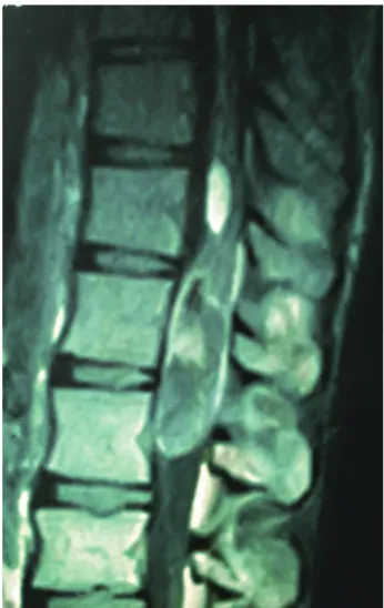
contrast and fat suppression studies was showing a well circumscribed lesion approximately 5.4×2.1×1.8 cm in the cauda equina inseparable from conus medullaris with fat droplets within the central spinal canal extending up to the medulla. The lesion was iso to hypo intense on T1 and hyperintense on T2 weighted images. There was no significant post contrast enhancement. T1 hyper intense signal is seen scattered throughout the central spinal ca- nal extending upto the medulla. MRI scan on T1 weight- ed (sagittal view) images showed a mass lesion contain- ing fat peripherally, at the level of conus medullaris and hyperintense fat content in the central canal extending in to the dorsal and cervical spinal cord up to the medulla secondary to rupture of the mass (Fig. 1).
2. Provisional preoperative diagnosis was ruptured in- traspinal dermoid
Patient was operated with D11-L2 laminectomy and near total excision of the lesion. The spinal cord was expanded and myelotomy was done at the conus medullaris. Intra- operatively, the lesion was intramedullary, white cheesy with hair inside. The capsule was densely adherent to the spinal cord, therefore near complete excision was achieved.
3. Lesion was reported as dermoid cyst on frozen sections Histopathological examination showed a fibro collag- enous cyst wall lined by stratified squamous epithelium.
Fig. 1. Magnetic resonance imaging. T1 weighted (sagittal view) images showing hyperintense fat content in the central canal extending up to the medulla secondary to rupture of the mass.
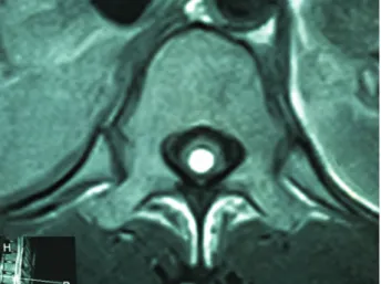
Fig. 2. Magnetic resonance imaging scan T1 weighted (axial view) images at D-12 vertebral level showing hyper intense fat content in the central canal.
in view of the presence of hair on gross examination the diagnosis of dermoid cyst was given (Fig. 5).
Patient’s low back pain had improved immediately fol- lowing the surgery. There was no neurological deficit in the lower limbs after the surgery. However patient had persistent urinary incontinence and on clean intermittent self catheterisation at the time of discharge.
At 6 month follow-up, patient was relieved of his low
back pain with improvement in the bladder functions and weaned off the urinary catheter.
Discussion
Dermoid tumors constitute 1.1% of intraspinal tumors [1]. They arise from the inclusion of ectopic embryonic rests of ectoderm within the spinal canal at the time of neural tube closure during embryonic development [2].
They occur predominantly in the lumbosacral region (60%), involving the cauda equina and conus medullaris [3], and are quite rare in the upper thoracic (10%) and cervical regions (5%) [3,4]. As in our case patient had dermoid cyst in the conus medullaris.
They are benign slow growing lesions which become clinically apparent during the second or third decade of
Fig. 3. Magnetic resonance imaging scan T1-weighted fat suppressed (sagittal view) images at conus medullaris extending in to the dorsal spinal cord demonstrates signal suppression of the fat content and no significant enhancement.
Fig. 4. Magnetic resonance imaging scan T1-weighted post-contrast (sagittal view) images at conus medullaris showing no significant post contrast enhancement.
life [5]. And males are more commonly affected, as in our case patient was 22-year-old male.
These may be associated with a dorsal dermal sinus in about 20% of the cases [6] along with bony malforma- tions and tethered cord [7].
The clinical history of our patient was related to the lo- calization of the lesion.
Dermoid tumors can reach rather large sizes without producing any symptoms or findings.
Although their nature is benign and development is slow, dermoid tumors have a high morbidity and mortal- ity risk, especially when rupture occurs. A dermoid cyst can rupture during surgery, after a trauma or spontane- ously [8-10].
Clinical symptoms of acute rupture are headache, nau- sea, vomiting, vertigo, vision problems, aseptic chemical meningitis, hemiplegia, mental changes, and coma [8,9].
Later on, the existence of fat droplets in the subarachnoi- dal and ventricular spaces may lead to arachnoiditis, ven- triculitis, and, consequently, mental disorder may occur.
MRI is the investigation of choice for detecting intra- spinal dermoid tumors and delineating their extent [10].
Computed tomography (CT) scan is very useful in iden- tifying areas of fat and calcification. The presence of fat, calcification, soft tissue and fluid gives a clue to the diag-
nosis of dermoid cyst.
MRI typically reveals heterogeneous signal intensity because of the presence of fat, calcium and fluidv [6].
High lipid content emanated from sebaceous glands causes high signal intensity on T1W spin echo images.
Bone and cartilage might be found in the tumor itself and sometimes calcification can be seen in their walls.
Dermoid tumors can be made up of 2 different pieces, one with a higher lipid content and the other with a solid or more liquid content, and this condition may lead to a liquid-liquid level [3].
In our case, on fat-suppressed sequences, fat content of the tumor was clearly demonstrated. The relatively high signal intensity from fat, with the bright signal on T1W images and fat suppression on fat suppression studies makes identification of the lipid component easy [7], as in our case.
Diagnostic accuracy of MR imaging will increase with the use of IV contrast medium, especially in determining meningeal inflammatory reaction related to the spreading out of tumoral contents
The differential diagnosis of spinal dermoid cysts in- cludes lipomas, teratomas, and spinal cord tumors (astro- cytoma, ependymoma).
The reported number of dermoid tumors is increasing due to the frequent use of MR imaging. Free fatty materi- al within the ventricular system and intracranial cisterns were reported in all cases, but fat particles in the dilated central spinal canal were reported in only few studies [8].
With the frequent use of MR imaging it was found that spontaneous dermoid rupture, which was previously thought of as a rather serious or fatal condition, is com- mon and usually only slightly symptomatic or asymptom- atic.
Histopathological findings are very classic of dermoid cyst with fibro collagenous cyst wall lined by stratified squamous epithelium with the presence of hair on gross examination as in our case.
Patients presenting with features of spinal cord com- pression or radiculopathy or neurological deficit require surgical intervention for tissue diagnosis and decompres- sion. In our case, we have done D11-L2 laminectomy and near total excision of the lesion with neurological improvement.
However, because of the paucity of the number of cases of the ruptured dermoid cyst in the central canal, the management of ruptured fat droplets into the central ca- Fig. 5. Histopathological examination showing a fibro collagenous cyst
wall lined by stratified squamous epithelium with hair follicles and sebaceous glands in the dermis suggestive of dermoid cyst (×10).
nal is still not definite.
The management consists of excision of the dermoid cyst as completely as possible. However if the wall of the lesion is densely adherent to cord, near total excision in such cases is acceptable [1,4].
Long term follow-up is also not available because of few numbers of cases.
Ruptured dermoid cyst in the central canal is a very rare entity, however it should be kept in the differential diagnosis if the patient becomes acutely symptomatic with headache, nausea, vomiting, either following trauma or spontaneously. MR imaging is the most important radiologic modality to diagnose, to determine the distri- bution of ruptured tumor content into the subarachnoid space, or into the central spinal canal, to determine com- plications such as hydrocephaly or meningitis after rup- ture, and for follow-up after surgery.
Surgical excision of the lesion for the diagnosis with close observation and follow up studies to detect compli- cations should result in significant neurological improve- ment
Conflict of interest
No potential conflict of interest relevant to this article was reported.
Acknowledgments
We would like to thank our Dean, Sir J.J. group of hospi- tals for allowing us to publish the hospital record.
References
1. Lunardi P, Missori P, Gagliardi FM, Fortuna A.
Long-term results of the surgical treatment of spi- nal dermoid and epidermoid tumors. Neurosurgery 1989;25:860-4.
2. Netsky MG. Epidermoid tumors. Review of the lit- erature. Surg Neurol 1988;29:477-83.
3. Graham DV, Tampieri D, Villemure JG. Intramedul- lary dermoid tumor diagnosed with the assistance of magnetic resonance imaging. Neurosurgery 1988;23:765-7.
4. Kumar S, Gulati DR, Mann KS. Intraspinal dermoids.
Neurochirurgia (Stuttg) 1977;20:105-8.
5. Egelhoff JC. Pediatric head and neck imaging. In:
Haaga JR, editor. CT and MR imaging of the whole body. 4th ed. London: Mosby; 2003. p.696.
6. McAdams HP. Mediastinum. In: Haaga JR, editor. CT and MR imaging of the whole body. 4th ed. London:
Mosby; 2003. p.959-60.
7. Cha JG, Paik SH, Park JS, Park SJ, Kim DH, Lee HK.
Ruptured spinal dermoid cyst with disseminated in- tracranial fat droplets. Br J Radiol 2006;79:167-9.
8. Scearce TA, Shaw CM, Bronstein AD, Swanson PD.
Intraventricular fat from a ruptured sacral dermoid cyst: clinical, radiographic, and pathological correla- tion. Case report. J Neurosurg 1993;78:666-8.
9. Barsi P, Kenez J, Varallyay G, Gergely L. Unusual ori- gin of free subarachnoid fat drops: a ruptured spinal dermoid tumour. Neuroradiology 1992;34:343-4.
10. Wilms G, Casselman J, Demaerel P, Plets C, De Haene I, Baert AL. CT and MRI of ruptured intracra- nial dermoids. Neuroradiology 1991;33:149-51.