J. of Korean Bone & Joint Tumor Soc.
Volume 15, Number 2, December, 2009
※통신저자: 박박 용용 구구
서울특별시 동대문구 회기동 1 경희대학교 의과대학 병리학교실
Tel: 02) 958-8742, Fax: 02) 957-1489, E-mail: [email protected]
양성 및 악성 연골 종양에서의 STAT3 활성화
한림대학교 의과대학 병리학교실, 경희대학교 의과대학 병리학교실*
박혜림∙박용구*
목적: STAT3는 주요 세포 진행과정을 조절하는 암유전자로, 활성화되면 여러 악성종양의 생물학적, 임상적 특징과 연관된다고 알려져 있고, 한편 배아줄기세포와 연관된 유전자이기도 하다. 본 연구에서는 연골종양의 발생에 STAT3 활성화가 관여하는지 살펴보았다.
대상 및 방법: 총 33예의 각종 양성 및 악성 연골종양에서 STAT3 활성화를 살펴보기 위해 활성화된 pSTAT3tyr705에 특이한 단클론성 항체를 이용한 면역조직화학법을 시행하였다.
결과: 통상적인 연골육종 17예 중, 조직학적 등급 3의 연골육종은 3예(50%)에서 pSTAT3 에 양성이었고, 등급 1 및 2 연골육종은 모두 음성이었다. 즉 pSTAT3 양성도는 조직학적 등 급과 통계학적으로 유의한 상관관계(p=0.0432)를 나타내었다. 또한 투명세포 연골육종 2예 (50%)도 pSTAT3에 양성이었다. 내연골종, 연골모세포종, 연골점액양섬유종 등 12예의 양 성 연골종양 중 6예(50%)에서 pSTAT3가 관찰되었다.
결론: STAT3 활성화는 통상적인 연골육종 중 고도의 조직학적 분화도가 나쁜 등급에서 주 로 발견된다. 배아줄기세포 표지자인 STAT3가 양성 및 악성 연골종양 일부에서 활성화되는 것으로 미루어보아 연골 기원 종양에서도 악성종양의 줄기세포 가설을 제안할 수 있다.
색인 단어: 연골종양, STAT3, 면역조직화학법
Introduction
Signal transducers and activators of tran- scription (STAT) are members of latent transcription factors that are activated/
phosphorylated in response to the binding of growth factors and polypeptide cytokines to receptors with tyrosine kinase activity26). Phosphorylation precipitates dimerization, which is stabilized by reciprocal phospho-
tyrosine SH2 interactions. STAT3 dimers then move to the nucleus, where they bind to specific DNA response elements in target gene promoters and enable gene transcrip- tion. Some target genes of STAT3 include those involved in apoptosis, cell cycle regula- tion, and induction of growth arrest such as Bcl-xL, cyclin D1, p21, WAF1/CIP1, and c- myc23).
Within this family, STAT3 is an oncogene
whose constitutive activation has been iden- tified in various human cancers, such as breast, lung, and others7,11,14,21)
. A previous study has demonstrated that STAT3 activa- tion directly contributes to the growth of neoplastic cells, and the abrogation of STAT3 activation leads to significant cell death16). Furthermore, STAT3 activation sta- tus has been found to correlate with a worse clinical outcome in anaplastic large cell lym- phoma, head and neck squamous cell carci- noma, and renal cell carcinoma10,11,17). On the other hand, there is also evidence that it behaves as a tumor suppressor or a marker for better outcome in some malignant tumors2,7,14). Because STAT3 plays such a pleomorphic role in signal transduction, its role as an oncogene or a tumor suppressor may be a function of the setting.
There is increasing evidence that cancer is a stem cell disease. In this model, cancers can be considered an abnormal organ in which the bulk of tumor growth is provided by a small population of cells, cancer stem cells (CSCs), that divide asymmetrically to produce more CSCs and a non-tumorigenic population of cancer cells1). This asymmetric division is thought to contribute to the het- erogeneity of solid tumors. In support of this, primitive sarcosphere colonies from osteosarcoma cell lines were identified and characterized using molecular and cytochem- ical techniques for embryonic stem cell markers. Expression of the embryonic stem cell-associated genes Nanog, Oct4 and STAT3 indicated a primitive phenotype25).
Recently, an antibody specific for the acti- vated (phospho-Tyr705) form of STAT3 has become available. This activated form may be a better probe for function than total STAT3. Although a high frequency of tumors contain constitutively activated
STAT3, its relationship to benign and malig- nant chondroid tumors has not been fully determined. Dobashi et al. studied the involvement of epidermal growth factor receptor (EGFR) and downstream molecules in bone and soft tissue tumors6). In their study, two cases of chondrosarcoma were included, and they revealed negative reac- tion with pSTAT3.
To further elucidate the role of STAT3 in benign and malignant chondroid tumors, we assessed STAT3 activation in various chon- droid bone tumors. In addition, the clinical significance of STAT3 activation in chon- drosarcomas was also discussed with litera- ture review.
Materials and Methods 1. Tumor samples
This study was conducted with the approval of the Institutional Review Board.
Tumor samples were available for 12 benign chondroid tumors (6 enchondromas, 3 chon- droblastomas, and 3 chondromyxoid fibro- mas). Seventeen cases of conventional chon- drosarcoma and four cases of clear cell chon- drosarcoma were also included. Among con- ventional chondrosarcomas, there were 6 cases of grade I tumors, 5 cases of grade II tumors, and 6 cases of grade III tumors. All the chondrosarcoma samples represented pretherapy tumors.
2. Immunohistochemistry
Monoclonal antibody reactive with pSTAT3tyr705 (D3A7, 1:100; Cell Signaling Technology, Beverly, MA, USA) was uti- lized. Immunostaining was performed on the automated Ventana Benchmark XT
immunostainer (Ventana Medical Systems, Tucson, AZ, USA) using the manufacturer’s deparaffinization, antigen retrieval, and detection reagents. Antigen retrieval was carried out using CC1-Standard. Positive and negative control sections were included.
Immunostains were interpreted by two pathologists (HRP and YKP). The percent- age of positively staining neoplastic nuclei was determined. Tumors were considered to be STAT3 active when ≥20% of neoplastic cells showed unequivocal, nuclear pSTAT3
tyr705
immunostaining, regardless of the stain- ing intensity.
3. Statistical analysis
All statistical analyses were carried out
with the DBSTAT program (version 4.1, Seoul, Korea). Mann Whitney U (Wilcoxon rank sum) test and Kruskal Wallis test were used to compare the expression of pSTAT3 between clinical variables (e.g., histologic grade). When the p value was less than .05, the statistical difference was regarded as significant.
Results
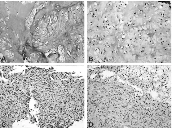
Using an antibody reactive with pSTAT3tyr705, we identified nuclear immunos- taining in 6 cases (50%) of 12 benign chon- droid tumors (Fig. 1). Four cases of enchon- droma (n=6) and one case each of chondrob- lastoma (n=3) and chondromyxoid fibroma (n=3) were pSTAT3 positive. Among conven-
Fig. 1. pSTAT3 expression in benign chondroid tumors. (A) Two cases (33.3%) of enchondroma were negative. (B) Four cases (66.7%) of enchondroma were positive for pSTAT3. Note the dark nuclear staining patterns in tumor cells. (C) One case (33.3%) of chondroblastoma was posi- tive. (D) One case (33.3%) of chondromyxoid fibroma was positive (IHC for pSTAT3, ×100).
C A
D
B
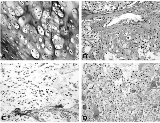
tional chondrosarcomas (n=17), three cases (50%) of grade III chondrosarcomas (n=6) were pSTAT3-positive (Fig. 2). All grade I (n=6) and II (n=5) chondrosarcomas were pSTAT3-negative. This pSTAT3 positivity according to the histologic grade was statisti- cally significant (p=0.0432). Two cases (50%) of clear cell chondrosarcomas (n=4) were pSTAT3-positive (Fig. 2). No correlations between the staining patterns and the pres- ence of malignancy or tumor type were observed (p=0.1306 and p=0.2655, respective- ly) (Table 1). Positive pSTAT3 staining in non-neoplastic tissue was observed in endothelial cells, used as internal controls.
Discussion
In this study, we assessed the STAT3 acti- vation status in chondroid tumors using a monoclonal antibody that recognizes the tyrosine-phosphorylated form of STAT3, pSTAT3tyr705. The concept that this antibody recognizes only the activated form of STAT3 is supported by several pieces of evidence.
First, it has been shown previously that pSTAT3tyr705 immunoreactivity correlates with levels of STAT3 DNA binding activity18). Second, Lai et al. reported that pSTAT3tyr705 immunoreactivity increases after interleukin stimulation15). Third, as shown in this study,
Fig. 2. pSTAT3 expression in malignant chondroid tumors. (A) All grade I conventional chondrosar- comas were negative. (B) All grade II conventional chondrosarcomas were negative. (C) Conventional chondrosarcoma, grade III, revealed characteristic diffuse nuclear immunostain- ing for pSTAT3 in the half of cases. (D) Two cases (50%) of clear cell chondrosarcoma were positive (IHC for pSTAT3, ×100).
C A
D
B
pSTAT3tyr705 immunoreactivity with tissue sec- tions is mainly confined to the nucleus.
Thus, this antibody is a useful tool to assess the activation status of STAT3.
Half of the grade III chondrosarcomas were STAT3-positive in our study. This was statistically significant compared to the neg- ativity of all grade I and II chondrosarco- mas. Half of the clear cell chondrosarcomas were also STAT3-positive. In another series of bone and soft tissue tumors, nuclear staining of pSTAT3 was observed in 25.8%
of sarcomas, that is, malignant fibrous his- tiocytoma, liposarcoma, synovial sarcoma, and chordoma6). However, two cases of chon- drosarcoma included were pSTAT3 negative.
Most STAT3 cases exhibited simultaneous activation of EGFR. The data from that series support the idea that STAT3 activa- tion is largely dependent on EGFR signaling.
Similarly, EGFR was found to play a role in STAT3 activation in skin and head and neck cancer pathogenesis3,21). However, our previ- ous data suggest that such a mechanism appears unlikely in chondroid tumors, as our data reveals that the amplification and over- expression of the HER-2/neu oncogene is absent or at least very rare in malignant cartilaginous tumors19).
Another mechanism leading to constitutive
STAT3 activation is the loss of physiological negative regulators, which include three families of proteins: suppressors of cytokine signaling (SOCS), protein inhibitors of acti- vated STAT (PIAS), and phosphotyrosine SH2-containing phosphatases (SHP). Loss of function of these negative regulators is increasingly recognized in conjunction with constitutive STAT3 activation in various types of cancers8,12,24). These pathways are potential candidates for our future investi- gations in bone and soft tissue tumors, including chondroid tumors.
Half of the benign chondroid tumors were pSTAT3tyr705-positive in our study. The posi- tivity of enchondromas seemed to be higher than chondroblastomas and chondromyxoid fibromas; however, this was not statistically significant. In one previous study, STAT3 was activated in one type of benign soft tis- sue tumor: giant cell tumor of tendon sheath6). This may imply that this molecule may also function in the differentiation or maintenance of their specific phenotypes.
STAT3 activation has been found to corre- late with a worse clinical outcome in several cancers10,11,13,18)
. For instance, Khoury et al.
reported that STAT3 activation was associat- ed with worse overall survival in patients with anaplastic large cell lymphoma11). In
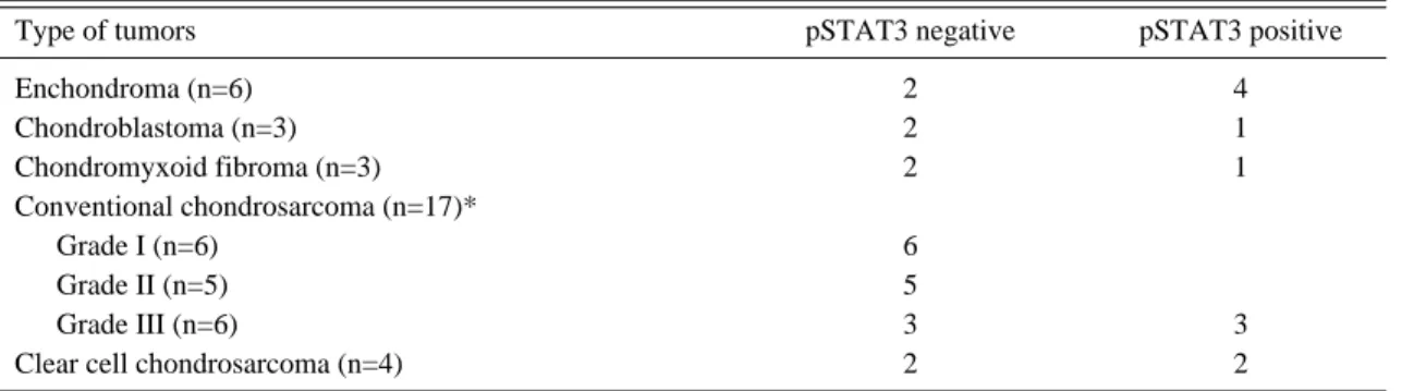
Table 1. Expression of pSTAT3 in chondroid tumors
Type of tumors pSTAT3 negative pSTAT3 positive
Enchondroma (n=6) 2 4
Chondroblastoma (n=3) 2 1
Chondromyxoid fibroma (n=3) 2 1
Conventional chondrosarcoma (n=17)*
Grade I (n=6) 6
Grade II (n=5) 5
Grade III (n=6) 3 3
Clear cell chondrosarcoma (n=4) 2 2
*p=0.0432 according to the histologic grade (Kruskal Wallis test)
this study, only grade III conventional chon- drosarcomas were STAT3-positive, compared to grade I and II tumors. Of course, the prognostic importance of grading chondrosar- comas is well known. Unfortunately, we were not able to obtain sufficient follow-up data to determine whether worse clinical outcomes were observed in these STAT3-pos- itive patients. In our previous study, EGFR and STAT3 expression were not correlated with metastasis or survival in osteosarcoma5). On the other hand, expression of pSTAT3 has also been reported to be a marker for improved overall survival in breast cancer and Ewing sarcoma7,14). The finding that activated STAT3 is associated with better outcomes in breast cancer is subject to numerous interpretations. Because STAT3 is known to be persistently activated in src- transformed lines22), it is not surprising to find it activated in a large fraction of the tumors. The fact that it is associated with better outcome may simply mean that tumors that activate these pathways are less aggressive than tumors that progress even in the absence of STAT3 activation. Alternatively, it may be that STAT3 plays a role as a tumor suppressor protein. Evidence that STAT3 plays a role in cellular differentiation and apoptosis may be consistent with better outcomes in breast cancer4).
Meanwhile, our results regarding STAT3 activation in chondrosarcomas may suggest the extension of the cancer stem cell hypothesis to include other tumors in the cartilaginous lineage. Bone sarcomas are a clinically and molecularly heterogeneous group of malignancies characterized by vary- ing degrees of mesenchymal differentiation.
Tumor stem cells have been recently impli- cated in the pathogenesis of other heteroge- neous, highly malignant tumors20). Gibbs et
al. demonstrated the existence of a small subpopulation of self-renewing bone sarcoma cells that are capable of forming suspended spherical, clonal colonies, also called “sarcos- pheres,” in anchorage-independent, serum- starved conditions9). These bone sarcoma cells, as well as tissue specimens, express activated STAT3 and the marker genes of pluripotent embryonic stem cells, Oct3/4 and Nanog. Despite the divergent origins of chondrosarcoma and osteosarcoma, the expression profiles and stem-like properties of the respective cell types were indistin- guishable. This suggests that the cellular machinery responsible for the maintenance of the cancer stem cell phenotype may like- wise be very similar in these tumors9). Assuming that the cancer stem cell theory is correct, cytotoxic chemotherapeutic agents, developed in part by assessing the gross response of the tumor mass, may not address this small proportion of tumorigenic cells. The cancer stem cell theory could explain the near-complete resistance of chon- drosarcoma to standard drug therapy.
Summary
In summary, we report STAT3-positivity in grade III conventional chondrosarcomas compared to grade I and II. However, we also detected STAT3-positivity in half of the benign chondroid tumors we examined. Our results suggest that STAT3 is activated in a subset of benign and malignant chondroid tumors and may support the extension of the cancer stem cell hypothesis to include tumors of cartilaginous lineage.
REFERENCES
01) Al-Hajj M and Clarke MF: Self-renewal and solid
tumor stem cells. Oncogene, 23: 7274-7282, 2004.
02) Bromberg J and Darnell Jr JE: The role of STATs in transcriptional control and their impact on cellular function. Oncogene, 19: 2468-2473, 2000.
03) Chan KS, Carbajal S, Kiguchi K, Clifford J, Sano S and DiGiovanni J: Epidermal growth fac- tor receptor-mediated activation of Stat3 during multistage skin carcinogenesis. Cancer Res, 64:2382-2389, 2004.
04) Chapman RS, Lourenco PC, Tonner E, et al.:
Suppression of epithelial apoptosis and delayed mammary gland involution in mice with a condi- tional knockout of Stat3. Genes Dev, 13:2604-2616, 1999.
05) Do SI, Jung WW, Kim HS and Park YK: The expression of epidermal growth factor receptor and its downstream signaling molecules in osteosarco- ma. Int J Oncol, 34: 797-803, 2009.
06) Dobashi Y, Suzuki S, Sugawara H and Ooi A:
Involvement of epidermal growth factor receptor and downstream molecules in bone and soft tissue tumors. Hum Pathol, 38: 914-925, 2007.
07) Dolled-Filhart M, Camp RL, Kowalski DP, Smith BL and Rimm DL: Tissue microarray analysis of Stat3 and phospho-Stat3 (Tyr705) in node-negative breast cancer shows nuclear localiza- tion is associated with a better prognosis. Clin Cancer Res, 9:594-600, 2003.
08) Flowers LO, Subramaniam PS and Johnson HM: A SOCS-1 peptide mimetic inhibits both con- stitutive and IL-6 induced activation of STAT3 in prostate cancer cells. Oncogene, 24: 2114-2120, 2005.
09) Gibbs CP, Kukekov VG and Reith JD: Stem-like cells in bone sarcomas: implications for tumorigen- esis. Neoplasia, 7: 967-976, 2005.
10) Horiguchi A, Oya M, Shimada T, Uchida A, Marumo K and Murai M: Activation of Stat3 in renal cell carcinoma: a study of incidence and its association with pathological features and clinical outcome. J Urol, 168: 762-765, 2002.
11) Khoury JD, Medeiros LJ, Rassidakis GZ, et al.:
Differential expression and clinical significance of tyrosine-phosphorylated STAT3 in ALK+and ALK- anaplastic large cell lymphoma. Clin Cancer Res, 9: 3692-3699, 2003.
12) Khoury JD, Rassidakis GZ, Medeiros LJ, Amin HM and Lai R: Methylation of SHP1 gene and
loss of SHP1 protein expression are frequent in sys- temic anaplastic large cell lymphoma. Blood, 104:1580-1581, 2004.
13) Kusaba T, Takayama T, Yamazumi K, et al.:
Expression of P-STAT3 in human colorectal adeno- carcinoma and adenoma; correlation with clinico- pathological factors. J Clin Pathol, 58:833-838, 2005.
14) Lai R, Navid F, Rodriguez-Galindo C, et al.:
STAT3 is activated in a subset of the Ewing sarco- ma family of tumours. J Pathol, 208: 624-632, 2006.
15) Lai R, Rassidakis GZ, Medeiros LJ, Leventaki V, Keating M and McDonnell TJ: Expression of STAT3 and its phosphorylated forms in mantle cell lymphoma cell lines and tumors. J Pathol 199: 84- 89, 2003.
16) Lee SO, Lou W, Qureshi KM, Mechraein-Ghomi F, Trump DL and Gao AC: RNA interference tar- geting Stat3 inhibits growth and induces apoptosis of human prostate cancer cells. Prostate, 60: 303- 309, 2004.
17) Masuda M, Suzui M, Yasumatu R, et al.:
Constitutive activation of Stat3 correlates with cyclin D1 overexpression and may provide a novel prognostic marker in head and neck squamous cell carcinoma. Cancer Res, 62: 3351-3355, 2002.
18) Mora LB, Buettner R, Seigne J, et al.:
Constitutive activation of Stat3 in human prostate tumors and cell lines: direct inhibition of Stat3 sig- naling induces apoptosis of prostate cancer cells.
Cancer Res, 62: 6659-6666, 2002.
19) Park HR, Kim YW, Jung WW, Kim HS, Unni KK and Park YK: Evaluation of HER-2/neu status by real-time quantitative PCR in malignant carti- laginous tumors. Int J Oncol, 24: 575-580, 2004.
20) Reya T, Morrison SJ, Clarke MF and Weissman IL: Stem cells, cancer, and cancer stem cells.
Nature, 414: 105-111, 2001.
21) Song L, Turkson J, Karras JG, Jove R and Haura EB: Activation of Stat3 by receptor tyrosine kinases and cytokines regulates survival in human non-small cell carcinoma cells. Oncogene, 22:
4150-4165, 2003.
22) Turkson J, Bowman T, Garcia R, Caldenhoven E, De Groot RP and Jove R: Stat3 activation by Src induces specific gene regulation and is required for cell transformation. Mol Cell Biol, 18: 2545- 2552, 1998.
23) Turkson J and Jove R: STAT proteins: novel mol- ecular targets for cancer drug discovery. Oncogene, 19: 6613-6626, 2000.
24) Wang LH, Yang XY, Zhang X and Farrar WL:
Nuclear receptors as negative modulators of STAT3 in multiple myeloma. Cell Cycle, 4: 242-245, 2005.
25) Wilson H, Huelsmeyer M, Chun R, Young KM,
Friedrichs K and Argyle DJ: Isolation and charac- terization of cancer stem cells from canine osteosar- coma. The Veterinary J, 175: 69-75, 2008.
26) Yu H and Jove R: The STATs of cancer-new mol- ecular targets come of age. Nature Rev Cancer, 4:
97-105, 2004.
STAT3 is Activated in a Subset of Benign and Malignant Chondroid Tumors
Hye-Rim Park, M.D., Yong-Koo Park, M.D.*
Department of Pathology, College of Medicine, Hallym University, Anyang, Korea, Department of Pathology and Medical Science and Engineering Research Center for Bioreaction to
Reactive Oxygen Species, College of Medicine, Kyung Hee University, Seoul, Korea*
Purpose: STAT3 is an oncogene that regulates critical cellular processes, and its constitutive activation has been demonstrated to correlate with biological and clinical features in many types of human malignancy.
Materials and Methods: In this study, STAT3 activation was assessed in variable benign and malignant chondroid tumors in bone by immunohistochemistry using a monoclonal antibody specific for tyrosine705-phosphorylated STAT3 (pSTAT3tyr705).
Results: Among conventional chondrosarcomas (n=17), three cases(50%) of grade III chon- drosarcomas were pSTAT3-positive. All grade I and II chondrosarcomas were pSTAT3-nega- tive. This pSTAT3 positivity according to the histologic grade was statistically significant (p=0.0432). Two cases(50%) of clear cell chondrosarcomas were pSTAT3-positive. Six cases (50%) among 12 benign chondroid tumors(6 enchondromas, 3 chondroblastomas, and 3 chon- dromyxoid fibromas) were also pSTAT3tyr705-positive.
Conclusion: In conclusion, STAT3 activation is associated with higher tumor grade in con- ventional chondrosarcomas. Our results suggest that STAT3 is activated in a subset of benign and malignant chondroid tumors, and may support the extension of the cancer stem cell hypoth- esis to include tumors of cartilaginous lineage.
Key Words: Chondroid tumors, STAT3, Immunohistochemistry
Address reprint requests to Yong-Koo Park, M.D.
Department of Pathology, Kyung Hee University Hospital,
#1 Hoeki-dong, Dongdaemun-ku, Seoul 130-702, Korea
TEL: 82-2-958-8742, FAX: 82-2-957-0489, E-mail: [email protected]
Abstract