410 Copyright © 2019 Korean Neurological Association
JCN
Open AccessIsolated Abducens Nerve Palsy due to a Dural Arteriovenous Fistula with Drainage into the Inferior Petrosal Sinus
Dear Editor,
Abducens nerve palsy is the most common type of ocular nerve palsy to occur in isola- tion, and it has several associated etiologies.1 It is important to identify the causative lesion to determine the etiology of abducens nerve palsy and optimize the treatment. In this con- text, magnetic resonance imaging (MRI) plays an important role in the management of ab- ducens nerve palsy.2 In rare cases, pathology involving Dorello’s canal, which is the osteofi- brous canal in the skull base that includes the abducens nerve and the inferior petrosal sinus (IPS), can cause abducens nerve palsy; however, this is often overlooked.3 Here, we present a rare case of a left transverse-sigmoid sinus dural arteriovenous fistula (dAVF) with drainage into the left IPS that presented with isolated left abducens nerve palsy presumably caused by venous hypertension in the left IPS within Dorello’s canal.
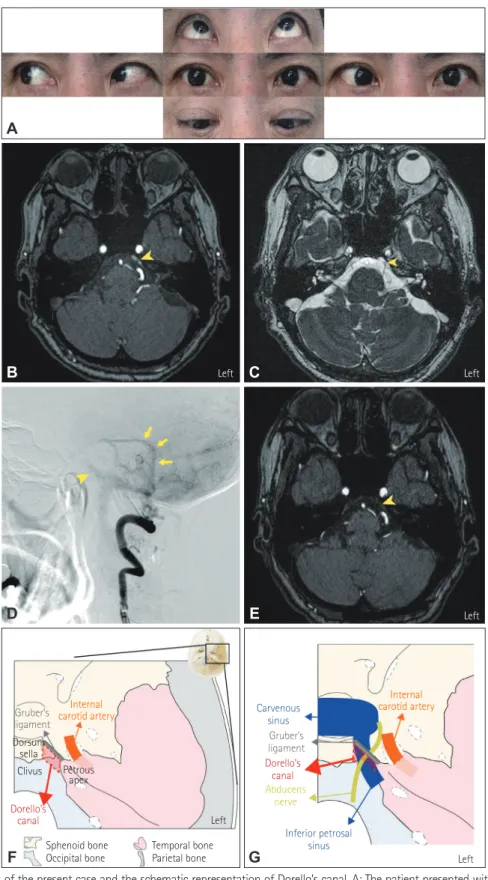
A previously healthy 64-year-old woman was referred to our hospital due to acute-onset painless diplopia without any associated symptoms, which exacerbated over the course of 1 week. A neurological examination revealed impaired abduction of the left eye with no other abnormal findings (Fig. 1A), which suggested isolated left abducens nerve palsy. Contrast- enhanced MRI did not reveal any causative tumor lesions or inflammatory lesions. However, time-of-flight magnetic resonance angiography (TOF-MRA) revealed venous enhancement of the left IPS at the left Dorello’s canal, indicating the presence of rapid retrograde venous drainage into the left IPS (Fig. 1B and C). Subsequent cerebral angiography confirmed a left transverse-sigmoid sinus dAVF allowing retrograde venous drainage into the left IPS (Fig. 1D).
Embolization of the dAVF alone resolved the enhancement of the left IPS at the left Dorel- lo’s canal in TOF-MRA (Fig. 1E) and, subsequently, the left abducens palsy.
Dorello’s canal, which was first described by Wenzel Leopold Gruber in 1859 and later by Primo Dorello in 1905, is the osteofibrous canal in the skull base located under Gruber’s liga- ment.4 Dorello’s canal is bordered by Gruber’s ligament superiorly, the dorsum sellae of the sphenoid bone medially, the clivus of the occipital bone inferiorly, and the petrous apex of the temporal bone laterally5 (Fig. 1F). Since the abducens nerve runs alongside the IPS with- in the canal4 (Fig. 1G), we should pay attention to Dorello’s canal in cases of abducens nerve palsy. A dAVF is an abnormal arteriovenous shunt between the dural arteries and dural ve- nous sinuses or meningeal veins, and presents with various symptoms caused by venous hypertension of the vein involved in the drainage.6 Based on the clinical course and radio- graphical findings in the present case, we believe that venous hypertension due to retrograde venous drainage into the IPS caused by the dAVF damaged the abducens nerve in Dorello’s canal. Isolated abducens nerve palsy due to compression of the abducens nerve by the IPS in Dorello’s canal is extremely rare, but has been reported in patients with IPS thrombosis6 and in those undergoing IPS sampling as part of a workup for Cushing’s disease.7 Neurologists should pay careful attention to Dorello’s canal when evaluating patients with isolated abdu- cens nerve palsy, and investigating this canal using MRI with detailed anatomical knowledge Kazuto Tsukitaa,b
Haruhi Sakamaki-Tsukitaa,b Toshihiko Suenagaa
a Department of Neurology, Tenri Hospital, Tenri, Nara, Japan
b Department of Neurology, Graduate School of Medicine, Kyoto University, Kyoto, Japan
pISSN 1738-6586 / eISSN 2005-5013 / J Clin Neurol 2019;15(3):410-412 / https://doi.org/10.3988/jcn.2019.15.3.410
Received February 19, 2019 Revised March 10, 2019 Accepted March 15, 2019 Correspondence Kazuto Tsukita, MD Department of Neurology, Tenri Hospital, 200 Mishima-cho, Tenri, Nara 632-8552, Japan Tel +81-743-63-5611 Fax +81-743-63-1530
E-mail [email protected]
cc This is an Open Access article distributed under the terms of the Creative Commons Attribution Non-Com- mercial License (https://creativecommons.org/licenses/by-nc/4.0) which permits unrestricted non-commercial use, distribution, and reproduction in any medium, provided the original work is properly cited.
LETTER TO THE EDITOR
www.thejcn.com 411
Tsukita K et al.
JCN
Fig. 1. Imaging findings of the present case and the schematic representation of Dorello’s canal. A: The patient presented with impaired abduction of the left eye. B: TOF-MRA image shows venous enhancement of the left IPS in the left Dorello’s canal (arrowhead). C: True fast imaging with steady-state precession shows the abducens nerve (arrowhead). D: Angiogram from a left lateral view of the left vertebral artery reveals a dural arteriovenous fistula (arrows) in the left transverse-sigmoid sinus with retrograde drainage into the left IPS (arrowhead). E: Follow-up TOF-MRA image shows resolution of the venous enhancement of the left IPS (arrowhead). F: Schematic representation of the axial view of the skull base shows the anatomical location of Dorello’s canal. G: The abducens nerve runs alongside the IPS in Dorello’s canal before entering the cavernous sinus. IPS: inferior petrosal sinus, TOF-MRA: time-of- flight magnetic resonance angiography.
Internal
carotid artery Carvenous
sinus
Inferior petrosal sinus Dorello’s
canal Abducens
nerve Dorello’s
canal Gruber’s ligament
Gruber’s ligament Dorsum
sella
Clivus Petrous apex
Sphenoid bone
Occipital bone Temporal bone Parietal bone
D
F
E
G
Internal carotid artery
Left
Left Left
Left
A
B C
Left
412 J Clin Neurol 2019;15(3):410-412
Isolated Abducens Nerve Palsy due to a dAVF
JCN
of the canal is particularly important.
Conflicts of Interest
The authors have no potential conflicts of interest to disclose.
Acknowledgements
We thank Dr. Takahiro Kamada for providing us with the inspiration to write the manuscript. Dr. Kamada died in January 2019, and we wish to dedicate this article to his memory. We also thank Dr. Tomoo Tokime and Dr. Yoshinori Akiyama for helping us to treat the patient.
REFERENCES
1. Patel SV, Mutyala S, Leske DA, Hodge DO, Holmes JM. Incidence, associations, and evaluation of sixth nerve palsy using a population- based method. Ophthalmology 2004;111:369-375.
2. Tamhankar MA, Biousse V, Ying GS, Prasad S, Subramanian PS, Lee MS, et al. Isolated third, fourth, and sixth cranial nerve palsies from
presumed microvascular versus other causes: a prospective study. Oph- thalmology 2013;120:2264-2269.
3. Mittal SO, Siddiqui J, Katirji B. Abducens nerve palsy due to inferior petrosal sinus thrombosis. J Clin Neurosci 2017;40:69-71.
4. Kshettry VR, Lee JH, Ammirati M. The Dorello canal: historical devel- opment, controversies in microsurgical anatomy, and clinical implica- tions. Neurosurg Focus 2013;34:E4.
5. Joo W, Yoshioka F, Funaki T, Rhoton AL Jr. Microsurgical anatomy of the abducens nerve. Clin Anat 2012;25:1030-1042.
6. Gandhi D, Chen J, Pearl M, Huang J, Gemmete JJ, Kathuria S. Intra- cranial dural arteriovenous fistulas: classification, imaging findings, and treatment. AJNR Am J Neuroradiol 2012;33:1007-1013.
7. Lefournier V, Gatta B, Martinie M, Vasdev A, Tabarin A, Bessou P, et al. One transient neurological complication (sixth nerve palsy) in 166 consecutive inferior petrosal sinus samplings for the etiological diag- nosis of Cushing’s syndrome. J Clin Endocrinol Metab 1999;84:3401- 3402.