CASE REPORT
pISSN 1738-2262/eISSN 2093-6729Korean J Spine 9(2):108-110, 2012 www.e-kjs.org
108
Copyright
© 2012 The Korean Spinal Neurosurgery SocietyHorner Syndrome associated with a Herniated Cervical Disc: A Case Report
Hyunjin Ma, Insoo Kim
Department of Neurosurgery, Dongsan Medical Center, College of Medicine, Keimyung University, Daegu, Korea
Horner syndrome (HS) occurs when there is interruption of the oculosympathetic pathway. The causes of HS are various, but HS originated from herniated cervical disc is very few. HS attributable to the lesion of the first-order neuron of cervical spinal cord is extremely rare. A 41-year old male was admitted for sudden onset of left ptosis and right side numbness.Neurological examination revealed ptosis, miosis and facial anhidrosis on the left side. MRI and CT scans demonstrated large left paramedian disc herniation with cord compression at the C4-5 level. The herniated disc was removed through anterior approach and his symptoms were improved after the operation.
Key Words: Horner SyndromeㆍSpinal CordㆍIntervertebral Disc
●Received: May 11, 2012 ● Revised: June 2, 2012
●Accepted: June 27, 2012
Corresponding Author: Insoo Kim, MD, PhD
Department of Neurosurgery, Dongsan Medical Center, Keimyung University, Dalseong-Ro, Jung-Gu, Daegu 700-712, Korea
Tel: +82-53-250-7730, Fax: +82-53-250-7356
E-mail: [email protected] Fig. 1. Patient examination reveals left ptosis, miosis, and anhid- rosis by starch test.
INTRODUCTION
Horner syndrome (HS) was characterized first in humans by Johann Friedrich Horner in 1869
11). HS results from inter- ruption of the oculosympathetic pathway at anywhere along its course between the hypothalamus and the orbit
1). HS is characterized by the classic triad of ipsilateral eyelid ptosis, miosis and facial anhidrosis. There are many causes of HS, but herniated cervical disc (HCD) is a very rare cause among them. HCD is a common cause of spinal cord compression, but HS associated with HCD has been described just in very few literatures. There were only two cases of herniated thora- cic disc at T1-2 as the causes of HS
2,6). In addition, the occur- rence of HCD as the etiology of HS has not been reported.
Thus, this study describes extremely rare case of HS associated with HCD.
CASE REPORT
A 41-year old male patient awoke to find sudden onset of left ptosis, right side numbness without identifiable history
of trauma or physical stress. He first visited local medical cen-
ter and had brain magnetic resonance imaging (MRI) taken,
however, no specific findings were found. When he was refer-
red to our neurosurgery department via emergent department,
neurological examination presented left side ptosis, miosis and
anhidrosis of left half of face which are typical symptoms of
HS, and there was numbness below right side of the T8 derma-
tome. We consulted department of ophthalmology and con-
firmed left ptosis and miosis (Fig. 1). In darkness, pupils meas-
ured 5 mm on the right eye and 3 mm on the left eye, and
the left pupil showed dilatation lag. There was 2 mm of left
ptosis. Sweat test showed only right side starch powder on the
face changed to dark brown color, which means left side anhi-
drosis (Fig. 1). The cervical spine MRI demonstrated an up-
ward migrated and large left paramedian disc herniation with
Horner Syndrome
Korean J Spine 9(2) June 2012 109
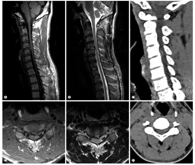
Fig. 2. T1-weighted sagittal (A) and axial (D), and T2-weighted sagittal (B) and axial (E) magnetic resonance image showing a large left paramedian C4-C5 disc herniation and severe compression of the spinal cord. Computed tomography (C, F) reveal some high density in the disc material.
severe unilateral spinal cord compression at the C4-C5 level which is high signal intensity with internal low signal intensity on T2WI and intermediate signal intensity on T1WI. And extru- ded disc revealed some high density in cervical spine computed tomography (Fig. 2). As large disc herniation with cartilagi- nous endplate fragments was doubted, we conducted a stand- ard microsurgical anterior approach to the C4-C5 interspace on 2 days after admission. Posterior longitudinal ligament had been ruptured and spinal cord was severely compressed espe- cially at left side. The extruded disc material was migrated to upward including part of the cartilaginous endplate. After complete decompression of neural structures, anterior cervical fusion was performed with a cage filled with an allograft bone chip. Postoperatively, right side numbness was improved but left miosis and ptosis remained until discharge. However, after two months from the operation, miosis, ptosis and anhidrosis
were completely recovered, too.
DISCUSSION
Horner syndrome, or oculosympathetic paralysis, was chara- cterized first in humans by Johann Friedrich Horner in 1869
11). It is resulted from interruption of the oculosympathetic path- way between the hypothalamus and the orbit. The clinical fin- dings of HS include ipsilateral miosis, ptosis, and facial anhid- rosis
1).
The oculosympathetic pathway begins with a first-order neu-
ron at the posterior lateral aspect of the hypothalamus and
through the brain stem extends down the spinal cord from
C8 to T2. Second-order (preganglionic) neuron, which is lo-
cated in the intermediolateral gray substance of the spinal cord
HJ Ma, et al.
110
www.e-kjs.org
at the level C8-T2 (Ciliospinal Center of Budge-Waller), then exit the spinal cord via the ventral roots and enter the para- vertebral sympathetic chain. The preganglionic pathway pass- es over the apex of the lung and ascends in the cervical sym- pathetic chain to the superior cervical ganglion. The third-or- der (postganglionic) neuron, which is superior cervical gan- glion located at the level of C2-C3, posterior to the carotid sheath and anterior to the longus colli muscle, follow the car- otid plexus into the skull, join with the ophthalmic nerve, and enter the orbit. HS may occur as a result of injury anywhere along this pathway
1,5,9).
Based on localization of the oculosympathetic pathway in- terruption, a HS is often classified as central (first-order neu- ron), preganglionic (second-order neuron) or postganglionic (third-order neuron).
The causes of central HS are brain stem ischemia, brain tumors, demyelinating diseases, syringomyelia and transverse myelitis
1,4). Preganglionic HS is resulting from thoracic or neck tumor, direct spinal cord trauma, herniated disc at C8-T1, iat- rogenic disruption of the sympathetic pathway from radical neck dissection or selective nerve root block, carotid angiog- raphy, stenting or endarterectomy, spontaneous carotid dis- section, and aortic aneurysm to various malignant conditions that directly or indirectly affect the normal sympathetic in- nervations
3,5,7-10,12,13). The preganglionic type is most often cau- sed by a tumor or trauma. The causes of postganglionic HS are vascular headaches, tumor or aneurysm in cavernous sinus, nasopharyngeal tumor, and trauma with basal skull fracture.
And most frequently it is seen as a consequence of carotid artery dissection or during cluster headache. However, many postganglionic lesions are idiopathic. Anhidrosis is rarely conspi- cuous, and in the postganglionic subtype, it is virtually absent
1,9). Central HS is uncommonly encountered in isolation. In the spinal cord, the fibers of first-order neurons travel in Budge’s center immediately lateral to the dorsal gray matter and syn- apse in the spinal cord gray matter
9).
The possible cause of HS in this case is that the HCD dire- ctly compresses spinal cord producing an insult to the first- order neuron of the sympathetic pathway within the spinal cord at the level of C4-C5.
MR imaging is the most reliable investigative procedure and should be accepted as an initial diagnostic tool for HS associa- ted with HCD.
Patients with HCD commonly have neck pain, cervical radi- culopathy, myelopathy and a combination of these symptoms.
However, when patients have no cervical symptoms initially, or completely lack cervical symptoms, this unusual presenta- tion can lead to a delayed or incorrect diagnosis in many wrong directions. In particular, as the patient had no cervical symp- toms initially, the physician suspected a cerebral stroke first.
Careful history-taking and detailed neurologic examinations are indispensable steps for early diagnosis of HS associated with HCD. For the treatment of it, early diagnosis and prompt surgical decompression is necessary because the significant cord compression will bring about further neurological deficits.
CONCLUSION
Although HS is well recognized, there are no reports of the HS following HCD. If physicians noted HS associated with HCD, it should be considered significant spinal cord compre- ssion and early surgical decompression for prevention of perma- nent complication of HS is urgently needed.
REFERENCES
1. Amonoo-Kuofi HS: Horner syndrome revisited: with an up- date of the central pathway. Clin Anat 12:345-361, 1999 2. Gelch MM: Herniated thoracic disc at T1-2 level associated
with Horner's syndrome. J Neurosurg 48:128-130, 1978 3. Kaplowitz K, Lee AG: Horner syndrome following a selective
cervical nerve root block. J Neuroophthalmol 31:54-55, 2011 4. Kerrison JB, Biousse V, Newman NJ: Isolated Horner's syn- drome and syringomyelia. J Neurol Neurosurg Psychiatry 69:
131-132, 2000
5. Lee JH, Lee HK, Lee DH, Choi CG, Kim SJ, Suh DC: Neu- roimaging strategies for three types of Horner syndrome with emphasis on anatomic location. AJR Am J Roentgenol 188:
W74-81, 2007
6. Lloyd TV, Johnson JC, Paul DJ, Hunt W: Horner's syndrome secondary to herniated disc at T1-T2. AJR Am J Roentgenol 134:184-185, 1980
7. Miura J, Doita M, Miyata K, Yoshiya S, Kurosaka M, Yama- moto H: Horner's syndrome caused by a thoracic dumbbell- shaped schwannoma: sympathetic chain reconstruction after a one-stage removal of the tumor. Spine 28(2):33-36, 2003 8. Montgomery DM, Brower RS: Cervical spondylotic myelo-
pathy. Clinical syndrome and natural history. Orthop Clin North Am 23:487-493, 1992
9. Reede DL, Garcon E, Smoker WR, Kardon R: Horner's synd- rome: clinical and radiographic evaluation. Neuroimaging Clin N Am 18:369-385, 2008
10. Russell JH, Joseph SJ, Snell BJ, Jithoo R: Brown-Sequard syn- drome associated with Horner's syndrome following a pene- trating drill bit injury to the cervical spine. J Clin Neurosci 16:975-977, 2009
11. van der Wiel HL: Johann Friedrich Horner (1831-1886). J Neurol 249:636-637, 2002
12. Walton KA, Buono LM: Horner syndrome. Curr Opin Oph- thalmol 14:357-363, 2003
13. Zhao CQ, Jiang SD, Jiang LS, Dai LY: Horner Syndrome due to a solitary osteochondroma of C7: a case report and review of the literature. Spine 32(16):471-474, 2007