• 검색 결과가 없습니다.

Reperfusion Therapy in Unclear-Onset Stroke Based on MRI Evaluation (RESTORE) - A Prospective Multicenter Study

N/A
N/A
Protected

Academic year: 2021

Share "Reperfusion Therapy in Unclear-Onset Stroke Based on MRI Evaluation (RESTORE) - A Prospective Multicenter Study"

Copied!
15
0
0

로드 중.... (전체 텍스트 보기)

전체 글

(1)

3278

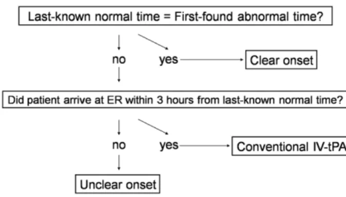
S troke occurs in the darkness as well as in daylight. Although most strokes develop suddenly, the onset time cannot be pinpointed in patients with wake-up strokes or unwitnessed day- time strokes causing aphasia or unconsciousness. An estimated 14% to 28% of ischemic strokes are wake-up strokes.

1

Including unwitnessed daytime strokes, the incidence of unclear-onset stroke may rise substantially. Unclear-onset strokes are gener- ally excluded from standard thrombolytic therapy, represent- ing an important unmet clinical need. For convenience and

conservatism, the last-known normal time is artificially defined as onset time, generally rendering this stroke population beyond the therapeutic time window for reperfusion therapy.

1

Wake-up strokes within 3 hours of symptom detection have comparable clinical and magnetic resonance imaging (MRI) characteristics as clear-onset strokes within 3 hours of symptom onset.

2,3

Thus, a substantial proportion of wake-up stroke patients may develop their strokes near the time of wake up. Recently, fluid-attenuated inversion recovery Background and Purpose—Unclear-onset strokes are generally excluded from time-based thrombolytic therapy. We

examined the safety and feasibility of magnetic resonance imaging-based reperfusion therapy in unclear-onset stroke.

Methods—This prospective, multicenter, single-arm study screened consecutive unclear-onset stroke patients within 6 hours of symptom detection. Patients with perfusion-diffusion mismatch >20% and negative or subtle fluid-attenuated inversion recovery changes were treated with intravenous tissue plasminogen activator, intra-arterial therapy, or a combination. The safety outcome was symptomatic intracranial hemorrhage within 48 hours after treatment. The primary efficacy outcome was a 3-month modified Rankin Scale score of 0 to 2. Controls were untreated unclear-onset stroke patients prospectively captured in stroke registries.

Results—Of 430 unclear-onset stroke patients, 83 (19.3%) received reperfusion therapy (mean age, 67.5 ± 10.4 years; males, 66.3%; median baseline National Institutes of Health Stroke Scale, 14). Symptomatic intracranial hemorrhage with any neurological decline developed in 5 patients (6.0%). Symptomatic intracranial hemorrhage with National Institutes of Health Stroke Scale worsening ≥4 developed in 3 patients (3.6%). Thirty-seven patients (44.6%) achieved modified Rankin Scale score of 0 to 2, and 24 (28.9%) had modified Rankin Scale score of 0 to 1. Female, baseline National Institutes of Health Stroke Scale score, no immediate or early recanalization, and more white blood cells were independent predictors of poor outcome. Compared with untreated controls, the treated group was significantly associated with good outcomes of modified Rankin Scale score of 0 to 2 after adjusting for age, sex, and baseline National Institutes of Health Stroke Scale in logistic regression analysis (odds ratio, 2.25; 95% CI, 1.14–4.49).

Conclusions—In unclear-onset stroke patients, magnetic resonance imaging-based reperfusion therapy was feasible and safe. Randomized controlled trials are warranted to confirm the benefit of reperfusion therapy for unclear-onset stroke. (Stroke. 2012;43:3278-3283.)

Key Words: acute stroke ◼ magnetic resonance imaging ◼ thrombolysis ◼ unclear-onset stroke ◼ wake-up stroke

Reperfusion Therapy in Unclear-Onset Stroke Based on MRI Evaluation (RESTORE)

A Prospective Multicenter Study

Dong-Wha Kang, MD*; Sung-Il Sohn, MD*; Keun-Sik Hong, MD; Kyung-Ho Yu, MD;

Yang-Ha Hwang, MD; Moon-Ku Han, MD; Jun Lee, MD; Jong-Moo Park, MD; A-Hyun Cho, MD;

Hye-Jin Kim, MD; Dong-Eog Kim, MD; Yong-Jin Cho, MD; Jaseong Koo, MD;

Sung-Cheol Yun, PhD; Sun U. Kwon, MD; Hee-Joon Bae, MD; Jong S. Kim, MD

Received September 03, 2012; final revision received September 12, 2012; accepted September 19, 2012.

From the Departments of Neurology (D.-W.K., H.-J.K., S.U.K., J.S.K.) and Clinical Epidemiology and Biostatistics (S.-C.Y.), Asan Medical Center, University of Ulsan College of Medicine, Seoul, South Korea; Keimung University Dongsan Medical Center, Daegu, South Korea (S.-I.S.); Inje University, Ilsan Paik Hospital, Goyang, South Korea (K.-S.H., Y.-J.C.); Hallym University Sacred Heart Hospital, Anyang, South Korea (K.-H.Y.); Kyungpook National University Hospital, Daegu, South Korea (Y.-H.H.); Seoul National University Bundang Hospital, Seongnam, South Korea (M.-K.H., H.-J.B.); Yeungnam University Hospital, Daegu, South Korea (J.L.); Eulji General Hospital, Eulji University, Seoul, South Korea (J.-M.P.); Catholic University, St Mary’s Hospital, Seoul, South Korea (A.-H.C., J.K.); Catholic University, Seoul St Mary’s Hospital, Seoul, South Korea (J.K.) and Dongguk University Ilsan Hospital, Goyang, South Korea (D.-E.K.).

*Drs Kang and Sohn contributed equally to this paper.

The online-only Data Supplement is available with this article at http://hyper.ahajournals.org/lookup/suppl/doi:10.1161/STROKEAHA.112.

675926/-/DC1.

Correspondence to Dong-Wha Kang, Department of Neurology, Asan Medical Center, University of Ulsan College of Medicine, 88 Olympic-ro, 43-gil, Songpa-gu, Seoul 138–736, South Korea. E-mail [email protected]

© 2012 American Heart Association, Inc.

Stroke is available at http://stroke.ahajournals.org DOI: 10.1161/STROKEAHA.112.675926

by guest on February 2, 2017 http://stroke.ahajournals.org/ Downloaded from by guest on February 2, 2017 http://stroke.ahajournals.org/ Downloaded from by guest on February 2, 2017 http://stroke.ahajournals.org/ Downloaded from by guest on February 2, 2017 http://stroke.ahajournals.org/ Downloaded from by guest on February 2, 2017 http://stroke.ahajournals.org/ Downloaded from by guest on February 2, 2017 http://stroke.ahajournals.org/ Downloaded from by guest on February 2, 2017 http://stroke.ahajournals.org/ Downloaded from by guest on February 2, 2017 http://stroke.ahajournals.org/ Downloaded from by guest on February 2, 2017 http://stroke.ahajournals.org/ Downloaded from by guest on February 2, 2017 http://stroke.ahajournals.org/ Downloaded from

(2)

Kang et al MRI-Based Thrombolysis in Unclear-Onset Stroke 3279

(FLAIR) changes have been proposed as surrogate markers for estimating the lesion age within the first few hours of stroke.

4

Positive diffusion-weighted imaging (DWI) and negative FLAIR changes reliably identify stroke within 3 or 4.5 hours.

4,5

Thus, multimodal MRI including DWI, perfusion- weighted imaging (PWI), and FLAIR may enable selection of target unclear-onset stroke patients who will benefit from thrombolytic therapy with acceptable safety.

Previous studies that investigated the safety and efficacy of thrombolysis in unclear-onset stroke have reported con- tradictory results (Supplemental Table 1).

6–11

We report here the results of Reperfusion Therapy in Unclear-Onset Stroke Based on MRI Evaluation (RESTORE). Our hypothesis was that MRI-based reperfusion therapy in unclear-onset stroke is feasible and beneficial with acceptable safety.

Methods Patients

This was a prospective, multicenter, single-arm study to test the fea- sibility and safety of reperfusion therapy in unclear-onset stroke. Six

university hospitals in South Korea participated and enrolled patients from September 2006 to June 2009. Four medical centers had previ- ous experience with MRI-based thrombolysis in >10 unclear-onset stroke patients; the other 2 centers had no prior experience.

We defined unclear-onset stroke if last-known normal time and first-found abnormal time were discordant. Consecutive unclear- onset stroke patients arriving at the emergency room within 6 hours of first-found abnormal time were screened. Patients arriving within 3 hours from last-known normal time were considered for standard intravenous tissue plasminogen activator (IV-tPA) therapy and were, therefore, excluded from the current study (Figure 1). This study was approved by the institutional review board of each center. Written in- formed consent was obtained from the legal guardians of each patient.

Magnetic Resonance Imaging

MRI examinations were performed with a 1.5-T or 3-T MRI unit (Supplemental Methods). The acute stroke MRI protocol in each cen- ter included DWI, PWI, FLAIR, gradient echo imaging, and intra- cranial and extracranial magnetic resonance angiography. PWI was acquired with a bolus of gadolinium-based MRI contrast agent, and maps of mean transit time were calculated using signal intensity-time curves analysis.

In addition to the standard eligibility criteria for IV-tPA, we used MRI criteria for thrombolysis decision making in unclear-onset stroke patients (Supplemental Methods). The MRI-specific inclu- sion criterion was a PWI-DWI mismatch >20%. MRI-specific exclu- sion criteria were (1) extensive early infarct defined as acute DWI lesions involving >1/3 of the middle cerebral artery territory or the entire anterior or posterior cerebral artery territory, or (2) no mis- match area between DWI and FLAIR lesions (Figure 2A). However, patients with subtle or negative FLAIR hyperintensity within acute DWI lesions were included (Figure 2B and 2C): subtle FLAIR hy- perintensity was defined as FLAIR changes that could not have been identified without reference to acute DWI lesions. MRI criteria were assessed by visual inspection. All MR images were centrally stored and posthoc analyzed by an independent investigator (A.-H.C) who was not affiliated with the centers enrolling patients.

Thrombolytic Treatment

For reperfusion therapy, IV-tPA, intra-arterial therapy, and a combina- tion were allowed. Based on the time interval of first-found abnormal Figure 1. Algorithm to determine unclear-onset stroke eligibility

for thrombolysis. ER indicates emergency room, IV-tPA, intrave- nous tissue plasminogen activator.

Figure 2. Representative figures showing diffusion-weighted imaging (DWI)-fluid-attenuated inversion recov- ery (FLAIR) mismatch and perfusion- weighted imaging (PWI)-DWI mismatch.

A, No DWI-FLAIR mismatch; B, subtle FLAIR changes (arrow) within an acute DWI lesion; C, no FLAIR changes. MRA, magnetic resonance angiography.

by guest on February 2, 2017 http://stroke.ahajournals.org/ Downloaded from

(3)

3280 Stroke December 2012

time to arrival and arterial occlusion status, we selected one of the following reperfusion therapies: (1) IV-tPA alone (0.9 mg/kg, 10%

as a bolus and the remainder over 60 minutes) for patients arriving within 3 hours of first-found abnormal time and having neither an arterial occlusion nor a catheter-accessible occlusion;

12

(2) combina- tion of IV-tPA (0.6 mg/kg, 10% as a bolus and the remainder over 30 minutes) plus intra-arterial therapy for patients arriving within 3 hours of first-found abnormal time and having a catheter-accessible large arterial occlusion (ie, the M1 segment of the middle cerebral, internal carotid, or vertebrobasilar artery);

13

or (3) intra-arterial ther- apy alone for patients arriving 3 to 6 hours of first-found abnormal time and having a catheter-accessible large arterial occlusion. For pa- tients arriving 3 to 6 hours of first-found abnormal time and without a catheter-accessible occlusion, no reperfusion therapy was provided.

For intra-arterial therapy, intra-arterial urokinase (10 000–20 000 IU/

min; maximum permissible dose, 1 000 000 IU; Green Cross Pharm., Seoul, Republic of South Korea), mechanical clot disruption, angio- plasty/stenting, or the combination of these was allowed at the dis- cretion of responsible neurointerventionists. Antithrombotics were prohibited during and 24 hours after the procedure.

Immediate or early recanalization was assessed by catheter or mag- netic resonance angiography/computed tomography (CT) angiogra- phy at the end of or shortly after thrombolysis and was defined as flow grade improvement from Thrombolysis In Myocardial Infarction grade

14

0 or 1 to grade 2 or 3. Recanalization was evaluated by an independent investigator (H.-J.K.) blinded to the clinical outcome.

Clinical Evaluation

Using a standardized case report form, we prospectively collected data of demographics, risk factors, time-to-door, time-to-imaging, time-to-treatment, and initial blood pressure, body temperature, and laboratory results in the emergency room. Stroke severity was mea- sured by the National Institutes of Health Stroke Scale (NIHSS).

Stroke subtypes were determined using the modified Trial of Org 10172 in Acute Stroke Treatment classification.

15

Clinical outcome was evaluated with the modified Rankin Scale (mRS) at 3 months.

Outcome Measures

The safety end point was symptomatic intracranial hemorrhage (SICH) defined as hemorrhagic transformation or parenchymal he- matoma on CT within 48 hours after thrombolysis, which caused any neurological decline

12

or increases of ≥4 in NIHSS scores.

16

The primary efficacy end point was a good clinical outcome defined as an mRS score of 0 to 2 at 3 months after treatment. The secondary efficacy end point was an excellent clinical outcome of mRS score of 0 to 1 at 3 months. Presence of SICH and stroke scales were assessed by investigators blinded to the clinical information.

Untreated Cohort

For comparative analysis, we collected data of untreated unclear- onset stroke patients using prospective stroke registries in 8 university hospitals during the same study period. These controls were not considered for thrombolysis because an emergent MRI study was unavailable. We identified 355 untreated unclear-onset stroke patients who arrived within 6 hours of first-found abnormal time. Of those, we finally selected 156 concurrent controls who met the clinical eligibility criteria of the RESTORE study. All centers participating in this study were operating acute stroke units or stroke centers that followed the stroke management guidelines provided by the Korean Stroke Society.

Data Analysis

Rates of SICH and good and excellent clinical outcomes were nu- merically compared with those of previous benchmark thrombolysis trials.

12,16–21

To identify independent predictors of a poor clinical out- come within the RESTORE population, logistic regression analysis was performed. Variables with a P value ≤0.10 in univariable analy- ses were candidates for the multivariable logistic regression model.

Quantitative variables were not categorized. A backward elimination process (P≤0.05 to retain) was used to develop the final multivariable model. We also compared the proportion of a good clinical outcome between the treated and untreated unclear-onset stroke patients. Odds ratios and 95% CIs were obtained. A 2-tailed P<0.05 was considered significant. All statistical analyses were performed using SPSS for Windows (version 12.0; SPSS Inc.).

Results General Characteristics

During the study period, 430 unclear-onset stroke patients were screened. Of these, 347 patients were excluded for the following reasons: 198 had ineligible imaging criteria, 152 had ineligible clinical criteria, 29 were unable to initiate throm- bolytic therapy within the time windows, 7 were unable to give informed consent, and 2 were unable to undergo the MRI examination. Forty-one patients had ≥2 reasons for exclusion.

Eighty-three patients (19.3%) were treated and included in the final analysis. Sixty-three patients had wake-up strokes and 20 had unwitnessed daytime strokes (Supplemental Table 2).

The baseline characteristics of patients enrolled in this study are described in Table 1. The median age was 67 years, and the median NIHSS score was 14. Sixty-four patients (77.1%) had a baseline NIHSS score of ≥10, and 74 (89.2%) had a major vessel occlusion. The most commonly administered treatment was intra-arterial therapy alone in 57 (68.7%), followed by combined IV-tPA plus intra-arterial therapy in 17 (20.5%), and IV-tPA alone in 9 (10.8%).

Table 1 Baseline Characteristics of Treated and Untreated Patients With Unclear-Onset Stroke

Variables Treated (n=83) Untreated (n=156)

Age, y 67 (61–75) 70 (61–77)

Sex (male) 55 (66.3%) 88 (56.4%)

Baseline National Institutes of Health Stroke Scale

14 (10–18) 12 (6.25–17)

Risk factors

Hypertension 54 (65.1%) 94 (60.3%)

Diabetes 23 (27.7%) 57 (36.5%)

Hypercholesterolemia 24 (28.9%) 40 (25.6%)

Smoking (current) 30 (36.1%) 54 (34.6%)

Previous history of stroke 19 (22.9%) 31 (19.9%) Stroke subtypes

Large artery atherosclerosis 46 (55.4%) 77 (49.4%)

Cardioembolism 31 (37.3%) 57 (36.5%)

Small vessel occlusion 0 (0%) 0 (0%)

Other or undetermined pathogenesis

6 (7.2%) 22 (14.1%)

Times

Last-known normal-to-door, h 8.6 (5.4–11.1) 7.8 (4.9–11.7) First-found abnormal-to-door, h 1.7 (0.9–2.7) 2.0 (1.0–3.6)

Door-to-MR imaging, min 57 (39–77) …

Door-to-treatment, min 155 (100–195) … First-found abnormal-to-treatment, h 4.6 (2.8–6.0) …

Numbers in parentheses are median (interquartile range) or number (%).

MR indicates magnetic resonance.

by guest on February 2, 2017 http://stroke.ahajournals.org/ Downloaded from

(4)

Kang et al MRI-Based Thrombolysis in Unclear-Onset Stroke 3281

Safety and Efficacy Outcomes

SICH with any neurological decline occurred in 5 patients (6.0%), and SICH with an increase of ≥4 in the NIHSS score was observed in 3 patients (3.6%). Thirty-seven patients (44.6%) achieved a good clinical outcome (mRS 0–2), and 24 (28.9%) had an excellent clinical outcome (mRS 0–1).

There was no difference in safety and efficacy outcomes between wake-up and unwitnessed daytime stroke patients (Supplemental Table 2). The current findings of efficacy and safety were comparable with those of earlier benchmark thrombolysis trials (Supplemental Table 3).

Data regarding immediate or early recanalization were available in 81 (97.6%) patients: immediate or early recana- lization was achieved in 41 (50.6%) patients. Immediate or early recanalization was significantly associated with good clinical outcomes (for mRS 0–2, 25/41 [61.0%] versus 11/40 [27.5%], P=0.004; for mRS 0–1, 17/41 [41.5%] versus 6/40 [15.0%], P=0.013).

On central review of MR images, MRI criteria were not met in 9 patients: 1 patient had no PWI-DWI mismatch, 3 had DWI lesion >1/3 of middle cerebral artery territory, and 8 had no DWI-FLAIR mismatch. Three patients had ≥2 reasons for discordance. The efficacy and safety outcomes were not different between concordant and discordant cases (mRS 0–2, 44.6% versus 44.4%, P>0.99; mRS 0–1, 28.4% versus 33.3%, P=0.71; SICH with any neurological decline, 5.4% versus 11.1%, P=0.45; SICH with ≥4 increase of NIHSS, 2.7%

versus 11.1%, P=0.29).

Predictors of Poor Outcome

Factors related to poor clinical outcome (mRS 3–6) in univariable analyses were female (P<0.001), older age (P=0.046), nonsmoker (P=0.003), higher baseline NIHSS score (P<0.001), distal internal carotid occlusion (P=0.057), no immediate or early recanalization (P=0.004), more white blood cells (P=0.024), lower hematocrits (P=0.062), higher glucose (P=0.086), and 2 less-experienced centers in throm- bolysis for unclear-onset stroke before participating in this study (P=0.037). Of the 10 patients treated at the 2 less-expe- rienced centers, only 1 had a good outcome (mRS 1). In back- ward, stepwise, multiple logistic regression analysis, female, higher baseline NIHSS score, no immediate or early recanali- zation, and more white blood cells were independently associ- ated with a poor clinical outcome (Table 2).

Comparison With the Untreated Group

Baseline characteristics, including age, sex, risk factors, his- tory of previous stroke, time-to-door, white blood cell count, and glucose levels, were comparable between the treated and untreated unclear-onset stroke patients (Table 1). However, initial stroke severity tended to be more severe in the treated group than in the untreated group (P=0.054). The treated group tended to achieve a good outcome of mRS score of 0 to 2 more frequently than the untreated group in univariable analysis (44.6% versus 32.7%, P=0.091). In multiple logis- tic regression analysis adjusting for age, sex, and baseline NIHSS score, reperfusion therapy significantly increased the incidence of good clinical outcomes in unclear-onset stroke patients (odds ratio, 2.25; 95% CI, 1.14–4.49; P=0.019).

Discussion

This prospective, multicenter study tested the feasibility and safety of MRI-based reperfusion therapy in unclear-onset stroke patients. After therapy, nearly 30% of patients had no or mini- mal disability, and roughly 45% were able to perform their usual activities without help. Only 1 of 17 treated patients experienced SICH with any neurological worsening, and 1 of 28 treated had ICH associated with a substantial worsening in NIHSS score.

These bleeding complication rates are probably acceptable con- sidering the potential benefit of reperfusion therapy.

The previous studies, mostly smaller retrospective single- center studies, have reported contradictory outcome results:

6–11

an excellent outcome of mRS score of 0 to 1 ranged from 10%

to 37.5%; a favorable outcome of mRS score of 0 to 2 ranged from 28% to 50%; and the rates of symptomatic ICH ranged from 0% to 13.6%. Although CT-based therapy may be more widely applicable in real-world practice than MRI-based therapy, the outcomes of plain-CT–based thrombolysis in wake-up strokes are worse than in a placebo group

7

or a stan- dard IV-tPA group.

8

Because the dynamics of ischemic lesion evolution can be better delineated with multimodal MRI than with CT, MRI may be a more useful tool for identifying unclear-onset stroke patients who are potential candidates for thrombolysis therapy.

Regarding MRI-specific eligibility criteria, we used 2 mis- match concepts: PWI-DWI mismatch and DWI-FLAIR mis- match. PWI-DWI mismatch has been proposed as an indicator of reperfusion therapy beyond 3 or 6 hours of symptom onset.

However, a PWI-DWI mismatch exists in a considerable pro- portion of patients up to 24 hours after symptom onset.

22

This suggests that the simple presence of PWI-DWI mismatch does not provide a high probability of early stroke onset, and, therefore, may not be sufficient for selecting unclear-onset stroke patients whose actual onset times are within the time window ensuring beneficial and acceptable safety of reperfu- sion therapy.

On the contrary, DWI-FLAIR mismatch may be more use- ful for narrowing down the window of actual onset time in unclear-onset stroke patients. Patients with a DWI-FLAIR mismatch are likely to be within a time window for throm- bolysis with high specificity and positive predictive value despite relatively low sensitivity and negative predictive value.

4,5,23

These observations form the basis for the use of Table 2 Independent Predictors of Poor Clinical Outcome (mRS 3–6) in the Treated Group

Odds Ratio (95% CI)

P Value

Female 6.79 (1.59–29.0) 0.01

Baseline NIHSS 1.14 (1.01–1.29) 0.03

No immediate or early recanalization 8.80 (2.30–33.71) 0.002 White blood cells (>8.25 × 10

9

/L) 6.62 (1.73–25.30) 0.006

Backward, stepwise, multiple logistic regression analysis was performed with variables having P≤0.10 with univariable analysis. White blood cells, hematocrits, and glucose levels were dichotomized when entered into multivariable models.

mRS indicates modified Rankin Scale; NIHSS, National Institutes of Health Stroke Scale.

by guest on February 2, 2017 http://stroke.ahajournals.org/ Downloaded from

(5)

3282 Stroke December 2012

DWI-FLAIR mismatch for patient selection in thrombolytic therapy in unclear-onset stroke patients.

5

However, defining DWI-FLAIR mismatch is another challenge, and no criteria to date have captured general acceptance: is only a negative lesion on FLAIR an indication for treatment? Is there a cer- tain threshold of DWI-FLAIR mismatch for optimal patient selection? Approximately 50% of patients within 3 hours of symptom onset had FLAIR-positive lesions,

4,22

which may explain the low sensitivity of FLAIR lesions for determin- ing lesion age.

4,23

In this context, we included patients with subtle FLAIR hyperintensity within acute DWI lesions, and excluded patients if the area of FLAIR changes was well matched with DWI lesions. It is also unclear whether qualita- tive visual assessment is sufficient or whether the practicality of visual analysis should be traded for a more sophisticated quantitative assessment. Discordance between initial MRI assessment and posthoc central review was largely attributable to the disagreement on DWI-FLAIR mismatch. Interobserver agreement for acute ischemic lesion visibility on FLAIR was modest (κ=0.569) in a recent large cohort study.

5

Therefore, in the future, the criteria of DWI-FLAIR mismatch should be more clearly defined for optimal patient selection for throm- bolysis in unclear-onset stroke.

Regarding the factors related to clinical outcome, baseline stroke severity

24

and early recanalization

25

are well-known predictors. Being female has also been reported to be associ- ated with poor clinical outcome,

26

although the causes of sex difference in functional outcomes have yet to be fully eluci- dated. Systemic inflammatory response, reflected by neutro- philia, has been reported to be correlated with larger infarct volume and stroke severity,

27

which may explain the associa- tion between more white blood cells and a poor outcome in this study.

We show that unclear-onset stroke patients treated with reperfusion therapy had significantly better outcomes com- pared with the untreated control group after adjusting for age, sex, and baseline stroke severity. However, a careful interpretation is required. The untreated group was selected based on the same clinical criteria but not imaging criteria as RESTORE, because acute MRI was not performed in this group. Further studies are needed to confirm this observation.

This study has certain limitations. This is a nonrandomized study. PWI-DWI and DWI-FLAIR mismatches were visually assessed. The discordance rate of initial and posthoc central MRI assessments was not negligible. Clearer definitions and pretrial training sessions are required to increase the reproduc- ibility and reliability of MRI selection criteria in future trials.

Thrombolytic methods were diverse including intravenous and endovascular treatments, thus limiting the generaliza- tion of our results. Methods of assessing recanalization were also varied. The delay in door-to-treatment time should be improved in future clinical trials.

IV-tPA therapy within a 3-hour window was the only proven therapy from acute ischemic stroke trials in the 20th century. During the first decade of the 21st century, progress was achieved by extending the IV-tPA window to 4.5 hours.

28

We need to take a step forward, and this study provides strong evidence for the feasibility and safety of reperfusion therapy in

patients with unclear-onset stroke based on clinical and DWI/

PWI/FLAIR criteria. It is time to launch a well-designed, ran- domized, controlled trial to confirm the benefit and safety of reperfusion therapy in this important (yet so far, neglected) group of patients.

Sources of Funding

This study was supported by a grant of the Korea Health Technology R&D Project, Ministry for Health & Welfare, Republic of Korea (A111578).

Disclosures

Drs Kang has received a research grant from Ministry for Health &

Welfare, Republic of Korea. The other authors report no conflicts of interest.

References

1. Kang DW, Kwon JY, Kwon SU, Kim JS. Wake-up or unclear-onset strokes: are they waking up to the world of thrombolysis therapy? Int J Stroke. 2012;7:311–320.

2. Fink JN, Kumar S, Horkan C, Linfante I, Selim MH, Caplan LR, Schlaug G. The stroke patient who woke up: clinical and radiological features, including diffusion and perfusion MRI. Stroke. 2002;33:988–993.

3. Serena J, Dávalos A, Segura T, Mostacero E, Castillo J. Stroke on awakening: looking for a more rational management. Cerebrovasc Dis.

2003;16:128–133.

4. Thomalla G, Rossbach P, Rosenkranz M, Siemonsen S, Krützelmann A, Fiehler J, Gerloff C. Negative fluid-attenuated inversion recovery imaging identifies acute ischemic stroke at 3 hours or less. Ann Neurol.

2009;65:724–732.

5. Thomalla G, Cheng B, Ebinger M, Hao Q, Tourdias T, Wu O, Kim JS, Breuer L, Singer OC, Warach S, Christensen S, Treszl A, Forkert ND, Galinovic I, Rosenkranz M, Engelhorn T, Köhrmann M, Endres M, Kang DW, Dousset V, Sorensen AG, Liebeskind DS, Fiebach JB, Fiehler J, Gerloff C; STIR and VISTA Imaging Investigators. DWI-FLAIR mis- match for the identification of patients with acute ischaemic stroke within 4·5 h of symptom onset (PRE-FLAIR): a multicentre observa- tional study. Lancet Neurol. 2011;10:978–986.

6. Cho AH, Sohn SI, Han MK, Lee DH, Kim JS, Choi CG, Sohn CH, Kwon SU, Suh DC, Kim SJ, Bae HJ, Kang DW. Safety and efficacy of MRI-based thrombolysis in unclear-onset stroke. A preliminary report.

Cerebrovasc Dis. 2008;25:572–579.

7. Adams HP Jr, Leira EC, Torner JC, Barnathan E, Padgett L, Effron MB, Hacke W; AbESTT-II Investigators. Treating patients with ‘wake-up’

stroke: the experience of the AbESTT-II trial. Stroke. 2008;39:3277–3282.

8. Barreto AD, Martin-Schild S, Hallevi H, Morales MM, Abraham AT, Gonzales NR, Illoh K, Grotta JC, Savitz SI. Thrombolytic therapy for patients who wake-up with stroke. Stroke. 2009;40:827–832.

9. Breuer L, Schellinger PD, Huttner HB, Halwachs R, Engelhorn T, Doerfler A, Köhrmann M. Feasibility and safety of magnetic resonance imaging-based thrombolysis in patients with stroke on awakening: initial single-centre experience. Int J Stroke. 2010;5:68–73.

10. Kim JT, Park MS, Nam TS, Choi SM, Kim BC, Kim MK, Cho KH.

Thrombolysis as a factor associated with favorable outcomes in patients with unclear-onset stroke. Eur J Neurol. 2011;18:988–994.

11. Aoki J, Kimura K, Iguchi Y, Shibazaki K, Iwanaga T, Watanabe M, Kobayashi K, Sakai K, Sakamoto Y. Intravenous thrombolysis based on diffusion-weighted imaging and fluid-attenuated inversion recovery mis- match in acute stroke patients with unknown onset time. Cerebrovasc Dis. 2011;31:435–441.

12. Tissue plasminogen activator for acute ischemic stroke. The National Institute of Neurological Disorders and Stroke rt-PA stroke study group.

N Engl J Med. 1995;333:1581–1587.

13. Lewandowski CA, Frankel M, Tomsick TA, Broderick J, Frey J, Clark W, Starkman S, Grotta J, Spilker J, Khoury J, Brott T. Combined intrave- nous and intra-arterial r-TPA versus intra-arterial therapy of acute isch- emic stroke: Emergency Management of Stroke (EMS) Bridging Trial.

Stroke. 1999;30:2598–2605.

14. Sheehan FH, Braunwald E, Canner P, Dodge HT, Gore J, Van Natta P, Passamani ER, Williams DO, Zaret B. The effect of intravenous

by guest on February 2, 2017 http://stroke.ahajournals.org/ Downloaded from

(6)

Kang et al MRI-Based Thrombolysis in Unclear-Onset Stroke 3283

thrombolytic therapy on left ventricular function: a report on tissue- type plasminogen activator and streptokinase from the Thrombolysis in Myocardial Infarction (TIMI Phase I) trial. Circulation.

1987;75:817–829.

15. Ay H, Furie KL, Singhal A, Smith WS, Sorensen AG, Koroshetz WJ. An evidence-based causative classification system for acute ischemic stroke.

Ann Neurol. 2005;58:688–697.

16. Hacke W, Kaste M, Bluhmki E, Brozman M, Dávalos A, Guidetti D, Larrue V, Lees KR, Medeghri Z, Machnig T, Schneider D, von Kummer R, Wahlgren N, Toni D; ECASS Investigators. Thrombolysis with alteplase 3 to 4.5 hours after acute ischemic stroke. N Engl J Med.

2008;359:1317–1329.

17. Albers GW, Bates VE, Clark WM, Bell R, Verro P, Hamilton SA.

Intravenous tissue-type plasminogen activator for treatment of acute stroke: the Standard Treatment with Alteplase to Reverse Stroke (STARS) study. JAMA. 2000;283:1145–1150.

18. Hill MD, Buchan AM; Canadian Alteplase for Stroke Effectiveness Study (CASES) Investigators. Thrombolysis for acute ischemic stroke:

results of the Canadian Alteplase for Stroke Effectiveness Study. CMAJ.

2005;172:1307–1312.

19. Wahlgren N, Ahmed N, Dávalos A, Ford GA, Grond M, Hacke W, Hennerici MG, Kaste M, Kuelkens S, Larrue V, Lees KR, Roine RO, Soinne L, Toni D, Vanhooren G; SITS-MOST investigators.

Thrombolysis with alteplase for acute ischaemic stroke in the Safe Implementation of Thrombolysis in Stroke-Monitoring Study (SITS- MOST): an observational study. Lancet. 2007;369:275–282.

20. The IMS Study Investigators. Combined intravenous and intra-arterial recanalization for acute ischemic stroke: The interventional management of stroke study. Stroke. 2004;35:904–911.

21. Furlan A, Higashida R, Wechsler L, Gent M, Rowley H, Kase C, Pessin M, Ahuja A, Callahan F, Clark WM, Silver F, Rivera F. Intra-arterial prourokinase for acute ischemic stroke. The PROACT II study: a ran- domized controlled trial. Prolyse in Acute Cerebral Thromboembolism.

JAMA. 1999;282:2003–2011.

22. Darby DG, Barber PA, Gerraty RP, Desmond PM, Yang Q, Parsons M, Li T, Tress BM, Davis SM. Pathophysiological topography of acute ischemia by combined diffusion-weighted and perfusion MRI. Stroke.

1999;30:2043–2052.

23. Ebinger M, Galinovic I, Rozanski M, Brunecker P, Endres M, Fiebach JB. Fluid-attenuated inversion recovery evolution within 12 hours from stroke onset: a reliable tissue clock? Stroke. 2010;41:250–255.

24. Kidwell CS, Saver JL, Carneado J, Sayre J, Starkman S, Duckwiler G, Gobin YP, Jahan R, Vespa P, Villablanca JP, Liebeskind DS, Vinuela F.

Predictors of hemorrhagic transformation in patients receiving intra- arterial thrombolysis. Stroke. 2002;33:717–724.

25. Labiche LA, Al-Senani F, Wojner AW, Grotta JC, Malkoff M, Alexandrov AV. Is the benefit of early recanalization sustained at 3 months? A pro- spective cohort study. Stroke. 2003;34:695–698.

26. Reeves MJ, Bushnell CD, Howard G, Gargano JW, Duncan PW, Lynch G, Khatiwoda A, Lisabeth L. Sex differences in stroke: epidemiol- ogy, clinical presentation, medical care, and outcomes. Lancet Neurol.

2008;7:915–926.

27. Buck BH, Liebeskind DS, Saver JL, Bang OY, Yun SW, Starkman S, Ali LK, Kim D, Villablanca JP, Salamon N, Razinia T, Ovbiagele B. Early neutrophilia is associated with volume of ischemic tissue in acute stroke.

Stroke. 2008;39:355–360.

28. Saver JL. Optimal end points for acute stroke therapy trials: best ways to measure treatment effects of drugs and devices. Stroke. 2011;42:2356–2362.

by guest on February 2, 2017 http://stroke.ahajournals.org/ Downloaded from

(7)

Jaseong Koo, Sung-Cheol Yun, Sun U. Kwon, Hee-Joon Bae and Jong S. Kim

Han, Jun Lee, Jong-Moo Park, A-Hyun Cho, Hye-Jin Kim, Dong-Eog Kim, Yong-Jin Cho, Dong-Wha Kang, Sung-Il Sohn, Keun-Sik Hong, Kyung-Ho Yu, Yang-Ha Hwang, Moon-Ku

Prospective Multicenter Study

Reperfusion Therapy in Unclear-Onset Stroke Based on MRI Evaluation (RESTORE): A

Print ISSN: 0039-2499. Online ISSN: 1524-4628

Copyright © 2012 American Heart Association, Inc. All rights reserved.

is published by the American Heart Association, 7272 Greenville Avenue, Dallas, TX 75231 Stroke

doi: 10.1161/STROKEAHA.112.675926

2012;43:3278-3283; originally published online October 23, 2012;

Stroke.

http://stroke.ahajournals.org/content/43/12/3278

World Wide Web at:

The online version of this article, along with updated information and services, is located on the

http://stroke.ahajournals.org/content/suppl/2012/10/23/STROKEAHA.112.675926.DC1

Data Supplement (unedited) at:

http://stroke.ahajournals.org//subscriptions/

is online at:

Stroke Information about subscribing to Subscriptions:

http://www.lww.com/reprints

Information about reprints can be found online at:

Reprints:

document.

Permissions and Rights Question and Answer process is available in the

Request Permissions in the middle column of the Web page under Services. Further information about this Once the online version of the published article for which permission is being requested is located, click

can be obtained via RightsLink, a service of the Copyright Clearance Center, not the Editorial Office.

Stroke in

Requests for permissions to reproduce figures, tables, or portions of articles originally published Permissions:

by guest on February 2, 2017 http://stroke.ahajournals.org/ Downloaded from

(8)

1

SUPPLEMENTAL MATERIAL

Reperfusion Therapy in Unclear-Onset Stroke Based on MRI

Evaluation (RESTORE): A Prospective Multi-center Study

(9)

2 Supplemental Methods

1. Detailed MRI parameters of participating centers

Clinical MRI scanners from two different manufacturers (GE Medical Systems, Milwaukee, Wis, or Philips Medical Systems, Netherlands) were used. DWI parameters in each center included a repetition time (TR) of 3109 – 8000 msec, echo time (TE) of 62.3 – 84.8 msec, a matrix number of 128 × 128 or 160 × 160, a field of view of 220 – 260 mm, two b-values of 0 and 1000 s/mm 2 , a slice thickness of 5 mm, and an interslice gap of 1 – 2 mm. Gradient echo T2*-weighted imaging parameters in each center were a TR of 400 – 694 msec, TE of 15 – 30 msec, a flip angle of 15 – 20°, a matrix number of 192 × 256, 160 × 512, or 256 × 512, a field of view of 200 – 397 mm, a slice thickness of 5 mm, and an interslice gap of 1 – 2 mm. FLAIR imaging was obtained using a fast-spin echo sequence having TR of 8000 – 11000 msec, TE of 97.5 – 160 msec, inversion time of 2200 – 2500 msec and a pixel matrix of 128 × 256, 160 × 256, 256 × 512, 224 × 325, 192 × 320, or 224 × 256. PWI was carried out by using a bolus of gadolinium-based contrast material for selected 13- to 17-section positions measured 40 times sequentially, with a slice thickness of 5 mm, an interslice gap of 1 – 2 mm, a field of view of 250 or 260 mm, and a 128 × 128 pixel matrix. Common MRA parameters included a flip angle of 20 – 40°, a matrix number of 160 × 320, 192 × 384, 256 × 416, 432 × 512, or 512 × 512, and a field of view of 150 – 330 mm. Three-dimensional time- of-flight MRA of the circle of Willis was performed with a TR of 18 – 35 msec, TE of 2.9 – 6.9 msec. Three-dimensional contrast-enhanced MRA from the aortic arch to the level of the central skull base was obtained with a TR of 4.5 or 6.9 msec and a TE of 1.6 or 2.2 msec. In some centers, time-of-flight MRA was performed for evaluation of extracranial arteries.

2. Inclusion and exclusion criteria for thrombolysis in RESTORE

Inclusion criteria

Any patient who meets all of the following criteria is eligible for inclusion in this study.

1) Informed consent has been obtained according to a procedure approved by the ethics committee.

2) The patient is male or female and age between 18 and 85 years.

3) The patient has UnCLOS.

4) Treatment of the patient can be initiated within 6 hours after first found abnormal time.

MRI-specific inclusion criterion

5) The patient has a distinctive penumbra (at least 20%), measured by MRI (PWI-DWI mismatch), related to middle cerebral, anterior cerebral, or posterior cerebral artery territory in a hemispheric distribution.

Exclusion criteria

Any patient who meets one or more of the following criteria cannot be included in the study.

1) The patient has minor neurologic deficits (NIHSS <4, except aphasia or hemianopia).

2) The patient has rapidly resolving neurological symptoms and the rate of improvement is projected to give the patient an NIHSS score <4 at the time of treatment.

3) The patient has a pre-stroke mRS score of >1 (indicating previous disability).

4) The symptoms of stroke are suggestive of subarachnoid hemorrhage.

5) Evidence of infective endocarditis or septic embolism

6) The patient has a history or clinical presentation of ICH, subarachnoid hemorrhage,

(10)

3 or arterio-venous malformation.

7) Serious head trauma within 6 weeks

8) Prior ischemic stroke in previous 6 weeks (except small infarct) 9) Myocardial infarction in the previous 3 weeks

10) Gastrointestinal or urinary tract bleeding in previous 21 days 11) Major surgery in the previous 14 days

12) History of biopsy of a parenchymal organ, trauma with internal organ injury or lumbar puncture within 14 days

13) Arterial puncture at a non-compressible site in the previous 7 days

14) Uncontrolled high blood pressure (systolic > 185 mmHg or diastolic > 110 mmHg on 3 separate occasions at least 10 min apart despite appropriate treatment)

15) Evidence of active bleeding or acute trauma (fracture) on examination

16) Current use of oral anticoagulants and a prolonged prothrombin time (INR >1.7) 17) The patient has been treated with heparin in the previous 48 hours with prolonged

activated partial thromboplastin time, except for low dose subcutaneous low- molecular weight heparin with doses recommended for deep vein thrombosis prophylaxis

18) Baseline platelet count < 100,000 mm 3 19) Baseline hematocrit < 25%

20) Blood glucose concentration < 50 mg/dL (2.7 mmol/L) in case of CT screening 21) Seizure at onset with postictal residual neurological impairments in case of CT

screening

22) The patient has a terminal illness.

23) The patient is, in the opinion of the investigator, unlikely to comply with the clinical study protocol or is unsuitable for any other reason.

MRI-specific exclusion criteria

24) The patient has extensive early infarction in any affected area defined as an infarcted core involving > 1/3 of middle cerebral artery territory or the entire anterior cerebral or posterior cerebral artery territory.

25) The patient has no DWI-FLAIR mismatch, which means well-developed parenchymal hyperintensity on FLAIR in the corresponding area of acute DWI lesions. (Subtle or negative FLAIR changes within acute DWI lesions are not exclusion criterion.)

26) The patient has a contraindication to the imaging techniques. (This means ferromagnetic objects for MRI, contraindications to contrast agent.)

27) The patient has imaging evidence of ICH, subarachnoid hemorrhage, arterio-venous

malformation, or brain tumor. (Incidental meningioma and microbleeds are not

exclusion criteria. Incidental unruptured aneurysm that is small (< 5mm) is not an

exclusion criterion.)

(11)

4 Supplemental Results

In the comparison between wake-up and unwitnessed daytime stroke patients, the proportion

of female was significantly higher in unwitnessed daytime stroke group (p=0.03), while the

time from last known normal to arrival to emergency room was longer in wake-up stroke

patients (p=0.009).

(12)

5

Supplemental Table S1. Previous studies on reperfusion therapy for patients with wake-up or unclear-onset strokes

Cho et al.

(2008) 1

Adams et al.

(2008) 2

Barreto et al.

(2009) 3

Breuer et al.

(2010) 4

Kim et al.

(2011) 5

Aoki et al.

(2011) 6 Study design Retrospective,

3 centers

Substudy of randomized controlled trial

Retrospective, single center

Retrospective, single center

Retrospective, single center

Prospective, single center

N 32 22 46 10 29 10

Age (mean, year)

67.0 68.6 62.0 73 (median) 66.9 84 (median) NIHSS score

(median)

14.5 10 16 10.5 13 14 Screening

imaging

DWI/PWI /FLAIR

CT CT CT/MRI CT/DWI/PWI DWI/FLAIR

Time windows 6h 3h 3h 6h 3h 3h

Reperfusion methods

IV-tPA, IV/IA or IA

IV abciximab IV-tPA, IV/IA or IA

IV-tPA IV-tPA, IV/IA

IV-tPA

mRS 0-1 37.5% 10.0% 14.0% 30.0% 27.6% 30.0%

mRS 0-2 50.0% 33.0% 28.0% 50.0% 44.8% 40.0%

SICH 6.3% 13.6% 4.3% 0% 10.3% 0%

NIHSS = National Institutes of Health Stroke Scale; mRS = modified Rankin Scale; SICH =

symptomatic intracranial hemorrhage; DWI = diffusion-weighted imaging; PWI = perfusion-

weighted imaging; FLAIR = fluid-attenuated inversion recovery; IV-tPA = intravenous tissue

plasminogen activator; IA = intra-arterial

(13)

6

Supplemental Table S2. Comparison between wake-up and unwitnessed daytime stroke patients.

Wake-up stroke (n=63) Unwitnessed daytime stroke (n=20)

Age (years) 67.4 ± 10.8 67.9 ± 9.2

Sex (female) 17 (27.0%) 11 (55.0%)

Baseline NIHSS 14 (10-17) 14.5 (9.25-18.75)

Risk factors

Hypertension 40 (63.5%) 14 (70.0%)

Diabetes 18 (28.6%) 5 (25.0%)

Hypercholesterolemia 20 (31.7%) 4 (20.0%)

Smoking (current) 23 (36.5%) 7 (35.0%)

Previous history of stroke 17 (27.0%) 2 (10.0%)

Stroke subtypes

Large artery atherosclerosis 38 (60.3%) 8 (40.0%)

Cardioembolism 20 (31.7%) 11 (55.0%)

Small vessel occlusion 0 (0%) 0 (0%)

Other or undetermined etiology 5 (7.9%) 1 (5.0%)

Times

Last known normal-to-door (hr) 9.0 (6.2-12) 5.4 (3.3-9.1) First found abnormal-to-door (hr) 1.6 (0.75-2.8) 1.9 (1.1-2.6)

Door-to-MR imaging (min) 58 (39-79) 54 (35.5-69.75)

Door-to-treatment (min) 155 (104-190) 156 (82.25-199.25) First found abnormal-to-treatment (hr) 4.5 (2.9-6.0) 4.9 (2.8-6.0)

SICH with any neurologic decline 3 (4.8%) 2 (10.0%)

SICH with increase of 4 or more 2 (3.2%) 1 (5.0%)

mRS 0-1 18 (28.6%) 6 (30.0%)

mRS 0-2 29 (46.0%) 8 (40.0%)

Numbers in parentheses are median (interquartile range) or number (%).

(14)

7

Supplemental Table S3. Comparison between this study and previous benchmark thrombolysis trials

RESTORE NINDS-

tPA 7

STARS 8 CASES 9 SITS- MOST 10

IMS 11 PROACT- II 12

ECASS- III 13

n 83 312 389 1135 6483 80 121 418

Age (mean) 68 68 69 70 68 64 64 65

NIHSS (median) 14 14 13 14 12 18 17 9

Time window <6h <3h <3h <3h <3h <3h <6h <4.5h Methods IV, IA, IV/IA IV IV IV IV IV/IA IA IV SICH with any decline 6.0% 6.4% 3.3% 4.6% 7.3% 6.3% - 7.9%

SICH with ≥4 NIHSS 3.6% - - - 10% 2.4%

mRS 0-2 44.6% 50.4% 43.2% 46% 55% 42.5% 39.7% 66.5%

mRS 0-1 28.9% 42.7% 34.6% 31.8% 39% 30% 26% 52.4%

NIHSS = National Institutes of Health Stroke Scale; IV = intravenous; IA = intra-arterial; SICH = symptomatic intracranial hemorrhage; mRS

= modified Rankin Scale

(15)

8 Supplemental References

1. Cho AH, Sohn SI, Han MK, Lee DH, Kim JS, Choi CG, et al. Safety and efficacy of mri-based thrombolysis in unclear-onset stroke. A preliminary report. Cerebrovasc Dis. 2008;25:572-579.

2. Adams HP, Jr., Leira EC, Torner JC, Barnathan E, Padgett L, Effron MB, et al.

Treating patients with 'wake-up' stroke: The experience of the AbESTT-II trial. Stroke.

2008;39:3277-3282.

3. Barreto AD, Martin-Schild S, Hallevi H, Morales MM, Abraham AT, Gonzales NR, et al. Thrombolytic therapy for patients who wake-up with stroke. Stroke.

2009;40:827-832.

4. Breuer L, Schellinger PD, Huttner HB, Halwachs R, Engelhorn T, Doerfler A, et al.

Feasibility and safety of magnetic resonance imaging-based thrombolysis in patients with stroke on awakening: Initial single-centre experience. Int J Stroke. 2010;5:68-73.

5. Kim JT, Park MS, Nam TS, Choi SM, Kim BC, Kim MK, et al. Thrombolysis as a factor associated with favorable outcomes in patients with unclear-onset stroke. Eur J Neurol. 2011;18:988-994.

6. Aoki J, Kimura K, Iguchi Y, Shibazaki K, Iwanaga T, Watanabe M, et al. Intravenous thrombolysis based on diffusion-weighted imaging and fluid-attenuated inversion recovery mismatch in acute stroke patients with unknown onset time. Cerebrovasc Dis. 2011;31:435-441.

7. Tissue plasminogen activator for acute ischemic stroke. The National Institute of Neurological Disorders and Stroke rt-PA stroke study group. N Engl J Med.

1995;333:1581-1587.

8. Albers GW, Bates VE, Clark WM, Bell R, Verro P, Hamilton SA. Intravenous tissue- type plasminogen activator for treatment of acute stroke: The standard treatment with alteplase to reverse stroke (STARS) study. JAMA. 2000;283:1145-1150.

9. Hill MD, Buchan AM. Thrombolysis for acute ischemic stroke: Results of the Canadian alteplase for stroke effectiveness study. CMAJ. 2005;172:1307-1312.

10. Wahlgren N, Ahmed N, Davalos A, Ford GA, Grond M, Hacke W, et al.

Thrombolysis with alteplase for acute ischaemic stroke in the safe implementation of thrombolysis in stroke-monitoring study (SITS-MOST): An observational study.

Lancet. 2007;369:275-282.

11. Combined intravenous and intra-arterial recanalization for acute ischemic stroke: The interventional management of stroke study. Stroke. 2004;35:904-911.

12. Furlan A, Higashida R, Wechsler L, Gent M, Rowley H, Kase C, et al. Intra-arterial prourokinase for acute ischemic stroke. The PROACT II study: A randomized controlled trial. Prolyse in acute cerebral thromboembolism. JAMA. 1999;282:2003- 2011.

13. Hacke W, Kaste M, Bluhmki E, Brozman M, Davalos A, Guidetti D, et al.

Thrombolysis with alteplase 3 to 4.5 hours after acute ischemic stroke. N Engl J Med.

2008;359:1317-1329.

수치

Figure 2.  Representative figures  showing diffusion-weighted imaging  (DWI)-fluid-attenuated inversion  recov-ery (FLAIR) mismatch and  perfusion-weighted imaging (PWI)-DWI mismatch
Table 1  Baseline Characteristics of Treated and Untreated  Patients With Unclear-Onset Stroke

참조

관련 문서

First, after the Group Art Therapy, the scale of hopelessness depression in the test group decreased significantly in comparison with that before the group

In addition, if the patients had a high MPV level (cut-off value of 7.95 fL) without low-dose aspirin therapy, they were at risk for ischemic stroke, especially in

This study was conducted under the hypothesis that a group art therapy program may increase emotional expression and decrease life event stress in patients with

Guidelines for the early management of adults with ischemic stroke: a guideline from the American Heart Association/American Stroke Association Stroke Council,

After analyzing their behavior and attitude before and after the program, it can be claimed that the group art therapy not only provides the elderly with

Based on the results above, this study demonstrates that the group art therapy program has a positive effect on decreased problematic behaviors and

The associations of hepatic steatosis and fibrosis using fatty liver index and BARD score with cardiovascular outcomes and mortality in patients with new‑onset type 2

Maintenance therapy with rituximab leads to a significant prolongation of response duration after salvage therapy with a combination of rituximab, fludarabine,