구순구개열 환자의 악교정 수술 후의 골조직 안정도 와 연조직 변화율
신혜경1·Yuh-Jia Hsieh2·Yu-Fang Liao2,3·Lun-Jou Lo3,4·조명수1
1동국대학교 의과대학 경주병원 성형외과학교실, 2대만장궁기념병원 두개안면센터 교정과,
3대만장궁기념병원 두개안면연구소, 4대만장궁기념병원 성형외과
Purpose: The objective of this retrospective study was to assess the skeletal stability after orthognathic surgery for patients with cleft lip and palate. The soft tissue changes in relation to the skeletal movement were also evaluated.
Methods: Thirty one patients with cleft received orthognathic surgery by one surgeon at the Craniofacial Center, Chang Gung Memorial Hospital, Taoyuan, Taiwan. Osseous and soft tissue landmarks were localized on lateral cephalograms taken at preoperative (T0), postoperative (T1), and after completion of orthodontic treatment (T2) stages. Surgical movement (T0–T1) and relapse (T1–T2) were measured and compared.
Results: Mean anteroposterior horizontal advancement of maxilla at point A was 5.5 mm, and the mean horizontal relapse was 0.5 mm (9.1%). The degree of horizontal relapse was found to be correlated to the extent of maxillary advancement.
Mean vertical lengthening of maxilla at point A was 3.2 mm, and the mean vertical relapse was 0.6 mm (18.8%). All cases had maxillary clockwise rotation with a mean of 4.4 degrees. The ratio for horizontal advancement of nasal tip/anterior nasal spine was 0.54/1, and the ratio of A’ point/A point was 0.68/1 and 0.69/1 for the upper vermilion/upper incisor tip.
Conclusion: Satisfactory skeletal stability with an acceptable relapse rate was obtained from this study. High soft tissue to skeletal tissue ratios were obtained. Two-jaw surgery, clockwise rotation, rigid fixation, and alar cinch suture appeared to be the contributing factors for favorable results.
Keywords: Orthognathic surgery, Cleft lip and palate, LeFort I osteotomy
Bony Stability and Soft Tissue Changes after Orthognathic Surgery on Patients with Cleft
Heakyeong Shin1, Yuh-Jia Hsieh2, Yu-Fang Liao2,3,Lun-Jou Lo3,4, Myoung-Soo Jo1
1Department of Plastic and Reconstructive Surgery, Dongguk University Gyeongju Hospital, Gyeongju, Korea;
2Department of Craniofacial Orthodontics,3Craniofacial Research Center, 4Department of Plastic and Reconstructive Surgery, Chang Gung Memorial Hospital, Chang Gung University, Taoyuan, Taiwan
INTRODUCTION
Orthognathic surgery is required in 25 percent of patients
with cleft lip and palate for correction of the dentofacial de- formity.
1It is because of the intrinsic embryological defect ex- isting in patients with cleft as well as the facial growth distur- bance restricted by scar tissues from surgical interventions.
As a consequence, disturbance of the growth of the jaws, mal- occlusion, and discrepancy in maxillomandibular skeletal alignment develop.
Difficult situations are present in orthognathic surgery for patients with cleft. These are related to the severity of scarring
Correspondence: Lun-Jou Lo
Department of Plastic and Reconstructive Surgery, Chang Gung Memorial Hospital, No. 5, Fu-Hsin St, Gueishan, Taoyuan, Taiwan
Tel: +886-3-3281200, ext 2855, 2430 / Fax: +886-3-3271029 E-mail: [email protected]
Received January 9, 2012 / Revised January 24, 2012 / Accepted February 14, 2012
Original Article
from previous repairs, the less predictable vascular supply, the extent of advancement, the fixation of transposed seg- ments, and the possibility of post–surgical relapse.
2In addi- tion, a mobile dysplastic premaxilla, misaligned hypoplastic lateral maxillary segment, residual oronasal fistula, bony de- fect and the absence of the maxillary incisor teeth are possible confounding factors rendering the difficulty on orthodontic treatment, planning and surgery.
Postoperative relapse is one of the most annoying prob- lems after orthognathic surgery in patients with cleft. Relapse of LeFort I advancement in cleft patients with maxillary hyp- oplasia is reported to be from 25% to 50%.
3,4Whereas the re- lapse for non–cleft patients is known to be 10%.
5Intensive scarring, muscle pull, tension in soft tissues, interference with the nasal septum, and instability of bony fragments were the potential causes of the relapse in cleft patients.
6These prob- lems are related to the cleft itself, several factors such as the type of cleft, surgical method, extent of advancement, method of fixation, neuromuscular adaptation and orthodontics may also contribute to the relapse. There have been numerous studies on long term stability or relapse of maxillary advance- ment surgery by LeFort I osteotomy.
2–6However, the results varied, and the majority of these studies were carried out on cases of Caucasian patients.
In this study, we sought to investigate the stability of max- illa after orthognathic surgery for Asian patients with cleft lip and palate performed by one surgeon. The associated soft tis- sue changes were also analyzed. The results from this study may serve to provide useful information for predictable guidelines of orthognathic surgery treatment plan for patients with cleft lip and palate.
MATERIALS AND METHODS
1. Patients
There were 57 patients with cleft lip and palate who had undergone orthognathic surgery by the senior surgeon (LJL) at the Craniofacial Center, Chang Gung Memorial Hospital, Taoyuan, Taiwan, between January 2007 and December 2009. The cephalometric evaluations of 31 patients whose orthodontic treatment had been finished were reviewed.
There were 18 male and 13 female patients, and the age ranged from 16 to 37 years with a mean of 27 years. Surgery was per-
formed on all patients after the growth spurt. In this study, there were 27 patients with unilateral cleft and plate and 4 pa- tients with bilateral cleft and palate. Two–jaw surgery was performed in 28 patients, and single–jaw surgery with LeFort I osteotomy was performed in 3 patients. All patients had re- ceived full orthodontic treatment before and after surgery in the center. The postoperative orthodontic treatment period ranged from 9 to 34 months, with a mean of 15 months.
2. Cephalometric measurement and statistical analysis
The radiographs were taken within a month before opera- tion (T0), one week after the orthognathic surgery (T1), and after completion of orthodontic treatment at debonding (T2).
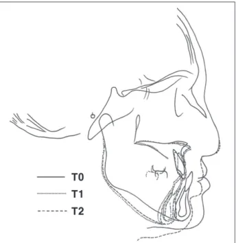
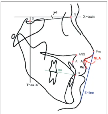
The tracing and cephalometric analysis of lateral cephalo- gram in T0, T1, and T2 stages were carried out by V–Ceph (Osstem, Seoul, Korea) (Fig. 1). The x–axis was determined by a line through nasion rotated 7° upwards from the sella–na- sion line. This line runs parallel to the Frankfort horizontal plane. And the y–axis was determined as a perpendicular line to the x–axis through sella (S). The cephalometric anatomic landmarks and reference lines are shown in Fig. 2. The cepha-
Fig. 1. The tracing of cephalogram in preoperative (T0), postopera- tive (T1), and debonding (T2) stage.
lometric variables in three stages (T0, T1, T2) were calculated by the computer program.
The amount of maxillary movement was determined by subtracting the values of T0 from T1, and the amount of re- lapse was calculated by subtracting T2 from T1. To avoid the errors due to postoperative edema, only the differences be- tween T2 and T0 in the analysis of soft tissue change were cal- culated. For maxillary movement and relapse, positive values reflect forward movement or clockwise rotation, and negative values for backward movement or counterclockwise rotation.
All cephalometric tracing and measurement were per- formed twice by the same examiner, who did not perform the surgery, to avoid observer bias. Paired t–test was used for the statistical analysis. T–test statistics with p values equal to or less than 0.05 were considered to be statistically significant.
Fig. 2. Cephalometric landmarks and reference lines. S, sella: the estimated center of the bony contour of the preoperative sella turcica;
N, nasion: the most anterior point of the frontonasal suture; A, A point: the deepest (most posterior) on the concave outline of the upper labial alveolar process; ANS, anterior nasal spine, most anteri- or limit of the floor of the nose, at the tip of the anterior nasal spine in the midsagittal plane; Is, incisivum superior: the tip of the crown on the long axis of the most prominent upper incisor; Prn, pronasale:
the most prominent point on the nose profile; A’, soft tissue A point (), the deepest concavity on the upper lip profile; Vu, vermilion bor- der of upper lip: the most anterior point on the convexity of the upper lip profile; E–line, the line is drawn from the tip of the nose to soft tissue pogonion; NLA, nasolabial angle: the angle is formed by two lines, namely, a columella tangent and upper lip tangent.
3. Surgical technique and orthodontic treatment For presurgical preparation, plaster model surgery and prediction tracing were performed and occlusal splint was made. The LeFort I osteotomy was done using a down–frac- ture technique and moved to the preplanned position by the model surgery and guided by an occlusal splint. The mobi- lized maxilla was fixed with four titanium miniplates at each side of zygomatic buttress and the pyriform region. If neces- sary, patients received segmentation osteotomy of maxilla for two–piece LeFort I surgery. Following this, a sagittal split os- teotomy of mandibular ramus was done with a setback of the mandible. After the maxillomandibular fixation using the oc- clusal splint, three bicortical screws were placed at the ramus area. The maxillomandibular fixation and occlusal splint were released and removed, and the relationship of the dental occlusion and condyle position were checked.
Bone grafting was not used in this group of patients. Inter- maxillary fixation was released and the occlusal splint was re- moved at completion of surgery. Alar cinch suture was done through the vestibular approach in order to prevent widening of the nose. Suction drains were inserted in the ramal wounds overnight. Patients resumed orthodontic treatment in a month after the surgery.
RESULTS
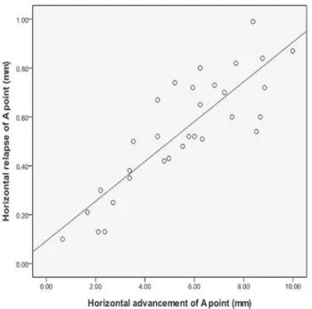
Because anterior nasal spine was often altered during the operation and the upper incisor inclination was changed by postoperative orthodontic treatment, point A was used to measure the surgical movement and relapse of maxilla. Table I shows surgical maxillary change and relapse. The mean hor- izontal advancement of maxilla (point A) was 5.5 mm, and the mean horizontal relapse was 0.5 mm (9.1%). Fig. 3 in pres- ent study are based on the reproducible slope of the scatter- gram (R
2=0.73). There were significant correlations between the degree of horizontal relapse and the extent of maxillary advancement. The mean vertical lengthening of maxilla (point A) was 3.2 mm, and the mean vertical relapse was 0.6 mm (18.8%).
The horizontal relapse of anterior nasal spine (ANS) was
0.5 mm (9.8%), similar to the relapse of point A. In contrast,
upper incisor tip was advanced a little bit more (1.9%) after
surgery. This was likely related to the orthodontic treatment
Table I. Cephalometric Variables (T0, T1, T2), Surgical Change and Relapse of Hard Tissue
T0 T1 T2 Surgical change Relapse
Mean SD Mean SD Mean SD T1-T0 SD T1-T2 (%) SD
Ax (mm) 56.8 3.4 62.3 6.3 61.8 6 5.5 2.4 0.5 (9.1) 0.2
Is-x (mm) 60.4 3.5 65.7 6.5 65.8 6.7 5.3 2.4 -0.1 (1.9) 0.3
ANS-x (mm) 60.2 3.6 65.3 5.8 64.8 6.4 5.1 2.1 0.5 (9.8) 2.3
Ay (mm) 61.9 5.1 65.1 3.6 64.5 3.8 3.2 2.0 0.6 (18.8) 1.5
SNA (°) 75.8 5.8 79.6 4.7 78.7 3.9 3.8 0.8 0.9 (23.7) 1.4
Occ. plane to SN (°) 10.6 5.2 15 5.6 15.2 4 4.4 3.0 -0.2 (-4.5) 2.2
T0, preoperative stage (within a month before operation); T1, postoperative stage (one week after the orthognathic surgery); T2, debonding stage (after comple- tion of orthodontic treatment); SD, standard deviation; Ax, distance from A point to Y-axis (mm); Is-x, distance from incisivum superior to Y-axis (mm);
ANS-x, distance from anterior nasal spine to Y-axis (mm); Ay, distance from A point to X-axis (mm); SNA, angle of N-S to N-A line; Occ. plane to SN, angle of occlusion plane to S-N line.
Table II. Cephalometric Variables (T0, T2) and Surgical Change of Soft Tissue
T0 T2 Surgical change
Mean SD Mean SD T2-T0 SD
Prn-x 89 4.5 91.5 6.3 2.5 0.9
A’-x 71.4 4.9 74.8 6.4 3.4 2.3
Vu-x 76.8 5.2 80.5 5.7 3.7 2.5
Upper lip E-plane -5.2 3 -1.4 2 3.8 1.2 Nasolabial angle 88.7 17.9 97.9 17 9.2 1.8 T0, preoperative stage (within a month before operation); T2, debonding stage (after completion of orthodontic treatment); SD, standard deviation;
Prn-x, distance from pronasale to Y-axis (mm); A'-x, distance from A' point to Y-axis (mm); Vu-x, distance from vermilion border of upper lip to Y-axis;
upper lip E-plane, the shortest distance from upper lip to E-plane; nasolabi- al angle, angle of columella tangent line to upper lip tangent line.
Table III. Correlation Ratio between Soft and Hard Tissue Movement Soft tissue
change Hard tissue
change Ratio
Prn-x/ANS-x 2.5 4.6 0.54
A’-x/A-x 3.4 5.0 0.68
Vu-x/Is-x 3.7 5.4 0.69
Prn-x, distance from pronasale to Y-axis (mm); ANS-x, distance from Anterior nasal spine to Y-axis (mm); A'-x, distance from A' point to Y-axis (mm); Ax, distance from A point to Y-axis (mm); Vu-x, distance from ver- milion border of upper lip to Y-axis; Is-x, distance from incisivum superior to Y-axis (mm).
Fig. 3. Significant relation between horizontal advancement and relapse of A point.
and orthodontic compensation to the skeletal relapse. The
mean SNA angle was changed from 75.8° to 79.6° and re-
lapsed by 0.9°. Maxillary rotation was assessed as the change
of the occlusal plane. All cases had clockwise rotation by
mean of 4.4° with a mean relapse of –0.2°. Mean soft tissue
changes are given in Table Ⅱ, and the ratios between soft and
hard tissue changes are shown in Table Ⅲ. The mean change
of soft tissue A’ point was measured 3.4 mm and the nasal tip
projection was increased by 2.5 mm. The vermilion border of
upper lip was advanced mean 3.7 mm and the mean upper lip
to E–line distance was increased 3.8 mm and became close to
esthetic line. And the average nasolabial angle increased from
88.7° to 97.9°. The ratio for horizontal advancement of nasal
tip (Prn)/anterior nasal spine (ANS) was 0.54/1 and the ratio
of A’ point/A point was 0.68/1 and 0.69/1 for the upper ver-
milion/upper incisor tip.
DISCUSSION
The treatment planning and operation of orthognathic surgery for cleft patients is more difficult than for non–cleft patients, because of further advancement, inadequate tissue quality and worse dental occlusion.
6In many previous studies on patients with cleft, the relapse rate of LeFort I osteotomy was reported to be between 25% and 50%.
3,4To reduce or pre- vent postoperative relapse in patients with cleft, multiple fac- tors were discussed when performing orthognathic surgery.
The amount of advancement for LeFort I osteotomy was con- sidered one of the major factors. Some reports showed that the relapse was influenced by the amount of advancement
6whereas the others could not find positive correlation be- tween the relapse and the amount of advancement.
7In our study, the positive correlation was found (Fig. 3). With this finding, it is rational to plan two–jaw surgery if excessive sin- gle maxillary advancement is expected. Other factors report- ed to be responsible for relapse included soft tissue scarring in palate and retromaxillary region.
4,6Tightness of the upper lip created by cheiloplasty is another factor maxillary growth re- straint and surgical relapse.
8Adequate degloving and release of restraint are advisable in order to fully mobilize and ad- vance the maxilla. Average horizontal relapse rate of 9.1%
demonstrated good stability as compared with that of previ- ous studies.
Several studies were done assessing the correlation be- tween the amount of vertical displacement and the relapse, and found that the postoperative vertical relapse is more than the horizontal relapse. There was a significant difference be- tween inferior repositioning and intrusion of maxilla. Most researchers agreed that maxillary intrusion was a stable movement but inferior repositioning of maxilla was more un- stable.
9It was explained that the inferior repositioning leads to reduction of bone contact at lateral portion of maxilla
10and increases soft tissue stretching resulting in movement of the screws during bone healing.
11The degree of vertical re- lapse in maxillary inferior repositioning has been reported to be variable,
9and the amount of relapse was roughly around 2 mm.
2Therefore, some authors recommend 2 mm overcor- rection in inferior repositioning.
12This is not observed in our study, with the amount of inferior repositioning 3.2 mm and relapse 0.6 mm. Although the vertical relapse rate was 18.8%
and greater than the horizontal relapse in our study, this ver-
tical relapse was lesser than that from previous studies.
Good stability was achieved without bone grafting in this study, but it should not be interpreted as negative function when there is a need in the situation of significant osseous gap. Bone grafting is recommended to promote stability.
7Be- cause it is regarded as a physical barrier against the relapse and can promote bone healing, bone grafting was recom- mended in cases of large advancement and inferior reposi- tioning of maxilla.
13Araujo et al.
14suggested that bone graft- ing should be used if the advancement of maxilla was more than 6 mm.
Another approach to avoid skeletal relapse is to obtain a good functional occlusion and clockwise rotation of the infe- riorly repositioned maxilla.
4Quejeda et al.
15reported that clockwise rotation plus inferior repositioning of maxilla pro- duced better stability than inferior repositioning parallel to the horizontal plane. Others reported that two–jaw surgery is more advantageous to achieve good occlusal plane, clockwise rotation and three dimensional correction of the maxillo- mandibular complex.
16Because LeFort I osteotomy may lead to autorotation of the mandible resulting in counterclockwise rotation of the occlusal plane, it is difficult to get a clockwise rotation for the maxillomandibular complex using LeFort I ostetomy alone.
17It is recommended to apply two–jaw sur- gery for cleft patients with class III malocclusion who require more than 10 to 12 mm maxillary advancement.
18There were only four patients with bilateral cleft in this
study, and therefore meaningful comparison of the relapse
rate between the unilateral and bilateral clefts could not be
made. It is assumed that relapse is more likely to occur in pa-
tients with bilateral cleft. Because of the problems with pre-
maxillary scarring, bilateral alveolar cleft, multiple missing
teeth, lip tension, unfavorable dental occlusion and unpre-
dictable premaxillary circulation, forward movement of the
maxilla in patients with bilateral cleft are likely to be associat-
ed with more difficulty and higher complication rate.
19While
some authors suspected that the degree of postoperative re-
lapse might be related to the cleft types,
6,7Heliovaara et al.
20reported that the skeletal stability and relapse were similar in
both cleft types although bilateral cleft patients had more
cleft–related problems and required more extent of advance-
ment. The maxillary relapse started immediately after surgery
and continued to be observed for 6 months.
2,7Surgeons and
orthodontists must direct their efforts to reduce the relapse
during the first 6 months after the orthognathic surgery.
In orthognathic surgery, bilateral sagittal split osteotomy (BSSO) is the preferred method for mandibular setback. The reasons include broad bony contact and stability for good bony healing, and it can be used for wide spectrum of defor- mity. Some authors studied skeletal stability after BSSO for mandibular advancement comparing two fixation methods, noncompressive bicortical screws and miniplates with uni- cortical screws, and concluded that the two methods did not differ significantly.
21On the contrary, Amano et al.
22found higher rigidity using screw fixation for BSSO setback, and that the postoperative morphological change continued in the plate fixation group for 1 years. They suggested that the plate fixation method might not be ideal for patients with Class III malocclusion and asymmetry who have a high risk of relapse. We prefer using the BSSO method for mandibular movement and fixing the segments by three bicortical screws for better rigidity.
23Subsequent to orthognathic surgery, the ultimate facial aesthetic appearance is a combination of dental, soft tissue and skeletal changes. Estimation of the resulting external soft tissue changes after the operation is as important as dental occlusion and skeletal changes. In previous studies, the ratio of soft to hard tissue change in nasal tip of cleft patients was reported to range from 0.25 to 0.53. It was a little higher ratio than that of non–cleft patients reported ranging from 0.13 to 0.29. However, the range of reported ratios was large and there were great individual variation. In our study, the ratio was 0.54 at nasal tip/anterior nasal spine level. The ratio at soft tissue A’ point or superior labial sulcus was reported between 0.48 and 0.66. Our data is higher, with the advancement ratio 0.68 at soft tissue A’ point/ A point. In several previous stud- ies, patients treated with maxillary advancement showed similar ratios of soft tissue to hard tissue change between 0.5 and 0.66:1 for upper lip to upper incisor change in cleft pa- tients and in non–cleft patients. But, the ratio of upper lip change in this study was 0.69, and it was higher as well. These ratios of soft tissue/hard tissue movement in cleft patients tend to be higher than those in non–cleft patients, possibly due to the scarred and less pliable soft tissues in the cleft pa- tients which catch up more closely with the underlying hard tissue. In our study, the further higher ratios of nasal tip/ante- rior nasal spine and A’/A point might be due to cinch sutures in particular.
CONCLUSION
In the present study, better skeletal stability after orthog- nathic surgery was obtained as compared with previous re- ports. The anteroposterior horizontal relapse was 9.1%, and the vertical relapse was 18.8%. The better skeletal stability might be due to the fact that majority of the patients received two–jaw surgery with clockwise rotation as well as rigid fixa- tion. Rigid fixation using bicortical screws is highly recom- mended. The ratios of soft tissue/hard tissue movement are 0.54:1 for nasal tip/anterior nasal spine, 0.68:1 for A’ point/A point, and 0.69:1 for upper lip/upper incisor tip. The ratios of nasal tip/anterior nasal spine and A’/A point are higher com- paring with those from previous reports, possibly due to the cinch suture on nasal base.
REFERENCES
1. Ross RB: Treatment variables affecting facial growth in complete uni- lateral cleft lip and palate. Cleft Palate J 24: 5, 1987
2. Welch TB: Stability in the correction of dentofacial deformities: a com- prehensive review. J Oral Maxillofac Surg 47: 1142, 1989
3. Kufner J: Four–year experience with major maxillary osteotomy for retrusion. J Oral Surg 29: 549, 1971
4. Freihofer HP Jr: Results of osteotomies of the facial skeleton in adoles- cence. J Maxillofac Surg 5: 267, 1977
5. Hoffman GR, Brennan PA: The skeletal stability of one–piece Le Fort 1 osteotomy to advance the maxilla; Part 2. The influence of uncontrol- lable clinical variables. Br J Oral Maxillofac Surg 42: 226, 2004 6. Hochban W, Ganss C, Austermann KH: Long–term results after max-
illary advancement in patients with clefts. Cleft Palate Craniofac J 30:
237, 1993
7. Posnick JC, Ewing MP: Skeletal stability after Le Fort I maxillary ad- vancement in patients with unilateral cleft lip and palate. Plast Recon- str Surg 85: 706, 1990
8. Turvey TA, Vig KW, Fonseca RJ: Maxillary advancement and contour- ing in the presence of cleft lip and palate. In: Turvey TA, Vig KW, Fon- seca RJ (eds): Facial clefts and craniosynostosis: principles and management. Philadelphia, WB Saunders, 1996, p 441
9. Wolford LM, Hilliard FW: The surgical–orthodontic correction of ver- tical dentofacial deformities. J Oral Surg 39: 883, 1981
10. Waite PD, Tejera TJ, Anucul B: The stability of maxillary advancement using Le Fort I osteotomy with and without genial bone grafting. Int J Oral Maxillofac Surg 25: 264, 1996
11. Proffit WR, Turvey TA, Phillips C: Orthognathic surgery: a hierarchy of stability. Int J Adult Orthodon Orthognath Surg 11: 191, 1996 12. Finn RA, Throckmorton GS, Bell WH, Legan HL: Biomechanical
considerations in the surgical correction of mandibular deficiency. J
Oral Surg 38: 257, 1980
13. Epker BN, Schendel SA: Total maxillary surgery. Int J Oral Surg 9: 1, 1980
14. Araujo A, Schendel SA, Wolford LM, Epker BN: Total maxillary ad- vancement with and without bone grafting. J Oral Surg 36: 849, 1978 15. Quejada JG, Bell WH, Kawamura H, Zhang X: Skeletal stability after
inferior maxillary repositioning. Int J Adult Orthodon Orthognath Surg 2: 67, 1987
16. LaBanc JP, Turvey T, Epker BN: Results following simultaneous mobi- lization of the maxilla and mandible for the correction of dentofacial deformities: analysis of 100 consecutive patients. Oral Surg Oral Med Oral Pathol 54: 607, 1982
17. Posnick JC, Taylor M: Skeletal stability and relapse patterns after Le Fort I osteotomy using miniplate fixation in patients with isolated cleft palate. Plast Reconstr Surg 94: 51, 1994
18. Epker BN, Fish LC: Simultaneous maxillary advancement and mandibular setback. In Epker BN, Fish LC, Stella JP (eds): Dentofacial deformities: integrated orthodontic and surgical correction. Vol. 1,
St. Louis, Mosby, 1986, p 526
19. Posnick JC, Tompson B: Modification of the maxillary Le Fort I os- teotomy in cleft–orthognathic surgery: the bilateral cleft lip and palate deformity. J Oral Maxillofac Surg 51: 2, 1993
20. Heliovaara A, Ranta R, Hukki J, Rintala A: Skeletal stability of Le Fort I osteotomy in patients with isolated cleft palate and bilateral cleft lip and palate. Int J Oral Maxillofac Surg 31: 358, 2002
21. Blomqvist JE, Isaksson S: Skeletal stability after mandibular advance- ment: a comparison of two rigid internal fixation techniques. J Oral Maxillofac Surg 52: 1133, 1994
22. Amano K, Yagi T, Iida S, Aikawa T, Yamashiro T, Takada K, Kogo M:
Facial frontal morphological changes related to mandibular setback osteotomy using cephalograms. J Craniomaxillofac Surg 37: 412, 2009
23. Ming–Yih L, Chun–Li L, Wen–Da T, Lun–Jou L: Biomechanical sta- bility analysis of rigid intraoral fixation for bilateral sagittal split osteot- omy. J Plast Reconstr Aesthet Surg 63: 451, 2010