Improvement of Neuronal Differentiation by PDE4 Inhibition in Human Bone Marrow-mesenchymal Stem Cells
Da Hee Jeong
1†, I-Seul Joe
1,2†and Goang-Won Cho
1,2*
1Department of Biology, College of Natural Science, Chosun University, Gwangju 501-759, Korea
2Department of Life Science, BK21-Plus Research Team for Bioactive Control Technology, Chosun University, Gwangju 501-759, Korea Received May 2, 2016 /Revised September 2, 2016 /Accepted September 5, 2016
Human
bone marrow mesenchymal stem cells (hBM-MSCs) can differentiate into various cell types including osteoblasts, adipocytes, chondrocytes, and myocytes. Previous studies, including our own, have shown that MSCs can also differentiate into neuron-like cells. However, their rate of neuronal differentiation is not sufficient for application to stem cell therapy, which requires well-defined cell types. For this purpose, we first examined the expression of neuronal lineage markers (GFAP, MAP-2, KCNH1, Nestin, NF-M, and Tuj-1) by real-time PCR, western blot, and immunocytochemical staining.
The expressions of the astrocyte marker GFAP and neuronal markers NF-M and Tuj-1 increased in neuronal differentiated MSCs (dMSCs). To improve the neuronal differentiation efficiency, PDE4, an important signaling intermediator in the progression of neuronal differentiation, was modulated using well-known inhibitors such as rolipram or resveratrol and then differentiated into neuronal cells (Roli- or RSV-dMSCs). The expressions of NF-M, Tuj-1 were increased while that of GFAP decreased in Roli- and RSV-dMSCs, which were examined by real-time PCR, western blot, and immunocytochemical staining. From these experiments, we have found that the neuronal differentiation efficiency can be ameliorated by the modulation of PDE4 activity.
Key words : hBM-MSCs, neuronal differentiation, PDE4 inhibition, resveratrol, rolipram
†Authors contributed equally.
*Corresponding author
*Tel : +82-62-230-6641, Fax : +82-62-230-6650
*E-mail : [email protected]
This is an Open-Access article distributed under the terms of the Creative Commons Attribution Non-Commercial License (http://creativecommons.org/licenses/by-nc/3.0) which permits unrestricted non-commercial use, distribution, and reproduction in any medium, provided the original work is properly cited.
Journal of Life Science 2016 Vol. 26. No. 12. 1355~1359 DOI : http://dx.doi.org/10.5352/JLS.2016.26.12.1355
서 론
신경계를 구성하는 세포는 크게 신경세포(neuron)와 신경 아교세포(glial cell) 두 가지 종류가 있고 이들은 모두 외배엽 에서 유래한다[2]. 신경세포는 신경계의 기본구조와 기능의 단 위로, 자극에 반응하고 자극을 전도하며 화학조절물질인 신경 전달물질(neurotransmitter)을 방출한다. 신경아교세포는 신 경세포의 기능을 돕는 세포로, 신경세포의 외부 환경을 조절 하는 데 도움이 되는 성상세포(astrocyte), 축삭을 싸고 있는 수초를 형성하는 희돌기교세포(oligodendrocyte) 등이 존재한 다. 신경 줄기 세포는 위와 같은 세포들의 공급을 책임진다.
Human mesenchymal stem cell (hMSCs)은 유래하는 조직의 세포뿐 아니라 뼈, 연골, 지방, 힘줄, 근육 그리고 골수 기질 등 여러 세포 유형으로 분화가 가능한 다능성 줄기세포이다[2, 16]. 최근 연구에 따르면 hMSCs가 신경세포(neuron–like cell)로 분화할 수 있음을 제시하였다[14].
Cyclic adenosine monophosphate (cAMP)는 축삭 재생과 신경 세포 보호를 촉진하는 중요한 신호전달 매체(signaling intermediator)이다[12, 13, 18]. 또한, 세포 내 cAMP의 증가는 신경표지단백질들(neuronal marker proteins)의 발현 향상을 통하여 신경분화(neuronal differentiation)을 촉진한다[20]. 세 포 내 cAMP를 조절하는 주요 인자인, phosphodiesterase-4 (PDE4)는 cAMP를 가수분해하여 5‘-AMP로 만들어 세포 내 cAMP를 감소시킨다. PDE4 억제자를 이용한 연구에서 우울 증(depression), 인식장애(cognitive deficit)과 알츠하이머병 (alzheimer’s disease) 등에서 효과를 관찰하였다[5].
Rolipram [11]은 PDE4 억제 화합물로 알츠하이머 병[5], 인 지장애[14], 헌팅턴무도 병(Huntington disease) [12], 외상적 뇌손상[1], 척수 손상[6], 호흡기 질환[7] 등 여러 질병모델에서 폭넓게 연구되었다. 천연 폴리페놀계 화합물인 resveratrol (RSV; 3, 4, 5'-tri-hydroxy-trans-stillbene, C
14H
12O) 역시 PDE4 를 억제 물질로 cAMP를 증가시킨다고 보고되었다[15]. Res- veratrol는 포도, 땅콩, 산딸기류 열매 등의 다양한 식물에서 발견되며[2, 15], 항산화(anti-oxidative), 항염증(anti-inflam- matory), 항노화(anti-senescence) 등의 효과가 보고되었다[3, 10, 17]. 또한, sirtuin의 활성을 자극하여 AMP-activated Kinase (AMPK) 신호경로를 통하여 미토콘드리아의 생합성, p53의 발현 등의 작용기전과 연관되어 있다[17].
본 연구에서는 PDE4억제 화합물인 rolipram과 resveratrol
을 이용하여 신경세포 분화 효율을 개선할 수 있음을 hBM-
MSCs에서 입증하였다.
재료 및 방법
Culture condition
hBM-MSCs (human bone marrow-mesenchymal stem cells)은 CEFO (Cell Engineering for Origin, Seoul, Korea)에 서 구매하였다. hBM-MSCs를 75T flask에 접종하여 10% FBS (fetal bovine serum), L-glutamine, penicillin, 그리고 strepto- mycin을 포함하는 DMEM low glucose 배양액을 이용하여 37°C, 5% CO
2조건에서 배양하였다. 본 연구의 모든 실험에서 7-passages의 hBM-MSCs가 이용되었다.
Compounds
본 실험에서 사용된 resveratrol (sigma Aldrich, R5010-100 mg)은 dimethyl sulfoxide (DMSO)에 녹인 뒤 50 mM로 희석 하여 사용 전까지 -20°C에 보관하였다. Rolipram (Tocris Bio- science, 0905-10 mg) 역시 DMSO에 녹인 뒤 25 mM로 희석하 여 사용 전까지 -20°C에 보관하였다.
Neuron differentiation
hBM-MSCs를 1 μM resveratrol, rolipram을 각각 처리하여 12시간 동안 배양한다. 이 후 DMEM, 10% FBS, 10 ng/ml ba- sic fibroblast growth factor (bFGF), 그리고 500 μM pre-in- duction media에서 24시간 동안 배양하고, 100 μM butylated hydroxyanisole (BHA)과 2% DMSO가 함유된 induction me- dia로 바꿔 5시간 30분 동안 배양하였다[19].
Real-time PCR
전체 RNA는 RNAiso (TAKARA, Japan)를 사용하여 추출 하였다. 추출한 RNA로 cDNA synthesis kit (TAKARA, Japan) 와 oligo-dT primer를 사용하여 제조사의 프로토콜에 따라 cDNA를 합성하였다. 합성된 cDNA는 SYBR Green PCR mas- ter mix (Applied Biosystems Inc., USA)와 함께 real-time PCR을 수행하였다. PCR에 사용된 primer은 사람의 NF-M (Sn; 5’-GTGAACCACGAGAAGGCTCA-3’, Asn; 5’-AGGTA GTCTTTGCGCTCCAC-3’), GFAP (Sn; 5’-TGGGAGCTTGA TTCTCAGCA-3’, Asn; 5’-CCTGGGCTTGACCTCTCTGTA- 3’), KCNH1 (Sn; 5’-TTGGAGATGTGTTCTGGAAGGAA-3’, Asn; 5’-AGGGCATCCCFCTTGATC-3’) Nestin (Sn; 5’-AGCC CTGACCACTCCAGTTT-3’, Asn; 5’-GCTGCTTACCACTTT GCCCT-3’), MAP-2 (Sn; 5’-TTGGTGCCGAGTGAGAAGAA- 3’, Asn; 5’-GGTCTGGCA GTGGTTGGTTAA-3’), β-actin (Sn;
5’-ATCCGCAAAGACCTGTACGC-3’, Asn; 5’-TCTTCATTG TGCTGGGTGCC-3’)을 이용하였다.
Immunocytochemical staining (ICC)
hBM-MSCs를 poly-L-lysine-coated coverslips (Fisher Sci- entific, Hampton, NH, USA)에서 키우고 신경세포로 분화를 유도하였다. NF-M과 GFAP (Santa cruz biotechnology, Dallas, TX, USA)를 Blocking buffer (5% Normal horse se- rum, sodium azide 0.02%가 포함 된 PBS용액)에 항체를 1:200 으로 희석하여 4°C에서 overnight하였다. 그 다음 Alexa488- conjugated donkey anti-goat IgG, Alexa555-conjugated don- key anti-rabbit IgG (1:400; Molecular Probes Inc., USA) 이차 항체와 Hoechst 33342 (1:10,000; Molecular Probes Inc., USA) 를 PBS-A에 희석하여 실온에서 1시간 동안 반응시켰다. 세포 는 Nikon Eclipse 80Ti microscopy (Nikon; Tokyo, Japan) 형 광 현미경으로 관찰하였다.
Western blot analysis
Protease와 dephosphatase inhibitors가 포함된 50 μl RIPA buffer (Santa Cruz Biotechnology, USA)를 처리한 뒤 4°C에 30분 동안 반응 시킨 후, 16,000× 4°C에서 20분간 원심 분리하 여 상층액에 있는 전체 단백질을 분리하였다. 30 μg의 sample 을 만들어 sodium dodecyl sulfate (SDS)-polyacrylamide gel 을 이용하여 전기영동을 한 후, nitrocellulose membrane (Mil- lipore, Germany)으로 전달시켰다. 단백질이 전이된 mem- brane을 blocking buffer (5% Normal horse serum, 0.1%
Tween-20, pH7.4)가 포함 된 TBS용액]에서 1시간 30분 동안 반응하였다. 1차 항체 β3 Tubulin (Tuj-1, 1:1,000; Santa Cruz Biotechnology, USA), Neurofilament-M (NF-M, 1:500; Santa Cruz Biotechnology, USA), Glial fibrillary acidic protein (GFAP, 1:500; Santa Cruz Biotechnology, USA), β-actin (1:5,000; Sigma, USA)는 blocking buffer 조건에 sodium azide 0.02%를 넣어 4°C에 overnight하였다. 2차 항체(Jackson Immuno Research Laboratories; West Grove, PA, USA)는 TBS-T (0.1% Tween-20)에 1:10,000으로 희석해 2시간 동안 상 온에서 반응시켰다. 반응 후 암실에서 ECL solution kit (Amersham Life Science, UK)를 사용하여 X-ray film에 노출 시켜 확인하였다.
결과 및 고찰
hBM-MSCs를 이용한 신경분화 유도
hBM-MSCs을 이용하여 “재료 및 방법”에 서술된 과정에 따라 신경분화(neuronal differentiation)를 유도하였다[8]. 신 경분화 여부를 검증하기 위하여 신경 표지자(neuronal mark- er)의 발현을 Real-time PCR법으로 확인하였다. 대조군(Ctrl- MSCs) 대비 유도군(dMSCs)에서 신경세포 표지자인 MAP-2,
NF-M, Nestin과 KCNH1의 발현이 증가함을 관찰하였다(Fig.1; t-test, *p<0.01, #p<0.05, mean ± SD, n = 5). 뿐만 아니라
Fig. 1. The expression of neuronal markers in neuronal differ- entiated hBM-MSCs. The expressions of neuronal genes in non-differentiated MSCs (Ctrl-MSCs) and differ- entiated MSCs (dMSCs) were examined by real-time PCR. All genes were significantly increased in dMSCs.
A
B
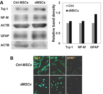
Fig. 2. The increased expression of both neuronal and glial markers in dMSCs. The MSCs (Ctrl-MSCs) and differ- entiated MSCs (dMSCs) were subjected to western blot analysis (A) and immunocytochemical staining (B) with specific antibodies for Tuj-1, NF-M, and GFAP. Tested proteins were up-regulated in dMSCs.
A
B
Fig. 3. The reduction of GFAP positive cells in PDE4 inhibited dMSCs. (A) Astrocyte specific marker GFAP was vi- sualized in non-differentiated MSCs (Ctrl-MSCs) and differenetiated MSCs with rolipram or resveratrol treat- ment (Roli-dMSCs, RSV-dMSCs). (B) GFAP positive cells were counted and shown in the graph.
성상세포 표지자인 GFAP의 발현 역시 증가함을 확인하였다 (Fig. 1; GFAP). 따라서 hBM-MSCs에 신경분화를 유도 하였을 때, 신경세포뿐만 아니라 성상세포로의 분화 또한 일어남을 확인하였다.
이러한 발현 증가를 단백질 수준에서 확인하기 위해 Wes- tern Blot을 수행하였고(Fig. 2A), 발현장소와 양을 확인하기 위해 Immunocytochemistry (ICC)를 수행하였다(Fig. 2B). 분 화를 유도한 dMSCs에서 신경세포 표지자인 Tuj-1, NF-M과 성상세포 표지자인 GFAP가 동시에 증가함을 확인하였다(Fig.
2). 이상의 실험을 통해 hBM-MSCs에서 신경분화를 유도하며 신경세포뿐만 아니라 신경아교세포의 한 종류인 성상세포로 의 분화도 일부 일어남을 확인하였다.
PDE4 억제를 통한 MSCs의 신경분화 개선효과
Rolipram이나 resveratrol은 PDE4 억제작용을 통하여 AMPK를 조절하는 것으로 알려져 있는데[15, 18], AMPK의 활성화는 신경분화에 중요한 신호전달계를 활성화한다[9]. 본 연구에서는 줄기세포의 신경분화 효율을 개선하기 위해 이 화합물을 이용하였다. 배양된 MSCs에 rolipram이나 resvera- trol를 각각 1 μM씩 12시간 동안 처리한 후 신경분화를 유도하 여 immunocytochemistry staining법을 이용하여 관찰하였다.
다수의 신경분화 줄기세포(dMSCs)에서 성상세포 표지자인 GFAP의 발현이 관찰되었지만, rolipram이나 resveratrol를 전 처리한 줄기세포(Roli-dMSCs, RSV-dMSCs)에서는 유의성 있 게 감소함을 확인하였다(Fig. 3A). 세포에서 GFAP의 발현율 을 보면, 신경분화 줄기세포(dMSCs)에서는 약 51.92%인 것이, rolipram 혹은 resveratrol을 처리하면, 각각 38.95% (Roli- dMSCs)와 34.73% (RSV-dMSCs)로 감소하였다(Fig. 3B). 이와 같은 실험을 통해 PDE4 억제자인 rolipram이나 resveratrol을 줄기세포에 전처리하였을 때 대조군에 비해서, 성상세포보다 신경세포로 분화하는 비율이 개선됨을 확인하였다.
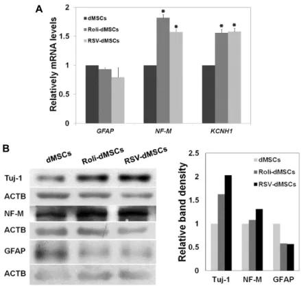
위와 같은 결과를 mRNA 수준에서 확인하기 위하여 Real-
A
B
Fig. 4. Decreased expression of GFAP in Roli- and RSV-dMSCs. (A) The expression of neuronal markers NF-M, KCNH1, and astrocyte marker GFAP were measured by real-time PCR (A) and western blot (B) in dMSCs, Roli- and RSV-dMSCs.
time PCR을 수행하였다. 대조군 대비 약물을 처리한 dMSCs 에서 신경세포 표지자인 NF-M와 KCNH1의 발현이 증가하였 고, 반면, 성상세포 표지자인 GFAP의 발현은 감소함을 확인하 였다(Fig. 4A; t-test, *p<0.001, mean ± SD, n = 5). 이 효과를 단백질 수준에서 검증하기 위해 Western Blot법을 수행하였 고, 마찬가지로 신경세포 표지자인 Tuj-1과 NF-M의 발현은 개선된 반면 성상세포 표지자인 GFAP의 발현은 감소하였다 (Fig. 4B; t-test, *p<0.01, #p<0.05, mean ± SD, n = 5). 이와 같이, PDE4 억제 화합물을 처리한 후 신경분화를 유도하면 신경아교세포와 같은 다른 lineage 세포로 분화를 억제하고 신경세포로 분화를 촉진하여 줄기세포의 신경분화 효율을 개 선함을 확인하였다.
인간의 BM-MSCs는 다양한 분화능력을 보유한 성체 줄기 세포(adult stem cells)이다. 선행 연구에 따르면, 이 MSCs를 이용하여 신경세포로 분화가 가능함을 보였다[8]. 본 연구에서 는 hBM-MSCs를 이용한 신경분화에서 신경세포뿐 아니라 신 경아교세포로도 분화가 가능함을 확인하였다. PDE4 억제자 인, rolipram, resveratrol을 이용하여, 신경분화에 중요한 신호 전달자인 PDE4-cAMP signaling pathway를 조절하여 신경아 교세포의 분화를 제안하고 신경세포의 분화를 촉진하였다. 이 상과 같은 방법은 줄기세포의 신경분화를 개선하고 조절하여 신경계 퇴행성질환과 같은 질환의 세포치료에 효과를 개선할 수 있으며, 인체 내 신경계에서 신경분화를 개선 하는 데에도 도움이 될 것으로 생각된다.
감사의 글
이 논문은 2016학년도 조선대학교 학술연구비의 지원을 받 아 연구되었음.
References
1. Atkins, C. M., Oliva, A. A. Jr., Alonso, O. F., Pearse, D. D., Bramlett, H. M. and Dietrich, W. D. 2007. Modulation of the cAMP signaling pathway after traumatic brain injury.
Exp. Neurol. 208, 145-158.
2. Caplan, A. I. 2007. Adult mesenchymal stem cells for tissue engineering versus regenerative medicine. J. Cell. Physiol.
213, 341-347.
3. Csiszar, A., Labinskyy, N., Pinto, J. T., Ballabh, P., Zhang, H., Losonczy, G., Pearson, K., de Cabo, R., Pacher, P., Zhang, C. and Ungvari, Z. 2009. Resveratrol induces mitochondrial biogenesis in endothelial cells. Am. J. Physiol. Heart Circ.
Physiol. 297, H13-20.
4. DeMarch, Z., Giampà, C., Patassini, S., Martorana, A., Ber- nardi, G. and Fusco, F. R. 2007. Beneficial effects of rolipram in a quinolinic acid model of striatal excitotoxicity.
Neurobiol. Dis. 25, 266-273.
5. García-Osta, A., Cuadrado-Tejedor, M., García-Barroso, C., Oyarzábal, J. and Franco, R. 2012. Phosphodiesterases as therapeutic targets for Alzheimer's disease. ACS Chem.
Neurosci. 3, 832-844.
초록:인간 골수유래-중간엽 줄기세포(hBM-MSCs)에서 PDE4 억제조절을 통한 신경세포 분화 효율 개선
정다희
1†․조이슬
1,2†․조광원
1,2*
(1조선대학교 생명과학과, 2조선대학교 BK21-Plus 연구팀)
인간 중간엽 줄기세포(hMSCs)는 신경세포(neuron-like cells)를 포함한 다양한 세포로 분화할 수 있는 능력을 지닌 성체 줄기세포(adult stem cells)이다. 본 연구에서는 인간의 골수유래-중간엽 줄기세포(bone marrow-mesen- chymal stem cells; hBM-MSCs)를 이용한 신경분화에서 신경세포 표지자(neuronal marker)인 NF-M, Tuj-1 뿐만 아니라 성상세포 표지자(glial marker)인 GFAP의 발현 역시 의미 있게 증가함을 real-time PCR, Western blot, and immunocytochemical staining법을 통하여 관찰하였다. 이를 개선하기 위하여, 신경분화에 중요한 신호전달 자(signal intermediator)인 PDE4를 억제한 후 신경분화를 유도하였다. PDE4 억제자인 rolipram 혹은 resveratrol 를 각각 처리하여 신경분화한 줄기세포(Roli- or RSV-dMSCs)에서 NF-M, Tuj-1의 발현이 증가하였고 반면, GFAP 의 발현은 감소함을 real-time PCR, Western blot, and immunocytochemical staining법을 통하여 관찰하였다. 본 연구를 통하여, PDE4를 조절하며 줄기세포의 신경분화를 개선할 수 있음을 보였다.
6. Hannila, S. S. and Filbin, M. T. 2008. The role of cyclic AMP signaling in promoting axonal regeneration after spinal cord injury. Exp. Neurol. 209, 321-332.
7. Huang, Z. and Mancini, J. A. 2006. Phosphodiesterase 4 in- hibitors for the treatment of asthma and COPD. Curr. Med.
Chem. 13, 3253-3262.
8. Jeong, S. G., Ohn, T., Kim, S. H. and Cho, G. W. 2013.
Valproic acid promotes neuronal differentiation by in- duction of neuroprogenitors in human bone-marrow mesen- chymal stromal cells. Neurosci. Lett. 554, 22-27.
9. Joe, I. S., Jeong, S. G. and Cho, G. W. 2015. Resveratrol-in- duced SIRT1 activation promotes neuronal differentiation of human bone marrow mesenchymal stem cells. Neurosci. Lett.
584, 97-102.
10. Khurana, S., Venkataraman, K., Hollingsworth, A., Piche, M. and Tai, T. C. 2013. Polyphenols: benefits to the car- diovascular system in health and in aging. Nutrients 5, 3779-3827.
11. Krause, W., Kuhne, G. and Sauerbrey, N. 1990. Pharmacoki- netics of (+)-rolipram and (-)-rolipram in healthy volunteers.
Eur. J. Clin. Pharmacol. 38, 71-75.
12. Neumann, S. and Woolf, C. J. 1999. Regeneration of dorsal column fibers into and beyond the lesion site following adult spinal cord injury. Neuron 23, 83-91.
13. Neumann, S., Bradke, F., Tessier-Lavigne, M. and Basbaum, A. I. 2002. Regeneration of sensory axons within the injured spinal cord induced by intraganglionic cAMP elevation.
Neuron 34, 885-893.
14. Normann, C. and Berger, M. 2008. Neuroenhancement: sta- tus quo and perspectives. Eur. Arch. Psychiatry Clin.
Neurosci. 258 Suppl 5, 110-114.
15. Park, S. J., Ahmad, F., Philp, A., Baar, K., Williams, T., Luo, H., Ke, H., Rehmann, H., Taussig, R., Brown, A. L., Kim, M. K., Beaven, M. A., Burgin, A. B., Manganiello, V. and Chung, J. H. 2012. Resveratrol ameliorates aging-related meta-bolic phenotypes by inhibiting cAMP phosphodiester-
ases. Cell 148, 421-433.
16. Pittenger, M. F., Mackay, A. M., Beck, S. C., Jaiswal, R. K., Douglas, R., Mosca, J. D., Moorman, M. A., Simonetti, D.
W., Craig, S. and Marshak, D. R. 1999. Multilineage poten- tial of adult human mesenchymal stem cells. Science 284, 143-147.
17. Price, N. L., Gomes, A. P., Ling, A. J., Duarte, F. V., Martin- Montalvo, A., North, B. J., Agarwal, B., Ye, L., Ramadori, G., Teodoro, J. S., Hubbard, B. P., Varela, A. T., Davis, J.
G., Varamini, B., Hafner, A., Moaddel, R., Rolo, A. P., Coppari, R., Palmeira, C. M., de Cabo, R., Baur, J. A. and Sinclair, D. A. 2012. SIRT1 is required for AMPK activation and the beneficial effects of resveratrol on mitochondrial function. Cell Metab. 15, 675-690.
18. Qiu, J., Cai, D., Dai, H., McAtee, M., Hoffman, P. N., Bregman, B. S. and Filbin, M. T. 2002. Spinal axon re- generation induced by elevation of cyclic AMP. Neuron 34, 895-903.
19. Woodbury, D., Schwarz, E. J., Prockop, D. J. and Black, I.
B. 2000. Adult rat and human bone marrow stromal cells differentiate into neurons. J. Neurosci. Res. 61, 364-370.
20. Zhang, L., Seitz, L. C., Abramczyk, A. M., Liu, L. and Chan, C. 2011. cAMP initiates early phase neuron-like morphology changes and late phase neural differentiation in mesen- chymal stem cells. Cell Mol. Life Sci. 68, 863-876.