Molecular Mechanisms through Which Peptidoglycan Induces IL-1β Expression in Monocytic Cells
Hyun-Cheol Seo, Sun-Mi Kim, Sae-A Lee, Byung-Yong Rhim and Koanhoi Kim*
Department of Pharmacology, Pusan National University School of Medicine, Yangsan 626-870, Korea Received October 16, 2012 /Revised November 21, 2012 /Accepted December 6, 2012
This study investigated the effects of PG on IL-1β expression and determined cellular factors involved in PG-mediated IL-1β up-regulation in mononuclear cells in order to understand the molecular mech- anisms underlying inflammatory responses associated with bacterial pathogen-associated molecular patterns in the diseased artery. Exposure of human monocytic leukemia THP-1 cells to PG resulted in enhanced secretion of IL-1β and also profound induction of the IL-1β gene transcript. These effects were abrogated by OxPAPC, an inhibitor of TLR-2/4. Pharmacological inhibitors such as U0126, SP6001250, Akti IV, rapamycin, and DPI also significantly attenuated PG-mediated IL-1β up-regulation. However, polymyxin B did not influence the IL-1β expression. This study indicates that PG contributes to vascular inflammation in atherosclerotic plaques by up-regulating expression of IL-1 β via TLR-2, Akt, mTOR, MAPKs, and ROS.
Key words : Interleukin-1β, peptidoblycan, THP-1 cell
*Corresponding author
*Tel:+82-51-510-8064, Fax:+82-51-510-8068
*E-mail : koanhoi@pusan.ac.kr
This is an Open-Access article distributed under the terms of the Creative Commons Attribution Non-Commercial License (http://creativecommons.org/licenses/by-nc/3.0) which permits unrestricted non-commercial use, distribution, and reproduction in any medium, provided the original work is properly cited.
Journal of Life Science 2012 Vol. 22. No. 12. 1637~1643 DOI : http://dx.doi.org/10.5352/JLS.2012.22.12.1637
서 론
Interleukin (IL)-1 family는 상당한 염기서열 상동관계를 공 유하고 β-pleated sheet구조(IL-1α, IL-1β, IL-1 receptor antag- onist [IL-1Ra], IL-18)를 포함하는 4가지 단백질로 구성된다.
IL-1β는 31 kDa leaderless procytokine으로 바뀌고 세포 내에 서 caspase-1에 의해 잘리어 10 kDa의 활성화된 형태로 된다 [8,4]. IL-1 receptor type 1 (IL-1R1)에 IL-1β가 연결되면 IL-1R 보조단백질은 수용체 복합체에 의해 모이고, 세포내 신호변환 은 p38 mitogen-activated protein kinase (MAPK) 활성인산화 cascade를 통해 실행된다. 신호 cascade는 전사인자 nuclear factor kappa B (NF-κB)와 activating protein-1 (AP-1)와 염증 성 유전자의 전사 후에 활성화되어 최고점에 달한다[2,24]. 광 범위한 신호 증폭 때문에 적은 양의 IL-1β로도 상당한 생물학 적 활동을 나타낸다.
IL-1 cytokine은 내피세포, 평활근세포 및 대식세포의 강한 염증성 효과로 인해 죽상동맥경화증의 혈관벽 염증전파수단 으로 간주된다[15,21,22]. 임상 및 동물연구에서 IL-1α 또는 그 수용체 IL-1R1의 발현은 죽상동맥경화에 연계된 것으로 나타 났다. 또한, 인간 죽상동맥경화 병변에 존재하는 대식세포는 IL-1β를 만든다[6,18]. IL-1β의 결핍은 apoE가 부족한 생쥐에
서 죽상동맥경화증의 심각성을 감소시킨다[11]. IL-1R1
–/–생 쥐에서 IL-1R 신호차단은 유전학, 식이요법, 감염노출을 포함 한 여러 활성 성향요인과 혈관병변의 악화를 일으키는 것으로 알려진 실험조건에서 죽상동맥경화의 진행을 감소시킨다[3].
따라서 IL-1β 발현의 조절을 이해하는 것은 죽상동맥경화와 긴밀한 관계가 있으므로 중요하다.
Peptidoglycan (PG)은 그람양성세균의 세포벽 구성성분이 고 평범한 사람의 장과 다른 점막의 식물군에 풍부하게 존재 한다. 점막이 아닌 곳에서 PG는 세균성 pathogen-associated molecular pattern (PAMP)로 선천적 면역 체계에 의해 인식되 므로 Toll-like receptors (TLR)를 통한 염증을 촉진한다[26,28].
또한 PG는 단핵세포에 의한 αmβ2-integrin의 발현을 유도하 고 단핵세포의 β2-integrin에 의존적인 이동(migration)을 증 가시킨다[19]. PG는 내피 세포에 의한 부착 분자의 발현과[5]
단핵세포와 대식세포에 의한 염증성 사이토카인과 케모카인 을 증가시킨다[13,27]. PG는 주로 대식세포가 풍부한 죽종부 위, 사람의 죽상동맥경화 병변에서 높은 비율로 존재한다[12].
따라서 병변에서 주가적인 염증성 요소로 추정된다. 따라서 PG가 염증반응을 유도하는 것을 통한 신호경로의 설명은 죽 상동맥경화증에서 세균성 PAMP의 역할의 현재의 지식에 기 여할 것이다. 그러나 PG가 IL-1β 발현을 유도하는 분자메커니 즘은 아직 밝혀지지 않았다.
본 실험에서 PG가 단핵세포와 대식세포에 의한 IL-1β의 분
비에 영향을 주는지를 조사하였다. PG가 THP-1 세포주, 사람
의 급성 단구계열의 백혈병 세포주에서 IL-1β의 분비를 상당
히 증가시키는 것을 발견하였다. 또한 본 실험에서는 PG와
매개된 사이토카인 발현에 관계된 세포분자를 확인하고, PG
(A) (B) (C)
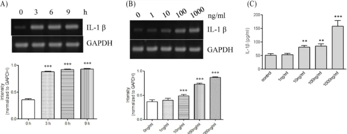
Fig. 1. Expression of IL-1β at the messenger and protein levels in response to PG. (A) THP-1 cells (1x10
6cells/ml) were incubated for indicated time periods with 1 μg/ml PG. IL-1β gene transcripts were amplified by RT-PCR. *** p <0.001 vs. 0 hr. (B) THP-1 cells were incubated for 9 hr with indicated amount of PG. IL-1β gene transcripts were amplified by RT-PCR. *** p <0.001 vs. 0 ng/ml. (C) THP-1 cells (1x10
6cells/ml) were stimulated for 9 hr with or without (control) PG (1 μg/ml). The amount of IL-1β released into the medium was measured by ELISA. Data are expressed as mean±SD (n=3 replicates/group). ** p <0.01 vs. control. *** p <0.001 vs. control.
와 매개된 IL-1β 상향조절에 TLR2, Akt, mammalian target of rapamycin (mTOR), MAPK, and reactive oxygen species (ROS)가 관여하는 것을 발견했다.
재료 및 방법
세포배양 및 시약
THP-1 세포는 American Type Culture Collection (ATCC, Manassas, VA, USA)에서 구매하고 제안한 대로 유지하였다.
황색포도상구균으로부터 분리한 PG인 polymyxin B와 oxi- dized 1-palmitoyl-2-arachidonosyl-sn-phosphatidylcholine (OxPAPC)는 InvivoGen (San Diego, CA, USA)에서 구입하였 다. 내독소가 없는 bovine serum albumin (BSA), LY294002, diphenyleneiodonium chloride (DPI), N-acetylcysteine (NAC), rapamycin과 SP600125는 Sigma-Aldrich Co. (St.
Louis, MO, USA)에서 구입하였다. U0126, SB202190, Akt in- hibitor IV (Akti IV)는 Cell Signaling Technology (Danvers, MA, USA)에서 구입하였다. 억제제 실험에서 THP-1세포를 앞의 화학물질들을 1시간동안 처리하고 PG (1 μg/ml)를 더해 9시간 동안 자극하였고, enzyme linked immunosorbent as- say (ELISA)로 배지에 분비된 IL-1β의 양을 측정하고 reverse transcription (RT)-polymerase chain reaction (PCR)로 IL-1β 유전자 전사체를 증폭시켰다.
IL-1β ELISA
분비된 IL-1β의 양을 상용되는 ELISA kit를 제조업자의 지
시에 따라 이용하여 측정하였다(BD Biosciences, San Diego, CA, USA). THP-1 세포는 0.1% BSA가 들어있는 RPMI me- dium 1640에 하룻밤 동안 배양하고, PG에 노출시킨 후 세포 배양배지를 모았다. 세포배양배지와 IL-1β의 표준을 IL-1β가 붙어있는 단일클론항체로 미리 코팅되어있는 microtiter plate 에 넣어주었다. 2시간 배양 후, plate를 씻고 IL-1β에 특정한 효소-복합 다 클론성 항체를 배양하였다. Plate를 씻고 기질을 첨가하고 배양한 다음 색의 강도를 측정하였다. 배지에 존재 하는 IL-1β의 양을 표준곡선에 따라 계산하였다. ELISA 결과 는 평균±표준편차로 나타내었다.
RT-PCR
THP-1 세포를 0.1% BSA가 들어있는 RPMI medium 1640 에 하룻밤 동안 배양하고 PG에 노출하였다. 전체 RNA를 세포 로부터 추출하고 Moloney murine leukemia virus reverse transcriptase를 이용하여 42°C에서 1시간 동안 역전사하였다.
Primer를 첨가하고 25 cycles (94°C for 30 sec, 55°C for 30
sec, 72°C for 30 sec)을 PCR을 실행하여 IL-1β 전사체를 증폭
시켰다. IL-1β의 primer로 5-GGACAAGGGAGGAAGATGC-
3 (forward)와 5-TCTTTCAACACGCAGGACAG-3 (reverse)
를 사용하고, glyceraldehyde-3-phosphate dehydrogenase
(GAPDH)의 primer로 5-AAGCTCTGCGTGACTGTCCT-3
(forward)와 5-GCTTGCTTCTTTTGGTTTGG-3 (reverse)를 사
용하였다. PCR 결과를 2% agarose gel에서 전기영동에 의해
분리하고 ethidium bromide 염색한 다음 이미지를 사진으로
찍었다. 각 band의 intensity를 IL-1β /GAPDH로 표시하였다.
통계처리
통계적인 분석은 GraphPad PRISM, version 5.0 (GraphPad Software Inc., San Diego, CA, USA)를 사용하여 실행하였고, p<0.05를 통계적으로 의미있는 것으로 간주된다.
결 과
전사체와 단백질단계에서 PG에 의한 IL-1β 발현의 상향조절 대식세포에서 IL-1β 발현에 PG의 영향을 알아보기 위해, IL-1β 발현의 전사수준을 RT-PCR로 조사하였다(Fig. 1A, B).
PG가 없을 때 THP-1 세포에서 IL-1β 전사체가 상당히 약하게 발견된 반면 PG가 존재하는 경우 IL-1β의 전사체의 발현을
Fig. 2. Effects of OxPAPC and polymyxin B on PG-mediated IL-1β up-regulation. (A) THP-1 cells were stimulated for 9 hr with or without PG (1 μg/ml) after treatment for 1 hr with OxPAPC (30 μg/ml) or polymyxin B (10 mg/ml). IL-1β gene transcript was amplified. *** p <0.001 vs. control. ### p <0.001 vs. PG. (B) The amount of IL-1β released into the medium was measured (n=3 repli- cates/group). *** p <0.001 vs. control. ### p <0.001 vs. PG.
유도하였다. IL-1β 전사체의 유도는 PG 처리 후 3시간 뒤부터 관찰되었고 9시간 까지 지속되었다. IL-1β 전사체의 발현은 PG 10 ng/ml이 존재했을 때 유도되었고 100과 1,000 ng/ml에 서는 더 분명해졌다. PG가 사이토카인의 방출에 어떠한 영향 을 주는지 아닌지를 조사하였다(Fig. 1C). THP-1 세포는 본질 적으로 적은 양의 IL-1β를 분비하였고, PG는 IL-1β 단백질 분 비를 상당히 증가시켰다. PG가 없는 control cell과 비교해보 면, IL-1β 분비물의 양은 PG의 양이 10, 100, 1,000 ng/ml 일때 각각 1.5-, 1.6- and 3.0-fold 증가하였다.
PG로 인한 IL-1β 발현에 TLR2/4의 억제가 미치는 영향 PG에 의한 IL-1β 발현에 TLRs의 관련성을 TLR2/4 억제제
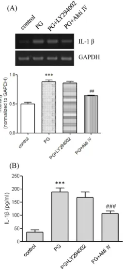
Fig. 3. Effects of LY294002 and Akti IV on PG-mediated IL-1β up-regulation. (A) THP-1 cells were stimulated for 9 hr with or without PG (1 μg/ml) after pretreatment with LY294002 and Akti IV (10 μM each). IL-1β gene tran- script was amplified. *** p <0.001 vs. control. ## p <0.01 vs.
PG. (B) The amount of IL-1β released into the medium was measured (n=3 replicates/group). *** p <0.001 vs.
control. ### p <0.001 vs. PG.
인 OxPAPC을 사용하여 조사하였다(Fig. 2A, B). OxPAPC은 PG와 매개된 IL-1β의 발현을 전사와 단백질 수준에서 억제시 켰다. PG에 의한 IL-1β의 분비는 OxPAPC의 존재 하에 거의 완전히 차단되어 대조군 세포만큼 줄어들고, PG에서 유도된 IL-1β 전사체의 발현도 OxPAPC의 존재 하에 억제되었다. PG 를 준비할 때 lipopolysaccharide (LPS)로 오염될 수도 있고, 오염된 LPS가 염증성 사이토카인과 케모카인의 발현을 증가 시킬 수 있다. 따라서, LPS의 강한 억제제인 polymyxin B를 사용하여 PG에 의한 IL-1β의 상향조절에 LPS가 기여하는지 아닌지를 조사하였다. Polymyxin B는 PG에 의한 IL-1β분비나 전사를 약화시키지 않았다(Fig. 2A, B).
PG로 유도된 IL-1β 발현에 mTOR과 Akt의 역할
PG는 Akt의 인산화를 증가시켜 Akt를 활성화시킨다[14].
Fig. 4. Effects of rapamycin on PG-mediated IL-1β up- regulation. (A) THP-1 cells were stimulated for 9 hr with or without PG (1 μg/ml) after pretreatment with rapa- mycin (100 nM). IL-1β gene transcript was amplified.
*** p <0.001 vs. control. (B) The amount of IL-1β released into the medium was measured (n=3 replicates/group).
*** p <0.001 vs. control. ### p <0.001 vs. PG.
PG에 의한 IL-1β 발현에 Akt가 관여하는 지를 LY294002와 Akti IV 사용하여 조사하였다(Fig. 3A, B). LY294002는 quer- cetin의 morpholine 유도체로 Akt를 활성화시키는 phos- phoinositide 3-kinases (PI3Ks)를 강력히 억제한다. Akti IV 는 Akt단백질 인산화효소의 억제제다. Akti IV는 IL-1β 발현 의 전사와 단백실 수준에 상당히 영향을 주었다. Akti IV는 PG에 의한 IL-1β유전자 발현의 유도를 현저히 감소시키고 IL-1β 분비를 상당히 감소시켰다. 그러나 LY294002는 PG와 매개된 IL-1β의 유전자 전사와 분비에 영향을 주지 않았다.
mTOR은 Akt의 하위분자이고 Akt의 생물학적 효과에 역할 을 담당하므로[7], mTOR의 억제제인 rapamycin을 사용하여 PG에 의한 IL-1β의 발현에 mTOR가 작용을 하는지 조사하 였다(Fig. 4A, B). Rapamycin은 IL-1β 발현에 영향을 주었다.
특히, rapamycin은 PG에 의한 IL-1β의 전사보다는 IL-1β의
Fig. 5. Effects of inhibitors of MAPKs on PG-mediated IL-1β
up-regulation. (A) THP-1 cells were stimulated for 9 hr
with or without PG (1 μg/ml) after pretreatment with
indicated MAPKs inhibitors (10 μM each). IL-1β gene
transcript was amplified. *** p <0.001 vs. control. # p <0.05
vs. PG. (B) The amount of IL-1β released into the me-
dium was measured (n=3 replicates/group). *** p <0.001
vs. control. ### p <0.001 vs. PG.
분비를 상당히 감소시켰다.
PG에 의해 유도된 IL-1β 발현에서 MAPKs의 역할 PG는 extracellular signal-regulated kinase (ERK), p38 MAPK, and c-jun N-terminal kinase (JNK)의 인산화를 증가 시킨다[14]. 이는 PG가 MAPKs를 활성화한다는 의미이다. PG 에 의해 유도된 IL-1β상향조절에서 MAPKs의 역할을 조사하 기 위해, 다음과 같은 MAPK 억제제 -SB202190 (p38 MAPK 억제제), SP600125 (JNK 억제제), U0126 (ERK 억제제)를 사용 하였다(Fig. 5A, B). U0126과 SP600125는 PG가 매개한 IL-1β 단백질의 분비 뿐만 아니라 PG가 유도한 IL-1β 전사체를 감소 시켰다. 반면, SB202190는 PG가 매개한 IL-1β유전자 전사와 단백질 분비를 억제하지 않았다.
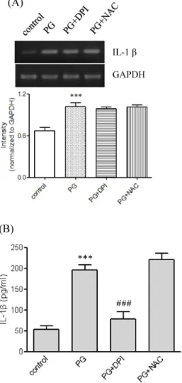
Fig. 6. Effects of ROS quenchers on PG-mediated IL-1β up-regulation. (A) THP-1 cells were stimulated for 9 hr with or without PG (1 μg/ml) after pretreatment with DPI (10 μM) or NAC (5 mM). IL-1β gene transcript was amplified. *** p <0.001 vs. control. # p <0.05 vs. PG (B) The amount of IL-1β released into the medium was meas- ured (n=3 replicates/group). *** p <0.001 vs. control.
### p <0.001 vs. PG.
PG로 유도된 IL-1β 발현에서 ROS의 역할
IL-1β의 발현에 ROS가 역할을 하는지를 NAC와 DPI를 사 용하여 조사하였다(Fig. 6A, B). NADPH산화효소의 억제제인 DPI는 PG가 매개한 IL-1β 분비를 상당히 억제하였고 PG가 매개한 IL-1β유전자 전사에는 약간의 영향을 주었다. 그러나 ROS의 직접적 scavenger인 NAC는 PG가 매개한 발현에 영향 을 주지 않았다.
고 찰
본 연구는 죽상동맥경화증 병변에 세균의 구성요소로 존재 하는 PG가 전사와 단백질 수준을 IL-1β 발현에 상향조절 한다 는 것을 대식세포주인 THP-1세포를 사용하여 증명하였다. 이 결과는 이전에 연구된 ribonuclease 보호분석을 사용하여 인 간의 혈액 단핵세포에서 PG와 LPS에 의한 IL-1α와 IL-1β 발현 이 유도된다는 Wang et al [27]의 결과와 유사하다. PG는 IL-1 β 유전자 전사의 발현을 유도시키고, PG가 없으면 IL-1β분비 의 증가는 감지되지 않는다. PG에 의한 IL-1β분비는 상당하긴 하지만, IL-1β전사 유도의 범위에서는 중요하게 고려되지 않 았다. 그것은 PG에 THP-1 세포가 노출되면 Dinarello et al [4]에 의해 가정된 것처럼 두번째 자극의 존재에서 활동적인 IL-1β의 더 많은 양의 분비를 할 수 있다는 것을 가정할 수 있다.
PG가 IL-1β의 발현을 유도하는 것이 분명하다 하더라도, IL-1β 발현에 관련된 세포분자는 아직 명확하지 않다. 본 연구 는 PG에 의한 IL-1β 발현에서 역할을 하는 세포인자를 확인하 였다. PG는 염증반응으로 이어지는 신호통로의 활성화를 유 도하는 TLR2에 의해 세균성 PAMP를 인식한다[1,10,26]. 따라 서 TLR2/4 저해제인 OxPAPC을 이용하여 IL-1β의 발현에 TLR2/4가 관여하는지를 조사하였다. OxPAPC은 IL-1β의 분 비와 전사를 완전히 차단한다. 또한 발현은 완전히 억제되고, polymyxin B는 L-1β 발현을 억제하지 않았다. 이 결과는 PG 에 의해 유도된 IL-1β 발현에 TLR4 보다는 TLR2가 관여한다 는 것을 의미한다.
PG는 Akt 인산화를 증가시키는데[14], 이 사실은 PG가 Akt
를 활성화시킴을 의미한다. 따라서 Akt와 Akt 활성인자인
PI3K [16]가 PG와 매개된 IL-1β 발현에 연관되는지를 조사하
였다. Akt 억제제는 IL-1β의 분비를 완전히 차단하는데 반하
여 PI3K 억제제는 IL-1β 발현을 억제하지 못했다. 이 결과는
PG가 매개한 IL-1β에 Akt가 필요하다는 것을 의미한다. Akt
는 mTOR을 포함한 protein target을 통해 생물학적 효과를
달성한다[7]. 따라서 mTOR가 PG에 의한 IL-1β 발현에 연관되
어 있는지를 조사하였다. mTOR의 억제제인 rapamycin은
IL-1β분비를 상당히 감소시키는 반면 유전자의 전사는 조금
감소시킨다. 이 결과는 rapamycin이 PG에 의한 IL-1β 발현을
유전자수준보다는 단백질 수준에서 억제시킴을 의미한다. 그
리고 이 결과는 단백질 합성에 mTOR가 책임이 있다는 것과 세포의 번역조직의 켜짐과 꺼짐에 영향을 준다는 사실과 일치 한다[20]. 떠라서 Akt/mTOR 경로가 PG와 매개된 IL-1β 상향 조절에 중요한 역할을 한다는 것을 확인하였다.
MAPKs는 serine/threonine-specific protein kinases로 세 포밖의 자극에 반응하고 다양한 세포의 활성을 조절하며 PG 에 의해 활성화 된다[14]. MAPKs는 TLR-2, -4, -9 신호에서 케모카인 생산에 중요하다[25]. 그러므로 IL-1β 발현에 MAPKs 가 역할을 하는지를 조사하였다. p38 MAPK는 아니지만 ERK 와 JNK의 선택적 억제는 IL-1β 발현을 상당히 감소시켰다.
이 결과는 PG에 매개된 IL-1β 발현에 MAPKs의 활성화(특히 ERK와 JNK)가 요구된다는 것을 가리킨다. TLR2는 PG의 반응 에서 ROS 생산에 영향을 준다는 보고[9]는 ROS가 TLR2보다 아래에서 작용하는 분자라는 것을 의미한다. 본 연구는 DPI와 NAC를 사용하여 ROS 또한 IL-1β 발현에 포함되는지 아닌지 를 조사하였다. DPI는 ROS를 생산하는 NADPH oxidase의 억제제이고, 이를 통해 ROS형성을 억제한다[17]. 세포 내 glu- tathione의 repletion을 위한 cysteine source 역할을 하는 thiol 복합체인 NAC는 ROS의 직접적인 scavenger 역할을 한다 [23]. DPI는 PG와 매개된 IL-1β의 상향조절을 상당히 감소시 켰다. 이 결과는 ROS가 PG에 의한 IL-1β 상향조절에 NADPH oxidase가 활발히 관여함을 나타낸다.
본 연구에서는 THP-1 세포가 PG에 노출되면 IL-1β 분비 증가와 IL-1β 유전자 전사의 유도에 대한 결과를 보여주었고, 그 과정에 TLR2, Akt, mTOR, ERK, JNK, ROS가 참여한다는 것을 밝혔다. 이후의 연구에서는 이 분자들의 신호 cascade의 전후관계를 나타내는 connections나 crosstalk의 유형을 밝힐 필요가 있다.
감사의 글
이 과제는 2011년도 부산대학교 교수국외장기파견지원비 에 의하여 연구되었음.
References