J Korean Soc Pediatr Nephrol 2012;16:32-37 http://dx.doi.org/10.3339/jkspn.2012.16.1.32
Copyright © 2012 The Korean Society of Pediatric Nephrology ISSN 1226-5292 (print) ISSN 2234-4209 (online)
방광요관역류를 가진 소아에서의 신초음파 소견
연세대학교 의과대학 소아과학교실*, 아주대학교 의과대학 소아과학교실†, 국민건강보험공단 일산병원 소아청소년과‡ 최민정* · 박세진*,† · 신재일* · 김기혁‡
Ultrasonographic Findings in Children with Vesicoureteral Reflux
Purpose: The aim of this study is to investigate the renal ultrasono graphic
findings in children with vesicoureteral reflux (VUR).Methods: We retrospectively reviewed the medical records of 83 patients who
were diagnosed with VUR and underwent ultrasono graphy at Ilsan hospital between January 2000 and December 2010.Results: Among 166 renal units, 108 (65.0%) were found to have vesicoureteral
reflux (VUR). Fifty-one (73.9%) had VUR in renal units with abnormal ultra- sonography (USG), whereas 57 (58.7%) had VUR in renal units with normal USG. Abnormal USG findings were independent risk factors for VUR (Odds ratio, 1.98; 95% CI, 1.01-3.89; P=0.045). In renal units with VUR, the number of normal USG finding was 52.8%, and the abnormal findings were as follows; increased cortical echogenicity 16.7%, hydronephrosis 17.6%, megaureter or ureter dilatation 8.3%, hydronephrosis and ureter dilatation 1.9%, duplication of ureter 1.9%, and atrophic kidney 0.9%. The prevalence of VUR was relatively higher in renal units with hydronephrosis (23/19, 82.6%), ureter dilatation (9/9, 100%), duplication of ureter (2/3, 66.6%), and atrophic kidney (1/1, 100%).Conclusion: Our study indicates that VUR was associated with abnormal USG
findings. When there are abnormal USG findings such as hydronephrosis, ureter dilatation, duplication of ureter, and atrophic kidney in children with UTI, VCUG is recommended to detect VUR after controlling UTI.Key Words: Vesicoureteral reflux, Renal ultrasonography, Children Min Jung Choi, M.D.*,
Se Jin Park, M.D.*
,†, Jae Il Shin, M.D.*,
and Kee Hyuck Kim, M.D.
‡The Institute of Kidney Disease, Department of Pediatrics*, Yonsei University College of Medicine, Severance Children’s Hospital, Seoul, Korea, Department of Pediatrics
†, Ajou University School of Medicine, Ajou University Hospital, Suwon, Korea, Department of Pediatrics
‡, National Health Insurance Corporation Ilsan Hospital, Goyang, Korea
Corresponding Author: Kee Hyuck Kim Department of Pediatrics, National Health Insurance Corporation Ilsan Hospital, Goyang, Korea
Tel: 031-900-0520, Fax: 031-900-0049 E-mail: [email protected]
Min Jung Choi and Se Jin Park contributed equally to this work
Received: 5 October 2011 Revised: 7 October 2011 Accepted: 1 December 2011
This is an open-access article distributed under the terms of the Creative Commons Attribu tion Non- Commercial License (http://crea tivecom mons.org/
licenses/by-nc/3.0/) which permits unrestricted non-commercial use, distribution, and reproduction in any medium, provided the original work is properly cited.
서론
방광요관역류는 소아 요로감염과 신반흔을 일으키는 중요한 원인 중의 하 나이며, 요로감염이 있는 소아의 약 25-50%에서 역류가 있으며, 방광요관역 류가 있는 소아의 30-49%에서 신반흔이 동반된다[1, 2]. 배뇨성 방광요도조
영술(voiding cystourethrography, VCUG)은 소아에서 요 로감염이 있을 때, 선천성 수신증, 후부요도판막, 방광게실 등 기타 선천성 신장질환 등이 있을 때 동반된 방광요관역 류를 확인하기 위해 시행하는 검사로[3] 미국 소아과학회 에서는 어린 소아(2개월에서 2세)에서 방광요관역류의 진 단을 위해 조기에 배뇨성 방광요도조영술을 시행할 것을 권고하고 있다[4]. 그러나 배뇨성 방광요도조영술은 침습 적인 도뇨관 삽입과 대량의 X-선에 대한 노출 등이 문제가 될 뿐 아니라 대부분의 방광요관역류는 자연 소실율이 높 은 I등급이나 II등급이라는 연구결과들이 있어서, ‘모든 요 로감염 소아에게 배뇨성 방광요도조영술이 반드시 필요한 가?’에는 논란이 있다[5]. 지금까지 침습적인 배뇨성 방광 요도조영술을 대체할 임상적, 영상학적, 혈액학적 예측 지 표에 대한 여러 가지 보고가 있었으나[5, 6] 아직까지는 소 아에서 방광요관역류의 진단을 위해 배뇨성 방광요도조영 술을 시행하는 것이 일반적인 방법이다[7]. Mahant 등[8]과 Alshamsan 등[9]은 신초음파가 첫 요로감염을 가진 소아 에서 방광요관역류를 예측하는데 크게 의미가 없다고 하였 으나, 최근 Ismaili 등[10]은 신초음파 소견과 중증 방광요관 역류사이의 연관성이 있다고 하였다.
본 연구는 방광요관역류가 있는 소아에서 신초음파 이상 유무와 방광요관역류와의 연관성을 알아보고, 신초음파 이 상 소견을 방광요관역류의 예측 지표로 사용할 수 있는지 알아보고자 시행하였다.
대상과 방법
2000년 1월부터 2010년 12월까지 일산병원 소아청소년 과에 내원하여 요로감염증으로 입원한 후 배뇨성 방광요 도조영술을 시행하여 방광요관역류를 진단받은 총 83명의 소아를 대상으로 후향적 분석을 시행하였다[11].
대상 소아들은 요로감염을 처음 진단받았으며, 영상 검 사로 신초음파(renal ultrasonography), 배뇨성 방광요도 조영술(voiding cystourethrography)를 시행하였다. 영상 검사는 영상의학과 의사와 핵의학과 의사 각각 1명에 의해 판독되었다. 신초음파 검사의 이상소견은 증가된 신피질 음영, 수신증, 요관확장 또는 거대요관, 수신증과 요관확장 이 같이 있는 경우, 중복요관, 신장 무형성, 손상된 위축신 으로 구분하였다.
배뇨성 방광요도조영술은 소변 배양 검사가 음성으로 확인된 후 시행하여 방광요관역류 여부를 검사하였고, 방 관요관역류의 세부등급은 국제소아역류연구회의 방광요관
역류 등급분류(International Reflux Study of Committee, 1981)에 따라 grade I-II를 경도(low grade), grade III를 증 등도(moderate grade), grade IV-V를 증증(high grade)으 로 분류하였다[5].
자료의 통계학적 분석은 SPSS for windows version 16 (Chicago, Illinois, USA)를 이용하였으며, 방광요관역류와 신초음파 이상소견과의 관계는 t-test, chi-square test, 로 지스틱 회귀모형(logistic regression anaylsis)을 이용하여 분석하였다. 통계적 유의수준은 P값이 0.05 미만인 경우를 유의한 것으로 정의하였다.
결과
1. 연구 대상 소아의 연령과 성별 분포
대상 소아는 모두 83명으로 전체 166 신단위(renal unit) 중 방광요관역류는 108신단위에서 관찰되었으며, 남아 가 43명(51.8%), 여아는 40명(48.2%)으로 전체 남녀 비는 1.1:1이었다. 이 중 1세 미만의 평균 나이는 3.1±2.56개월 (median 2개월, range 1-12개월)이었으며, 1세 이상의 평 균 나이는 58.9±43.01개월(median 32개월, range 13-170 개월)이었다.
2. 방광요관역류의 빈도
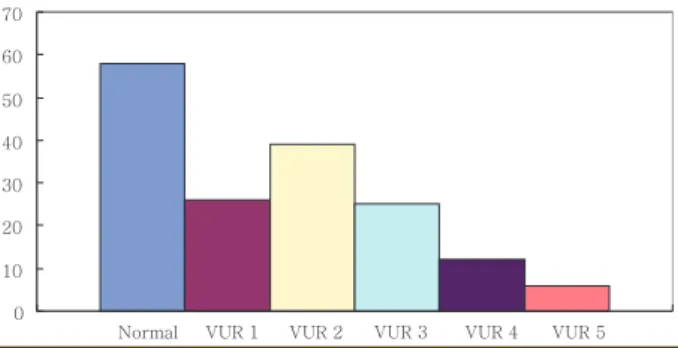
방광요관역류를 5단계로 나누어 보았을 때, 정상소견을 보이는 경우는 58신단위(34.9%), 1단계 방광요관역류는 26 신단위(15.7%), 2단계 방광요관역류는39 신단위(23.5%), 3 단계 방광요관역류는 25 신단위(15.1%), 4단계 방광요관 역류는 12 신단위(7.2%), 5단계 방광요관역류는 6 신단위 (3.6%)로 각각 관찰되었다(Fig. 1).
0 10 20 30 40 50 60 70
VUR 5 VUR 4 VUR 3 VUR 2 VUR 1 Normal
Fig. 1. Prevalence according to the grade of vesicoureteral reflux.
3. 나이, 성별과 방광요관역류와의 관련성
방광요관역류가 없는 58 신단위에서의 평균 나이는 41.8±
46.8개월이었고, 방광요관역류가 있는 108 신단위에서의 평 균 나이는 37.0±41.7개월이었으며, 두 그룹은 통계적으로 유 의한 차이가 없었다(P=0.502). 또한 심한 방광요관역류가 있 는 18신단위에서의 평균 나이는 24.1±34.19개월 이었고, 심 한 방광요관역류가 없는 90 신단위에서의 평균 나이는 39±
42.74개월이었으며, 두 그룹은 통계적으로 유의한 차이가 없 었다(P=0.151).
남아에서 방광요관역류 소견이 없는 신단위와 방광요관역 류를 보이는 신단위는 각각 32.5% (28/86), 67.4% (58/86)였 고, 여아에서 방광요관역류 소견이 없는 신단위와 방광요관 역류를 보이는 신단위는 각각 37.5% (30/80), 62.5% (50/80) 이었다. 역류가 있는 남녀 신단위에서 두 그룹은 통계적으 로 유의하지 않았다(P=0.519). 또한 심한 방광요관역류가 있 는 남아의 신단위는 22.4 % (13/58), 여아의 신단위는 10%
(5/50)이었으며 두 그룹은 통계적으로 유의한 차이가 없었다 (P=0.12).
4. 신초음파 이상소견과 방광요관역류의 유무
신초음파 이상소견을 보이는 경우 방광요관역류가 있는 신 단위는 73.9% (51/69), 신초음파 정상소견인 경우 방광요관 역류가 있는 신단위는 58.7% (57/97)이었다(Table 1). 신초음 파 이상소견 유무에 따른 두 그룹간에는 통계적으로 유의한
차이가 있었다(P=0.049).
신초음파 이상소견이 방광요관역류를 예측하는 독립인 자가 되는지 알아보기 위해 로지스틱 회귀분석을 시행하였 을 때, 신초음파 이상소견이 있으면 방광요관역류가 있을 확 률은 약 2배 가량 증가하였으며 이는 통계적으로 유의하였 다(Odds ratio 1.98; 95% confidence interval 1.01-3.89;
P=0.045).
방광요관역류가 있는 신단위에서 초음파 정상소견을 보 이는 경우는 52.8% (57/108), 피질음영증가 16.7% (18/108), 수신증 17.6 % (19/108), 요관확장 8.3% (9/108), 수신증을 동반한 요관확장 1.9% (2/108), 중복요관 1.9% (2/108), 신 장 무형성 0% (0/108), 손상된 위축신 0.9% (1/108)이었다 (Table 2). 이 중 수신증(57/97, 82.6%), 요관확장(9/9, 100%), 중복요관(2/3, 66.6%), 손상된 위축신(1/1, 100%)이 있는 경 우 방광요관역류가 발견될 빈도가 상대적으로 높았다.
고찰
소아 요로감염의 진단에 방사선 검사가 중요한 이유는 요 로감염에 선행되는 선천성 기형, 신반흔 그리고 기능적 이상 등을 찾아내는데 큰 역할을 하기 때문이다. 요로감염은 소아 기의 흔한 세균성 감염으로 성인과 달리 요로계의 선천 기 형을 동반하는 경우가 많다. Hsieh 등[12]은 130명의 첫 발 열성 요로감염 소아들을 대상으로 한 연구에서 81.6%에서 비뇨기계 기형을 발견하였다고 보고하였으며, 이를 바탕으 로 첫번째 요로감염 소아들에게서 신속한 영상의학적 평가 가 중요하다고 언급하였다. 영유아의 요로감염은 요로 기형 동반 문제 외에도 10-30%의 소아에서 급성 신우신염과 신 반흔을 일으키게 되며, 추후 고혈압과 만성 신부전까지도 초 래할 수 있으므로 요로감염 발생시 영상의학적 조기 진단과 치료가 매우 중요하다[5].
방광요관역류는 요관방광 이행부의 판막 기능부전으로 인
Table 1. Results of Vesicoureteral Reflux in Relation to Ultrasono-
graphic Findings
Findings VUR (-) VUR (+) Total renal units Abnormal USG 18 (26.0%) 51 (73.9%) 69 (100%) Normal USG 40 (41.2%) 57 (58.7%) 97 (100%) Abbreviation: VUR, vesicoureteral reflux
Table 2. Ultrasonographic Findings in Renal Units with VUR
USG findings VUR (-) VUR (+) Total renal units
Normal 40 (41.2%) 57 (58.7%) 97 (100%)
Increased cortical echogenicity 10 (35.7%) 18 (64.2%) 28 (100%)
Hydronephrosis 4 (17.4%) 19 (82.6%) 23 (100%)
Megaureter or Ureter dilatation 0 (0%) 9 (100%) 9 (100%)
Hydronephrosis + Ureter dilatation 2 (50%) 2 (50%) 4 (100%)
Duplication of ureter 1 (33.3%) 2 (66.6%) 3 (100%)
Renal agenesis 1 (100%) 0 (0%) 1 (100%)
Atrophic kidney 0 (0%) 1 (100%) 1 (100%)
Abbreviation: VUR, vesicoureteral reflux
해 소변이 방광에서 요관, 신우로 역류되어 나타나는 질환 으로, 신손상의 위험성이 있는 발열성 요로감염 소아에서 방광요관역류를 확인하는 것은 매우 중요한 문제이나, 방 광요관 검사와 관련되어 일부 연구에서는 시술 후 소변 배 양 검사를 실시한 후 요로 감염, 전신적 알레르기 반응, 접 촉성 피부염, 요도관 꼬임 등의 발생을 보고 하였다[13, 14].
또한, Zerin 등[15]에 의하면 배뇨성 방광요관조영술 시행 후 일부 소아에서 방광요관 검사와 관련되어 배뇨곤란, 보 챔, 발열, 빈뇨 등의 증상을 보였다. 따라서 요로감염 환자, 특히 처음으로 요로감염에 이환된 소아에서 일률적으로 침습적인 배뇨성 방광요도조영술을 시행해야 할 것인가에 대한 검토가 요구된다. 또한 첫 발열성 요로감염 소아에게 발생한 신손상에 대한 영상의학적 평가에 대해 지속적인 연구가 이루어지고 있으나, 아직 공통되는 진단방법은 확립 되어 있지 않은 상태로, 1999년 미국 소아과 학회에서는 첫 발열성 요로감염 소아에게 복부 초음파와 배뇨성 방광요 도조영술을 시행하는 것을 권고하였으나[4], Hansson 등 [16]은 복부 초음파와 99mTc-DMSA 신스캔을, Kass 등[17]
복부 초음파, 배뇨성 방광요관조영술과 99mTc-DMSA 신스 캔을 시행하도록 권장하였다.
과거 영아 요로감염에서 방광요관역류를 예측하는데 있 어 컬러 도플러 초음파가 유용하다는 연구결과[18]와 첫 발열성 요로감염 소아에서 방광요관역류를 예측하는데 배 뇨 중 신초음파가 유용하다는 연구[19]가 있었으나 이후 에는 신초음파가 방광요관역류를 예측하는데 도움이 되 지 않는다는 의견이 대부분이었다. Foresman 등[20]은 영 아 요로감염에서 급성기의 신우신염에 시행한 신초음파는 방광요관역류의 유무나 정도와 상관성이 떨어진다고 하였 고 Mahant 등[8], Alshamsam 등[9], Zamir 등[21]도 첫 발 열성 요로감염의 소아에서 방광요관역류를 예측하는데 신 초음파의 민감도와 특이도가 낮다고 하여 신초음파가 방광 요관역류를 예측하는데 있어서 그 효용성이 떨어진다고 보 았다. 뿐만 아니라 Kang 등[22]은 1개월 이상 3개월 미만의 남아(30명)에서 초음파 소견이 정상인 경우 방광요관역류 가 있을 위험도가 유의하게 낮았다고 하였다.
그러나 방광요관역류를 예측하기 위한 신초음파의 효용 성 연구는 지속되었으며 신초음파 소견 중 원위요관 확장 이나 신배의 간헐적인 확장은 방광요관역류의 중요한 예측 인자가 될 수 있다는 연구결과가 있었다[23, 24]. 최근 Lee 등[6]은 첫 발열성 요로감염으로 진단받은 220명의 소아 를 대상으로 한 후향적 연구에서 신초음파를 통한 경도 의 방광요관역류 예측도는 41.7%, 중등도의 방광요관역류 예측도는 86%이라고 하였으며, Ismaili 등[10] 역시 신초음파
를 통한 중등도의 방광요관역류 민감도는 97%, 특이도는 94%로 경도의 방광요관역류를 제외한 방광요관역류를 진 단하는데 신초음파가 유용하다고 하였다. Hannula 등[25]
도 첫 발열성 요로감염 소아에서 신초음파는 방광요관역 류를 진단하기 위한 일차적 선별검사로서 이용될 수 있다 고 하였으며, 더 나아가 간접 배뇨 초음파(indirect voiding US)와 요관 제트 도플러 파형 분석(ureteral jet Doppler waveform analysis)은 배뇨성 방광요도조영술에 비해 방 사선 노출이 적고 덜 침습적으로 3세 이상의 소아에서 배 뇨성 방광요도조영술을 대신할 수 있는 비교적 안전하고 유용한 검사라고 하였다[26].
신초음파는 비침습적이고 방사선에 대한 노출이 없으며 비용이 비교적 적게 들어 수신증 등의 폐색성병변이 있을 때 요로계의 구조와 이상 유무를 비교적 손쉽게 알아볼 수 있는 장점이 있으나[5], 방광요관역류나 신실질의 이상을 정확히 평가하기에 어렵다는 단점이 있다[7].
본 연구에서는, 나이, 성별은 방광요관역류 와 4단계 이 상의 심한 방광요관역류와 연관성을 보이지 않았고, 신초 음파 이상이 있는 신단위에서 방광요관역류의 빈도가 상대 적으로 높았다(73.9% vs. 58.7%; P=0.049). 또한 신초음파 이상소견은 방광요관역류을 예측하는 독립적 인자였다. 그 러므로 신초음파 이상소견이 방광요관역류의 중증도까지 예측하는 것은 가능하지 않으나 어느 정도 방광요관역류 자체를 예측하는 것은 가능하였다. 또한 방광요관역류가 있는 신단위의 경우, 나타나는 신초음파 이상소견으로는 수신증 17.6% (19/108), 피질음영증가 16.7% (18/108), 요관 확장 8.3% (9/108) 순으로 많아 이러한 신초음파 이상소견 이 보일 때 배뇨성 방광요도조영술의 시행이 필요할 것으 로 사료된다.
요약
목적: 본 연구는 방광요관역류를 보이는 소아에서 신초 음파 이상소견에 대해 알아보고자 하였다.
방법: 2000년 1월부터 2010년 12월까지 일산병원 소아 청소년과에 내원하여 요로감염증으로 입원한 후 배뇨성 방 광요도조영술을 시행하여 방광요관역류를 진단받은 83명 의 소아를 대상으로 총 166 신단위를 후향적으로 분석하 였다.
결과: 대상 소아 중 1세 미만의 평균 나이는 3.1±2.56개 월이었고, 1세 이상의 평균 나이는 58.9±43.01개월이었다.
신초음파 이상소견을 보이는 신단위에서 방광요관역류가
있는 경우는 73.9%였고, 신초음파 정상소견을 보이는 신단 위에서 방광요관역류가 있는 경우는 58.7%였으며 이는 통 계적으로 유의한 차이를 보였다(P=0.049). 로지스틱 회귀 분석에서 신초음파 이상소견이 있을 경우 방광요관역류가 있을 확률은 약 2배 증가하였다. 방광요관역류가 있는 신단 위에서 신초음파 정상소견을 보이는 경우는 52.8%, 피질음 영증가 16.7%, 수신증 17.6%, 요관확장 8.3%, 수신증을 동 반한 요관확장 1.9% 등이었다. 이 중 수신증(82.6%), 요관 확장(100%), 중복요관(66.6%), 손상된 위축신(100 %)이 있 는 경우 방광요관역류의 빈도가 상대적으로 높았다.
결론: 방광요관역류가 있는 신단위에서 신초음파 이상소 견으로 방광요관역류의 중증도를 예측하는 것은 가능하지 않으나 방광요관역류의 존재를 예측하는 것은 가능하다.
그러므로 수신증, 요관확장 같은 신초음파 이상소견이 있 을 시, 배뇨성 방광요도조영술의 시행이 필요할 것이다.
References