YD-10B에서 Cisplatin과 백작약의 병용처리에 의한 항암 효과
Anticancer Effects of Cisplatin in Combination with Paeonia Japonica in YD-10B Cells
상지대학교 보건의료과학대학 임상병리학과김은정
Eun-Jung Kim([email protected]) 요약
본 연구에서는 시스플라틴과 백작약 에틸아세테이트 분획물의 병용 처리에 의한 암세포 성장억제 및 PMA 에 의해 유도된 MMP-2 및 MMP-9 암전이 억제 효과를 조사하였다. 세포생존율 측정은 MTS법에 의해 조사 하였고, MMP-2/-9의 유전자발현과 활성은 RT-PCR과 Zymography법을 통하여 확인하였다. 결과에 의하 면, 백작약, 시스플라틴의 농도가 증가함에 따라 세포 성장억제 효과가 증가함을 보였다. 또한, 단독 처리에 비해 200 μM의 시스플라틴과 50 μg/ml의 백작약 병용 처리에 의해서는 YD-10B 세포의 성장이 50% 감소 하였다. PMA 처리된 YD-10B 세포에서 50 μg/ml의 백작약과 200 μM의 시스플라틴을 병용 처리하였을 때, MMP-2 및 MMP-9의 mRNA 발현과 단백질 활성들이 모두 유의하게 억제하였다. 그러므로 본 연구에서 는 시스플라틴과 백작약의 병용 처리는 시스플라틴 단독 처리보다 구강암의 암 침윤을 억제할 수 있는 효과적 인 항암제로서의 가능성을 기대할 수 있다.
■ 중심어 :∣병용처리∣시스플라틴∣백작약∣항암효과∣구강암세포주∣
Abstract
The present study investigated the anti-proliferate and anti-invasive of Phorbol 12-myristate 13-acetate (PMA)-induced matrix metalloproteinase (MMP-2) and MMP-9 activities of combined treatment with cisplatin and ethyl acetate fractions of Paeonia japonica. Cell Proliferation was detected by the MTS assay and the activity and mRNA expression of MMP-2/-9 were examined by zymography and RT-PCR. As results, cisplatin or p. japonica treatment of YD-10B cells resulted in a dose-dependent inhibition of cell growth. Also, the viability of YD-10B cells treated with combination of 200 μM cisplatin and 50 μg/ml p. japonica was inhibited to 50% in compared with the cisplatin alone.
In PMA-treated YD-10B cells, co-treatment of 200 μM cisplatin with 50 μg/ml p. japonica significantly inhibited mRNA expression and protein activation of MMP-2/-9. Therefore, This study suggest that the combination treatment of cisplatin and p. japonica potentiates a promising anti-invasive agent and has more potential anti-cancer drug for oral cancer therapy than cisplatin alone.
■ keyword :∣Combination∣Cisplatin∣Paeonia Japonica∣Anticancer∣YD-10B Cells∣
* 본 연구는 2018년도 상지대학교 교내연구비 지원을 받아 수행되었습니다.
접수일자 : 2020년 05월 07일
수정일자 : 2020년 05월 27일 심사완료일 : 2020년 05월 28일
교신저자 : 김은정, e-mail : [email protected]
I. 서 론
Cisplatin(CDDP;cis-diammineplatinum(II) dichloride)은 백금원자를 포함하고 있는 화합물로서, DNA 이중가닥 내 구아니딘(guanidine) 염기와 공유 결합하여 DNA손상을 유도하고, 결국에는 DNA복제가 억제됨으로 암세포 성장과 증식을 억제시키는 작용을 한다. 화학항암제 중에서 다양한 고형암 치료제로 50년 이상 널리 사용되어져 오고 있으며[1][2], 구강암을 치 료하는데 있어서도 가장 효과적이어서 단독 또는 병용 으로 가장 흔하게 사용되고 있다[3-5]. 그러나 cisplatin은 약제 내성 및 신장기능 감소 등 여러 가지 심각한 부작용이 나타나므로 이를 줄이기 위한 연구가 꾸준히 진행되고 있다[6-8].
천연물에 함유된 화합물은 항산화 효과, 항염증 효과 [9-11] 및 항암 효과등 다양한 약리활성을 나타내는 것 으로 보고되고 있다[12-15]. 최근 천연물로부터 개발된 물질들이 기존 항암제에 비해 독성이 적은 안전한 화학 물질로서, 항암효능을 갖고 있어 천연물에 대한 항암작 용에 대한 연구가 증대되고 있다. 최근 오랫동안 한약 재로 사용되어 온 백작약(p. japonica)이 항산화, 암전 이 억제 및 암세포사멸 능력이 있음을 보고하였다[16].
하지만 아직까지는 백작약에 대한 항암효과에 대한 연 구는 부족한 실정이다.
구강암의 발병률은 전체 악성종양의 2~5%, 사망률 은 전체 암의 2~3%로 보고되고 있으며, 구강암의 가장 흔한 형태는 80~90%를 차지하고 있는 구강편평세포암 종(oral squamous cell carcinoma)이다[17][18]. 구 강암을 치료하는 방법으로는 외과적 절제 수술, 방사선 요법, 화학항암제 요법 및 면역요법 등이 사용되어져 오고 있으며, 진행 및 전이 암에 대해서는 외과적 및 방 사선 또는 화학항암제와의 병용요법을 통해 생존율을 높이고 있다[3][4][19]. 하지만 이들 치료의 가장 큰 문 제점은 고형화된 종양을 치료표적으로 두고 있어 심각 한 치료부작용을 초래한다는 것이며, 재발 또는 전이성 종양에 대한 치료법은 여전히 미흡한 실정이다. 그러므 로 최근에는 종양치료의 한계를 극복하기 위해, 항암효 과를 나타내는 천연물에 대한 관심이 증가되고 있다 [20-22]. 따라서 본 연구에서는 구강암세포주를 사용하
여 다양한 고형암 치료를 위해 많이 사용되고 있는 항 암제인 cisplatin과 항암효과를 가진 백작약을 저농도 로 병용 투여하여 암세포증식 및 암전이 억제 효과를 조사하고자 한다.
II. 재료 및 방법
1. 시료 및 시약
백작약 에틸아세테이트(ethyl acetate) 분획물은 기 존연구에서 사용된 동일한 시료를 사용하였다[16].
Cisplatin은 (Sigma-Aldrich, St. Louis MO, USA)에 서 구입하여 사용하였다.
2. 세포배양
YD-10B 세포, human oral squamous carcinoma cells는 한국세포주은행(Korea Cell Line Bank, Seoul, Korea)에서 분양 받아 사용하였다. 세포 는 RPMI-1640 (Gibco-BRL, Life technologies Inc., Grand Island, New York, USA)배지에 10%
fetal bovine serum (Gibco-BRL), 1%
penicillin/streptomycin (Gibco-BRL)을 첨가하여 37℃, 5% CO2조건의 배양기에서 계대배양 하였다.
3. 세포생존율 측정
세포생존율을 조사하기 위해, cell counting kit-8 (CCK-8; Dojindo Laboratories, Kumamoto, Japan)을 이용하여 측정하였다. YD-10B 세포를 96-well plate에 well (200 μl)당 2 × 10⁴개의 수로 seeding하고 12시간 동안 배양한 후, 백작약과 cisplatin을 단독 또는 병용 처리하였다. 그리고 추가 적으로 37℃ 배양기에서 24시간 그리고 48시간 동안 반응시킨 후, CCK-8 용액을 세포배양액에 10 μl/well 을 첨가하고 1시간 동안 37℃ 배양기에서 반응하였다.
반응 후, microplate reader (Molecular Devices, Sunnydale, CA, USA)를 이용하여 450 nm에서 흡광 도를 측정하였다.
4. RT-PCR
세포로부터 TRIzol 시약(Invitrogen Co., Carlsbad, CA, USA)을 사용하여 총 RNA를 추출하였 다. 1 μg의 RNA를 가지고 ReverTra ACE PCR RT master mix kit (TOYOBO Co., Osaka, Japan)을 이용하여 PCR를 수행하였다. 본 실험에서는 MMP-2 (matrix metalloproteinase-2), MMP-9 (matrix metalloproteinase-9) 및 GAPDH (glyceraldehyde-3-phosphate dehydrogenase) primers[Table 1]을 사용하였고, housekeeping 유 전자인 GAPDH 유전자를 internal control로 사용하 였다. 증폭된 PCR 생성물은 ethidium bromide가 포 함된 1.5% agarose gel에 전기영동 하여 UV light상 에서 확인하였다.
Table 1. Primers for RT-PCR
Gene Sequence (5’-3’) Annealing temperature
product PCR size MMP-2 GCGACAAGAAGTATCGCTTC TGCCAAGGTCAATGTCAGGA 58℃ 390bp MMP-9 CCATTTCGACGATGACGAGTTCTTGTCGCTGTCAAAGTTCGAG 58℃ 530bp GAPDH GAAGGTGAAGGTCGGAGT GAAGATGGTGATGGGATTTC 58℃ 226bp
5. Gelatin zymography
백작약과 cisplatin을 단독 또는 병용 처리한 YD-10B (5 × 10⁵) 세포를 24시간 동안 배양한 후, 배지를 모아 centriprep YM-10 (Millipore, Billerica, MA, USA)을 사용하여 농축하였다. 20 μg 농도의 단백질은 non-reducing sample buffer (0.5 M Tris-Cl pH 6.8, 5% SDS, 20% glycerol, 1%
bromphenol blue)와 함께 혼합하여 0.1% gelatin이 포함된 10% SDS-PAGE에서 전기영동 하였다. 전기영 동 후, washing buffer (50 mM Tris-Cl, pH 7.5, 10 mM CaCl2, 2.5 % triton X-100, 1.0 uM ZnCl2)로 SDS을 제거하고 incubation buffer (50 mM Tris-Cl, pH 7.5, 10 mM CaCl2,150 mM NaCl, 0.02 % NaN3)로 37℃에서 18시간 동안 반응하였다.
Gel은 coomassie brilliant blue (7% glacial acetic acid, 40% methanol, 0.25% coomassie blue)로 1
시간 동안 염색한 후, destaining solution (7%
glacial acetic acid, 40% methanol)으로 탈색하여 white band을 확인하였다.
6. 통계 분석
세포독성효과, RT-PCR 그리고 gelatinase 활성 분 석은 Student’s t-test를 실시하였다. 실험결과는 3회 반복 실험을 통하여 mean ± SD로 나타내었으며, 각 실험군 간의 통계학적 분석은 유의한 결과를 얻었다.
III. 결과 및 토론
1. YD-10B세포주에서 백작약 단독 처리에 의한 생존율 분석
YD-10B 세포에서 백작약 에틸아세테이트 유기용매 분획물을 0, 25, 50 및 100 μg/ml의 다양한 농도로 처리하여 24, 48시간 동안 배양한 후, cell proliferation assay (MTS) 방법을 이용하여 세포의 생존율을 분석하였다. 대조군과 비교한 결과, 25 μ g/ml에서는 108.71% 그리고 103.22%, 50 μg/ml에 서는 94.14% 그리고 90.18%, 100 μg/ml에서는 68.27% 그리고 49.42%의 생존율을 보였다. 이와 같은 결과를 통해 백작약은 50 μg/ml의 농도까지는 거의 독성을 나타내지 않음을 알 수 있었다. 그리고 100 μ g/ml 고농도로 처리했을 때 높은 세포성장 억제를 나 타냈다[Fig. 1].
Fig. 1. In vitro cytotoxicity effects of p. japonica in YD-10B cells
The cells were treated with p. japonica at different concentrations for 24h and 48 h. Data represent the mean ± S.D. of three independent experiments. (*; p<0.05 compared with untreated control, ***; p<0.001 compared with untreated control).
2. YD-10B세포주에서 cisplatin의 단독 처리에 의한 생존율 분석
YD-10B 세포에서 cisplatin을 0, 100, 200, 400 및 600 μM의 다양한 농도로 처리하여 24, 48시간 동 안 배양한 후 세포의 생존율을 분석하였다. 대조군과 비교한 결과, 100 μM에서는 100.30% 그리고 93.68%, 200 μM에서는 88.57% 그리고 82.61%, 400 uM에서는 41.39% 그리고 30.74%, 600 μM에서는 25.77% 그리고 19.99%의 생존율을 보였다. 이와 같은 결과를 통해 cisplatin은 200 μM의 농도까지는 거의 독성을 나타내지 않음을 알 수 있었다. 그리고 400 μM 과 600 μM 고농도로 24시간 또는 48시간 동안 처리 한 결과에서는 높은 세포성장 억제를 나타냈다[Fig. 2].
Fig. 2. In vitro cytotoxicity effects of cisplatin in YD-10B cells
The cells were treated with cisplatin at different concentrations for 24h and 48 h. Data represent the mean ± S.D. of three independent experiments. (*; p<0.05 compared with untreated control, **; p<0.01 compared with untreated control, ***; p<0.001 compared with untreated control).
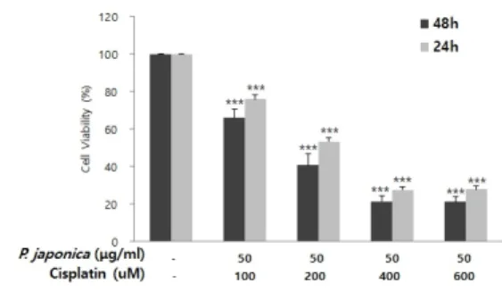
3. YD-10B세포주에서 백작약과 cisplatin의 병용 처리에 의한 생존율 분석
YD-10B 세포에서 10%의 생존율 감소를 보이는 50 μg/ml의 백작약 에틸아세테이트 분획물과 100, 200, 400 그리고 600 μM의 cisplatin을 병용 처리하여 24, 48시간 동안 배양한 후 세포의 생존율을 조사하였다.
대조군과 비교한 결과, 100 μM cisplatin과의 병용처 리에 의해서는 75.94%와 65.86%, 200 μM cisplatin 과의 병용처리에 의해서는 53.36%와 40.72%, 400 μ M cisplatin과의 병용처리에 의해서는 27.45%와 21.45%, 그리고 600 μM cisplatin과의 병용처리에 의해서는 27.80%와 21.16%의 생존율을 보였다. 거의 독성을 나타내지 않은 50 μg/ml 백작약과 200 μM
cisplatin을 병용 처리했을 때 대조군과 비교하여 50%
이상의 세포 생존율 감소를 보였다. 이와 같은 결과를 통하여, 병용 처리에 의한 암세포성장억제 효과가 상승 하였음을 확인할 수 있었다[Fig. 3].
Fig. 3. In vitro cytotoxicity effects of cisplatin in combination with p. japonica in YD-10B cells The cells were treated with 50 μg/ml p. japonica and cisplatin at different concentrations for 24h and 48 h. Data represent the mean ± S.D. of three independent experiments. (***; p<0.001 compared with untreated control).
4. YD-10B세포주에서 백작약과 cisplatin의 단 독 또는 병용 처리에 의한 MMP-2와 MMP-9 의 유전자 발현을 통한 암전이 억제 효과
YD-10B 세포에서 50 μg/ml 농도의 백작약 에틸아 세테이트 분획물과 200 μM 농도의 cisplatin을 단독 또는 병용 처리하여 MMP-2 및 MMP-9의 유전자발현 을 확인하였다. 대조군과 비교한 결과, cisplatin 단독 처리에서는 9.4%와 45.0%, 백작약 단독처리에서는 16.9%와 13.5%, cisplatin과 백작약의 병용처리에서 는 85.8%와 97.0%로 뚜렷한 유전자 발현억제를 나타 냈다[Fig. 4].(A)
(B)
Fig. 4. Effect of cisplatin in combination with p.
japonica on mRNA expression of MMP-2/9 in PMA-treated YD-10B cells
YD-10B cells were treated with the indicated concentration of cisplatin or p. japonica 2 hours prior to PMA (0.5 μM) stimulation. (A) 24 hours later, the levels of MMP-2/9 mRNA were determined by RT-PCR.
GAPDH were used as the internal control. (B) The relative expressions of MMP-2/9 mRNA were analyzed the band intensity using a GelQuant.NET program.
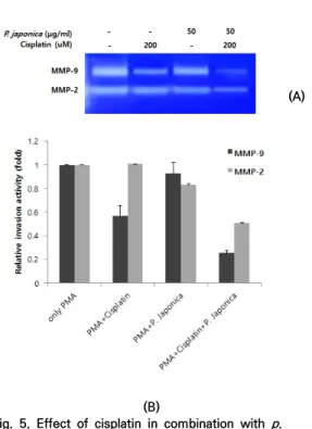
5. YD-10B세포주에서 백작약과 cisplatin의 단독 또는 병용 처리에 의한 MMP-2와 MMP-9의 단백질 활성을 통한 암전이 억제 효과
YD-10B 세포에서 백작약과 cisplatin의 병용처리에 의한 암 침윤 및 MMPs 활성에 미치는 효과를 조사하 기 위하여, 50 μg/ml 농도의 백작약 에틸아세테이트 분획물과 200 μM 농도의 cisplatin을 단독 또는 병용 처리하여 YD-10B세포를 혈청 없는 배지에서 배양하 였다. 그리고 24시간 동안 배양한 후, 배지의 상층액을 gelatin zymography법을 통해 MMP-2 및 MMP-9 단백질의 활성을 분석하였다. MMP-9 활성억제 효과 는 대조군과 비교한 결과, cisplatin 단독처리에서는 43.1%, 백작약 단독처리에서는 7.5%, cisplatin과 백 작약의 병용처리에서는 74.7%를 보였다. 그리고 MMP-2의 활성억제 효과를 대조군과 비교한 결과, cisplatin 단독처리에서는 변화가 없었고, 백작약 단독 처리에서는 17.1%, cisplatin과 백작약의 병용처리에 서는 49.3%를 나타냈다. 이러한 결과를 통하여 아무것 도 처리하지 않은 세포에 비해, 병용 처리한 세포에서 는 MMP-9과 MMP-2 단백질 활성이 크게 감소하였으 며 무엇보다 MMP-9 단백질 활성에 대한 억제 효과가 가장 뚜렷함을 확인할 수 있었다[Fig. 5].
(A)
Fig. 5. Effect of cisplatin in combination with (B) p.
japonica on the activities of MMP-2/9 in PMA-treated YD-10B cells
YD-10B cells were treated with the indicated concentration of cisplatin or p. japonica 2 hours prior to PMA (0.5 μM) stimulation. (A) 24 hours later, the activities of MMP-2/9 protein in the conditioned media were determined by gelatin zymography. (B) The relative expressions of MMP-2/9 protein were analyzed the band intensity using a GelQuant.NET program.
IV. 결론
Cisplatin은 화학항암제 중에서도 구강암, 난소암, 폐암, 간암, 대장암, 전립선암 및 유방암등의 다양한 고 형암 치료제로 가장 많이 사용되고 있다. 그러나 항암 제 내성 및 독성 부작용의 증가로 여전히 문제가 되고 있다. 이와 같은 항암제 내성 및 독성 부작용들을 극복 하기 위해 cisplatin과 함께 천연물을 병용 투여하는 연구가 꾸준히 진행되고 있다. 최근의 연구에 의하면, Hederagenin은 cisplatin에 내성을 가지는 두경부편 평세포암 세포주에서 Nrf2-ARF경로 억제를 통해 apoptosis가 유도되었고[23], Epigallocatechin gallate(EGCG)은 cisplatin에 내성을 가지는 CAR 구 강암세포주에서 AKT/STAT경로를 활성화하여 apoptosis와 autophagy를 유발하였다[24]. crocetin
과 cisplatin의 병용은 KYSE-150 세포주에서 p53/p21 경로를 통하여 cisplatin의 효과를 높였다 [25].
본 연구에서는 백작약 에틸아세테이트 분획물과 cisplatin의 YD-10B 구강암세포주에서의 단독 및 병 용처리에 의한 암세포 성장억제와 암전이 억제능력을 확인하였다. 백작약과 cisplatin의 병용처리는 50%이 상의 높은 암세포성장억제 효과를 나타냈으며 또한, YD-10B에서 PMA 처리에 의해 증가된 MMP-2 및 MMP-9의 유전자발현 및 단백질 활성들이 높은 암전 이 억제 활성을 나타냈다. 이와 같은 연구 결과를 바탕 으로 cisplatin과 백작약의 병용처리는 구강암 세포에 서 cisplatin에 대한 항암효과를 증진시킴을 알 수 있 었으며, cisplatin과 백작약 에틸아세트분획물의 병용 치료는 cisplatin의 효과를 극대화하여 적은 용량의 cisplatin에서도 약물내성과 부작용을 감소시킬 수 있 을 것으로 기대된다. 하지만 이들 효과에 대한 전임상 의 추가적인 연구가 필요할 것으로 생각된다.
참 고 문 헌
[1] X. Chen, Y. Wu, C. Y. Zhang, and Y. Zhang,
“Platinum based agents for individualized cancer treatment,” Curr. Mol. Med., Vol.13, pp.1603-1612, 2013.
[2] S. Dilruba and G. V. Kalayda, “Platinum based gruds: past, present and future,” Cancer chemother. Pahrmacol., Vol.7, pp.1103-1124, 2016.
[3] N. Toshiki, O. Akinobu, O. Takayuki, K.
Hiroyuki, F. Akifumi, O. Yukinobu, Y. Yoichi, H. Yoshitaka, and K. Yoshiaki, “Combined arsenic trioxide-cisplatin treatement enhances apoptosis in oral squamous cell carcinoma cells,” Cell Oncol, Vol.37, pp.119-129, 2014.
[4] L. Xin, G. Shu, X. K. Xiong, B. Y. Peng, J. M.
Huang, M. F. Cehn, and F. Y. Wang,
“Combination of quercetin and cisplatin emhances apoptosis in OSCC cells by downregulating xIAP through the NF-kB
pathway,” J. Cancer, Vol.10, pp.4509-4521.
2019.
[5] 서종천, 성일용, 김종렬 “구강편평세포암종에서의 cisplatin 유도 아폽토시스에서의 NF-kB의 활성화,”
대구외지, Vol.32, pp.94-100, 2006.
[6] L. Kelland, “The resurgence of platinum-based cancer chemotherapy,” Nat. Rev. Cancer, Vol.7, pp.573-584, 2007.
[7]. K. Tao, Y. Yin, Q. Shen, Y. Chen, R. Li, and W.
Chang, “Akt inhibitor MK-2206 enhances the effect of cislatin in gastric cancer cells,”
Biomed Rep., Vol.4, pp.365-368, 2016.
[8] P. Baharuddin, N. Satar, K. S. Fakiruddin, N.
Zakaria, M. N. Lim, and N. M. Yusoff,
“Curcumin improves the efficacy of cisplatin by targeting cancer stem like cells through p21 and cyclinD1-mediated tumor cell inhibition in non-small cell lung cancer cell lines,” Oncol Rep., Vol.35, pp.13-25, 2016.
[9] P. Ciganovic, K. Jakimiuk, M. Tomczyk, and M.
K. Zovko, “Glycerolic Licorice Extracts as Active Cosmeceutical Ingredients: Extraction Optimization, Chemical Characterization, and Biological Activity,” Antioxidants, Vol.8, pp.445-459, 2019.
[10] A. B. Kocka, N. Vorobets, M. Chrząscz, W.
Pietrzak, and K. Szewczyk, “Polyphenol Composition of Extracts of the Fruits of Laserpitium Krapffii Crantz and Their Antioxidant and Cytotoxic Activity,”
Antioxidants, Vol.8, pp.363-382, 2019.
[11] L. Bordoni, D. Fedeli, C. Nasuti, F. Maggi, F.
Papa, M. Wabitsch, R. D. Caterina, and R.
Gabbianelli, “Antioxidant and anti-inflammatory properties of nigella sativa oil in human pre-adipocytes,” Antioxidants, Vol.8, pp.51-63, 2019.
[12] H. R. Park, E. J. Ju, S. K. Jo, U. Jung, S. H. Kim, and S. T. Yee, “Enhanced antitumor efficacy of cisplatin in combination with HemoHIM in tumor-bearing mice,” BMC Cancer, Vol.9, pp.85-90, 2009.
[13] J. K. Kim and H. O. Yang, “The cytotoxic
effect of chaga mushroom water extract on HepG2 hepatoma cells,” J. Exp. Biomed Sci., Vol.17, pp.253-260, 2011.
[14] 김태희, 김안근, “MCF-7에서 Cisplatin과 타우린의 병용처리로 인한 항암효과 및 관련 기전,” 약학회지, Vol.57, pp.18-23, 2013.
[15] H. Song, L. Xiaolin, X. Rongrong, Y. Lingyun, K. Hui, Z. Xiaoning, W. Hong, and X. Weiping,
“The synergistic effect of resveratol in combination with cisplatin on apoptosis via modulating autophagy in A549 cells,” Acta.
Biochim. Biophys. Sin., Vol.48, pp.528-535, 2016.
[16] E. J. Kim and J. H. kim, “Evaluation of Anti-oxidative, Anti-thrombin, Anti-invasive and Pro-apoptotic Activities of Paeonia japonica,” Korean. J. Plant. Res., Vol.31, pp.16-23, 2018.
[17] O. Kujan, A. M. Glenny, J. Duxbury, N.
Thakker, and P. Sloan, “Evaluation of screening strategies for improving oral cancer mortality: a Cochrane systematic review,” J.
Dent. Educ., Vol.69, pp.255-265, 2005.
[18] L. Muzio, A. Santarelli, V. Panzarella, G.
Campisi, M. Carella, D. Ciavarella, M. Cosola, N. Giannone, and A. Bascones, “Oral squamous cell carcinoma and biological markers: an update on the molecules mainly involved in oral carcinogenesis,” Minerva. Stomatol., Vol.56, pp.341-347, 2007.
[19] C. Klug, D. Berzaczy, M. Voracek, and W.
Millesi, “Preoperative chemoradiotherapy in the management of oral cancer,” J.
Craniomaxillofac. Surg., Vol.36, pp.75-88, 2008.
[20] C. Y. Ng, H. Yen, H. Y. Hsiao, and S. G. Su,
"Phytochemicals in skin cancer prevention and treatment: an updated review," int. J. Mol. Sci., Vol.19, pp.941-965, 2018.
[21] C. Y. Sun, Q. Y. Zhang, G. J. Zheng, and B.
Feng, "Phytochemicals: current strategy to sensitize cancer cells to cisplatin," Biomed Pharmocather., Vol.110, pp.518-527, 2019.
[22] Y. Yang, Q. Zhang, Y. Chen, C. L. Liang, H.
Liu, and F. Qiu, "Antitumoreffects of immunity enhancing traditional chinese medicine,"
Biomed Pharmocather., Vol.121, pp.1-9, 2020.
[23] E. H. Kim, S. Baek, D. Shin, J. Lee, and J. L.
Roh, "Hederagenin induces apoptosis in cisplatin-resistant head and neck cancer cells by inhibiting the Nrf2-ARE antioxidant pathway," Oxid. med. Cell. Longev., Vol.2017, pp.1-12, 2017.
[24] C. H. Yuan, C. T. Horng, C. F. Lee, N. N.
Chiang, F. J. TSai, C. C. Lu, J. H. Chiang, Y. M.
Hsu, J. S. Yang, and F. A. Chen,
"Epigallocatecnin gallate sensitizes cisplatin-resistant oral cancer CAR cells apoptosis and autophagy through stimulating AKT/STAT3 pathway and suppressing multig\drug resistance 1 signaling," Envrion Toxicol., Vol.32, pp.845-855, 2017.
[25] S. Li, X. Y. Shen, T. ouyang, Y. Qu, T. Luo, and H. Q. Wang, "Synergistic anticancer effect of combined crocetin and cisplatin on KYSE-150 cells via p53/p21 pathway," Cancer Cell int., Vol.17, pp.1-18, 2017.
저 자 소 개
김 은 정(Eun-Jung Kim) 정회원
▪1996년 2월 : 연세대학교 임상병 리학(보건학사)
▪2000년 8월 : 고려대학교 생명공 학원 분자생물학(이학석사)
▪2009년 8월 : 연세대학교 응용생 명과학과 종양생물학 및 병리학(이 학박사)
▪2014년 4월 ~ 현재 : 상지대학교 임상병리학과 교수 <관심분야> : 종양생물학, 분자진단학, 병리학