Ankle Evertor Strength of Healthy Subjects in Different Ankle and Toe Positions
Sun-hee Ahn12, MSc, PT, Hyun-a Kim12, PhD, PT, Jun-hee Kim12, BPT, PT, Kyung-tae Kwak12, BPT, PT, Oh-yun Kwon23, PhD, PT
1Dept. of Physical Therapy, Graduate School, Yonsei University
2Laboratory of Kinetic Ergocise based on Movement Analysis
3Dept. of Physical Therapy, College of Health Science, Yonsei University
Abstract
1)Background: Ankle evertor muscles are important for preventing lateral ankle sprain. Since, the evertor muscles cross the ankle and toe joints, the position at which the ankle evertor muscle strength is measured is important. However, no studies have previously investigated the effect of ankle and toe positions on the strength of the ankle evertor muscle.
Objects: This study is aimed to determine the effect of various ankle and toe joint positions on the strength of the ankle evertor muscles in healthy subjects.
Methods: Eighteen healthy subjects participated in this study. Isometric ankle evertor strength of the dominant leg was determined in each subject in different ankle and toe positions (dorsiflexion (DF) with toe extension (TE), DF with toe flexion (TF), plantar flexion (PF) with TE, and PF with TF). A 2 by 2 repeated analysis of variance (ANOVA) was used to determine the difference in the evertor strength between the ankle positions (PF and DF) and toe positions (TE and TF).
Results: The results indicate that there was no significant ankle position by toe position interaction effect (p=.83). However, the ankle evertor strength was significantly increased in the ankle DF position than in the PF position (p<.01), and the ankle evertor strength during eversion with TE was significantly higher than eversion with TF (p<.01).
Conclusion: The findings of this study suggest that clinicians should consider the ankle and toe positions when measuring the muscle strength and during performance of selective muscle strengthening exercises of the ankle evertor muscles.
Key Words: Ankle evertor; Extensor digitorum longus; Isometric strength; Measurement position.
Introduction
Lateral ankle sprain is one of the most common injuries in young athletes, with symptoms, such as pain, swelling, dysfunction, and instability of the an- kle joint (Hertel 2000; Lorenzo-Sánchez-Aguilera et al, 2018; Nelson et al, 2007; Willems et al, 2002). The majority of the individuals who experience lateral an- kle sprain, approximately 70∼80%, progress to chron- ic ankle instability with symptoms persisting for more than 1 year (Santilli et al, 2005; Schmidt et al, 2011; terrier et al, 2017). Ankle biomechanics during
an ankle sprain show increased ankle inversion and internal rotation (Fong et al, 2009; Kristianslund et al, 2011). Additionally, previous studies investigated the biomechanics at the moment of ankle injury. They reported that ankle injury was found in dorsiflexion (DF) position in contrast to the theoretical hypothesis that ankle injury occurs in plantar flexion (PF) posi- tion (Fong et al, 2009; Kristianslund et al, 2011).
Appropriate control of the ankle muscles is neces- sary to prevent sudden ankle movement. Researchers have focused on the ankle evertor muscles in partic- ular, a group of muscles that are involved in a Corresponding author: Oh-yun Kwon kwonoy@yonsei.ac.kr
movement that is antagonistic to inversion. Willems et al (2002) and Donnelly et al (2017) reported that ankle evertor strength was significantly decreased in individuals with chronic ankle instability compared to healthy subjects (Donnelly et al, 2017; Willems et al, 2002). However, several other studies found no sig- nificant difference in ankle evertor strength in sub- jects with chronic ankle instability compared to healthy control subjects (Sierra-guzmán et al, 2018;
Terrier et al, 2017). Despite the many studies con- ducted on ankle evertor muscles, the effect of ankle evertor strength on lateral ankle injury is still undetermined.
The muscles that cross the lateral side of ankle joint can contribute to eversion movement. The main muscles involved in ankle eversion are the peroneus longus (PL) and peroneus brevis (PB) (Donnelly et al, 2017; Webster and Nussbaum 2016). These mus- cles produce eversion and PF movements because they cross the ankle joint posterolaterally (Muscolino 2016). Other muscles that cross the lateral side of the ankle joint are the peroneus tertius and extensor digitorum longus (EDL) (Muscolino 2016; Rourke et al, 2007; Yammine and Eric 2017). The peroneus ter- tius is sometimes missing and homologous to the EDL. The main function of the EDL is ankle DF and toe extension (TE), but it can also contribute to ev- ersion as this muscle crosses the ankle joint ante- rolaterally (Muscolino 2016). As the PL and PB cross the ankle joint while the EDL crosses the ankle joint, toe metatarsophalangeal (MTP) joint, and in- terpalangeal (IP) joint, the length of the muscles varies according to the position of the ankle and toes (Muscolino 2016).
Due to a length-tension relationship, the length of the muscles affects the force produced by their contraction. Therefore, the position at which muscle strength is measured can affect the outcome of mus- cle strength measurements (Ahn et al, 2018).
Donnelly et al (2017) studied ankle evertor strength in the neutral and PF positions (Donnelly et al, 2017). His results indicates that evertor strength was
higher in the neutral ankle position than PF position.
However, there have been no studies that in- corporated the positioning of both the toes and the ankle during ankle evertor strength measurements.
Investigating how ankle evertor strength of a healthy subject is affected by ankle and toe joint positions may result in improved clinical guidelines for strength measurement and training of ankle evertor muscles. Therefore, the purpose of this study was to investigate the effect of various ankle and toe joint positions on ankle evertor strength in healthy subjects. Although the main function of EDL is DF and TE, EDL also function as evertor. Therefore, the hypothesis of this study is that the ankle evertor strength would be higher in the DF position than in the PF position, and the evertor strength in the TE position would be higher than that in the TF posi- tion due to the action of EDL.
Methods
Subjects
The G*Power software ver. 3.0.10 (Franz Faul, Kiel University, Kiel, Germany) was used to estimate sample size in a pilot study of 3 subjects. From a priori analysis, a power of .95, set of .05 and the ef- fect size of .56 indicated that at least 14 subjects were needed. Eighteen healthy subjects (males: 12, females: 6) participated in this study (age=23.6±2.2 years; height=170.2±7.5 ㎝; weight=68.8±9.8 ㎏). The subjects recruited were healthy, and those with prior experience of ankle sprain, prior surgery in the lower extremities, limited ankle joint range of motion and neurological disorders were excluded (Cho et al, 2018). All subjects who volunteered to participate in this study signed a written consent form describing the procedure and the purpose of this study before the commencement of the experiments.
Instrumentation
The Smart KEMA strength sensor (KOREATECH,
Figure 1. Ankle evertor measurement position and the Smart KEMA sensor position.
A B C D
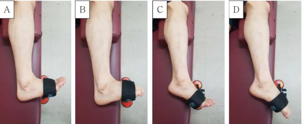
Figure 2. Ankle and toe positions for measuring the ankle evertor strength ( A:
dorsiflexion with toe extension, B: dorsiflexion with toe flexion, C: plantarflexion with toe extension, D: plantarflexion with toe flexion).
Inc., Seoul, Korea) was used to measure the iso- metric ankle evertor strength. This sensor is a tensi- ometer that measures the force at which both ends are pulled and the measuring range is 0∼1960 N (Ahn et al, 2018; Kim et al, 2017). To measure mus- cle strength, a 5-㎝ wide strap, which is attached to one end of the sensor is applied on the subject’s forefoot, and the length-adjustable belt attached to the opposite side of the sensor is fixed to the floor (Ahn et al, 2018). For all subjects, the tension at the starting position was adjusted to 2 kgf in order for all subjects to start at the same tension (Ahn et al, 2018). The Smart KEMA strength sensor has a high intra-rater reliability (ICC=.85∼.98) (Kim et al, 2017).
The measured data were collected at a sampling fre- quency of 10 ㎐ and transmitted via Bluetooth to the Smart KEMA application on a tablet. The trans- mitted strength data can be monitored in real time and analyzed through the application (Ahn et al, 2018; Kim et al, 2017).
Procedure
Evertor strength of the dominant foot was meas- ured in each subject. The dominant leg was the pre- ferred leg for kicking a ball (Webster and Nussbaum 2016). During the measurement, the subject took a side-lying position on the opposite of the measured ankle. Then the measuring leg was pushed forward by flexing the hip and knee, allowing the forefoot of the measured leg to come out of the table. Pillow support was applied under the knee to prevent pelvic, femur and tibia rotation. The investigator fixed the subject’s distal tibia to prevent the compensation of tibia external torsion and instructed the subject to ev- ert the ankle with maximum effort for 5 seconds (Figure 1). The subjects everted their ankle in the following four ankle and toe positions: (1) dorsiflexion with toe extension (DFTE) (Figure 2A); (2) dorsi- flexion with toe flexion (DFTF) (Figure 2B); (3) plan- tarflexion with toe extension (PFTE) (Figure 2C); and (4) plantarflexion with toe flexion (PFTF) (Figure
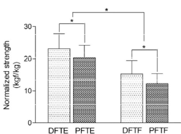
Figure 3. Ankle evertor strength in different ankle and toe positions (DFTE: dorsiflexion with toe extension, PFTE: plantarflexion with toe extension, DFTF: dorsiflexion with toe flexion, PFTF: plantarflexion with toe flexion,
*p<.01).
2D). The ankle DF position was set at 10˚ while the ankle PF position was set at 50˚ (Donnelly et al, 2017;
Kisner and Colby 2012). The investigator confirmed the ankle position using a standard goniometer at the starting position. Before the measurement, the subjects practiced to maintain the ankle angle in starting posi- tion while measuring evertor strength. In addition, the investigator monitored that if there is any DF or PF during measurement. The angle of toe flexion and ex- tension were set at the maximum possible angle of the subject. The order of measurement of the four positions was randomized using www.randomized.com.
Before the strength measurement, the subjects prac- ticed performing eversion with maximum force in each ankle and toe position, and then took a 5-minute rest before commencing with the experiment. The evertor strength was measured twice in each position, with a 3-minute rest period between measurements to pre- vent muscle fatigue.
Data analysis
The measured ankle evertor strength was proc- essed through the Smart KEMA application. The mean value of the middle 3 seconds of the collected 5-second ankle evertor strength data was calculated.
The average of the two measured values was calcu-
lated and saved. The strength data of each subject was divided by the subject’s body weight for nor- malization (Donnelly et al, 2017).
Statistical analysis
Statistical analysis was conducted using SPSS ver. 25.0 (IBM Corp., Armonk, NY, USA). The Kolmogorov-Smirnov test was used to confirm the normal distribution of the data. A 2 by 2 repeated model analysis of variance (ANOVA) was used to determine the difference in evertor strength between the ankle position (DF and PF) and toe position (TE and TF). The significance level was set at p=.05. If any significant interaction or effect was observed between groups and factors, the simple effect was compared by paired t-test or independent t-test us- ing Bonferroni corrections (p=.05/4=.013).
Results
All data were normally distributed. The 2 by 2 repeated ANOVA showed that there was no sig- nificant interaction effect between the ankle position and toe position (F=.05; p=.83) on the ankle evertor strength. However, there was a significant difference in the main effect of each ankle position and toe position. The strength of the ankle evertor was higher in the ankle DF (DFTE=23.1±4.7kgf/㎏;
DFTF=20.4±3.8kgf/㎏) than in the PF position (PFTE=15.3±4.2kgf/㎏; PFTF=12.3±3.0kgf/㎏) (F=24.0;
p<.01). The strength of the evertor muscles during eversion with TE was significantly higher than that during eversion with TF (F=44.55; p<.01) (Figure 3).
Discussion
This study investigated the effect of several ankle and toe joint positions on the ankle evertor strength in healthy subjects. The results indicate that the ev- ertor strength during eversion in the DF position
was significantly higher compared to eversion in the PF position while the evertor strength during ever- sion with TE was significantly higher compared to that during eversion with TF.
A previous study investigating the effect of the ankle positions on the evertor strength (Donnelly et al, 2017) showed that the evertor strength in a neu- tral position was significantly higher compared to that in the PF position (PF position: 1.73 Nm/㎏;
neutral: 2.10 Nm/㎏) (Donnelly et al, 2017). The re- sults of this study also showed that the evertor strength in the ankle DF position was higher than the PF position as in the previous study. This is re- lated to the functions of PL and PB, the prime mov- ers of eversion. Both PL and PB act as evertor and plantarflexor. Thus the PL and PB muscles are shortened in the PF position during eversion due to the direction of muscle action (Muscolino 2016;
Santilli et al, 2005). It is difficult for shortened mus- cles to generate maximal tension due to active in- sufficiency caused by an overlap of actin and myosin (Jeon et al, 2016; Sahrmann 2001). Therefore, the evertor strength may have increased in the DF posi- tion where the PL and PB can produce evertor force effectively compared to PF position.
Some of the muscles that contribute to eversion also cross the toe joints, meaning the position of the toes can also affect the evertor strength. However, previous studies on the ankle evertor strength did not consider the impact of toe positions on strength measurements. The results of this study showed that evertor strength during TE was significantly stron- ger than TF. The EDL muscle contributes to the eversion motion in the TE position. Thus, TE led to an increase in the evertor strength (Muscolino 2016).
In contrast, the flexor digitorum longus (FDL) and flexor hallucis longus (FHL) are synergistically in- volved in ankle inversion in the TF position because they cross the medial side of the ankle joint. While maintaining the toe flexion position during the meas- urement of evertor strength, FDL and FHL may act as an antagonist to evertors causing a decrease in
evertor strength in the TF position (Muscolino 2016).
In addition, since EDL activation is inhibited at the TF position, the contribution of the EDL to eversion is reduced, thus, affecting the evertor strength.
Many previous studies have investigated the rela- tionship between the ankle evertor weakness and chronic ankle instability. However, the results of these studies are still conflicted. Several studies have reported the evertor muscle weakness in individuals with chronic ankle instability, but some other studies have reported no significant difference between in- dividuals with ankle injury and healthy subjects (Donnelly et al, 2017; Terrier et al, 2017; Willems et al, 2002). The evertor muscles cross the ankle joint, and some of the evertor muscles also cross the toe joint. In addition, the ankle joint has a complex ar- ticular surface and the movement in the ankle joint occurs on three axes. For this reason, muscles across the ankle joint simultaneously contribute to the various movements of the ankle. Therefore, the ankle and toe joint positions and length of the mus- cles involved should be considered when measuring the ankle evertor muscle strength in clinics and on sports fields. In this study, we examined the change of evertor strength according to joint position of the ankle and toes of healthy subjects, and there was a significant difference in the strength of the evertor muscle according to the ankle and toe position.
Therefore, based on these results, we conclude that it is necessary to consider the ankle and toe posi- tions when measuring ankle muscle strength and during selective muscle strengthening exercise. In particular, clinicians apply evertor strength exercises for intervention to patients with lateral ankle sprain.
At this time, we recommend eversion in ankle PF with TF position to inhibit the compensation of EDL and selectively strengthen injured PL and PB based on the results of this study.
There are several limitations of this study. The generalization of the results is limited because only the young population were samples in this study.
However, because lateral ankle sprains are more
common in young and active individuals (Hertel 2000; Lorenzo-Sánchez-Aguilera et al, 2018), the re- sults of this study would help effective intervention for most patients with lateral ankle sprain. Further study is needed to investigate the effect of the ankle and toe position changes on the ankle evertor mus- cles in subjects with lateral ankle sprain. Activation of the muscles involved in ankle eversion was not measured. Hence, further studies are needed to de- termine the effect of the ankle and toe position on the ankle evertor muscle activation.
Conclusion
This study measured the effects of various ankle and toe positions on the strength of evertor muscles in healthy subjects. The results indicate that the ev- ertor strength during eversion in DF was sig- nificantly higher compared to eversion in PF while the evertor strength during eversion with TE was significantly higher compared to eversion with TF.
The muscles that contribute to the ankle eversion cross the ankle and toe joints; therefore, changes in the position of the ankle and toe joints can cause a change in evertor strength. Therefore, based on these results, clinicians will need to consider the ankle and toe joint positions when measuring the ankle evertor strength and providing selective training for patients.
References
Ahn SH, Hwang UJ, Jung SH et al. Hip external rotator strength and compensatory movement in three different positions. Health. 2018;10(01):
132-144.
Cho BK, Park JK, Choi SM et al. The effect of per- oneal muscle strength on functional outcomes after the Modified Broström Procedure for chronic ankle instability. Foot Ankle Int. 2018;
39(1):105-112. https://doi.org/10.1177/1071100717
735838
Donnelly L, Donovan L, Hart JM et al. Eversion strength and surface electromyography measures with and without chronic ankle instability meas- ured in 2 positions. Foot Ankle Int. 2017;38(7):
769-778. https://doi.org/10.1177/1071100717701231 Fong DT, Hong Y, Shima Y et al. Biomechanics of
supination ankle sprain: a case report of an ac- cidental injury event in the laboratory. Am J Sports Med. 2009;37(4):822-827. https://doi.org/
10.1177/0363546508328102
Hertel J. Functional instability following lateral an- kle sprain. Sports Med. 2000;29(5):361-371.
https://doi.org/10.2165/00007256-200029050-00005 Jeon IC, Kwon OY, Weon JH et al. Comparison of
psoas major muscle thickness measured by so- nography during active straight leg raising in subjects with and without uncontrolled lumbo- pelvic rotation. Man Ther. 2016;21:165-169.
https://doi.org/10.1016/j.math.2015.07.006
Kim HA, Hwang UJ, Jung SH et al. Comparison of shoulder strength in males with and without myofascial trigger points in the upper trapezius.
Clin Biomech (Bristol, Avon). 2017;49:134-138.
https://doi.org/10.1016/j.clinbiomech.2017.09.001 Kristianslund E, Bahr R, Krosshaug T. Kinematics
and kinetics of an accidental lateral ankle sprain.
J Biomech. 2011;44(14):2576-2578. https://doi.org/
10.1016/j.jbiomech.2011.07.014
Kisner C, Colby LA. Therapeutic exercise: Foundation and techniques 6. F.A. Davis Company. 2012:854.
Lorenzo-Sánchez-Aguilera C, Rodríguez-Sanz D, Gallego-Izquierdo T et al. Neuromuscular me- chanosensitivity in subjects with chronic ankle sprain; A cross-sectional study. Pain Med. 2019 [Epub ahead of print]
Muscolino JE. The muscular system manual: The skeletal muscle of the human body. Mosby, St.
Louis. 2016:598-603,622-629
Nelson AJ, Collins CL, Yard EE et al. Ankle injuries among United States high school sports athletes, 2005-2006. J Athl Train. 2007;42(3):381-387.
This article was received August 12, 2019, was re- viewed August 12, 2019, and was accepted September 11, 2019.
Rourke K, Dafydd H, Parkin IG. Fibularis tertius: re- visiting the anatomy. Clin Anat. 2007;20(8):946- 949. https://doi.org/10.1002/ca.20500
Sahrmann S. Diagnosis and treatment of movement impairment syndromes. Mosby, St. Louis. 2001:45.
Santilli V, Frascarelli MA, Paoloni M et al. Peroneus longus muscle activation pattern during gait cycle in athletes affected by functional ankle instability:
a surface electromyographic study. Am J Sports Med. 2005;33(8)):1183-1187. https://doi.org/10.1177/
0363546504274147
Schmidt H, Sauer LD, Lee SY et al. Increased in-shoe lateral plantar pressures with chronic ankle instability. Foot Ankle Int. 2011;32(11):
1075-1080. https://doi.org/10.3113/FAI.2011.1075 Sierra-guzmán R, Jiménez F, Abián-vicén J.
Predictors of chronic ankle instability: Analysis of peroneal reaction time, dynamic balance and isokinetic strength. Clin Biomech (Bristol. Avon).
2018;54:28-33. https://doi.org/10.1016/j.clinbiomech.
2018.03.001
Terrier R, Degache F, Fourchet F et al. Assessment of evertor weakness in patients with chronic ankle instability: Functional versus isokinetic
testing. Clin Biomech (Bristol. Avon). 2017;41:
54-59. https://doi.org/10.1016/j.clinbiomech.2016.
12.002
Webster CA, Nussbaum MA. Localized ankle fatigue development and fatigue perception in adults with or without chronic ankle instability. J Athl Train. 2016;51(6):491-497. https://doi.org/10.4085/
1062-6050-51.9.02
Willems T, Witvrouw E, Verstuyft J et al. Proprio- ception and muscle strength in subjects with a history of ankle sprain and chronic instability. J Athl Train. 2002;37(4):487-493.
Yammine K, Eric M. The Fibularis (peroneus) tertius muscle in humans: a meta-analysis of anatomi- cal studies with clinical and evolutionary implications. Biomed Res Int. 2017;2017:6021707.
https://doi.org/10.1155/2017/6021707