pISSN 1225-6552, eISSN 2287-7630 http://dx.doi.org/10.7853/kjvs.2012.35.4.289
< Original Article >
Veterinary Service
Available online at http://kjves.org
*Corresponding author: Okjin Kim, Tel. +82-63-850-6668, Fax. +82-63-850-7308, E-mail. [email protected]
A newly developed consensus polymerase chain reaction to detect Mycoplasma species using 16S ribosomal RNA gene
Sunhwa Hong
1, Sang-Ho Park
2, Yung-Ho Chung
3, Okjin Kim
1,4*
1
Center for Animal Resource Development, Wonkwang University, Iksan 570-749, Korea
2
Korea DNA Valley Co. Ltd, Iksan 570-749, Korea
3
Department of Companion Animal and Animal Resources Science, Joongbu University, Geumsan 312-702, Korea
4
Institute of Animal Experiment & Efficacy Evaluation, Wonkwang University, Iksan 570-749, Korea (Received 22 October 2012; revised 26 November 2012; accepted 29 November 2012)
Abstract
Mycoplasmas are highly fastidious bacteria, difficult to culture and slow growing. Infections with Mycoplasma species can cause a variety of problems in living organisms and in vitro cell cultures. In this study, we investigated the usefulness of a genus-specific consensus PCR analysis method to detect Mycoplasma species. The developed consensus primer pairs MycoF and MycoR were designed specifi- cally to amplify the 16S ribosomal RNA gene (rRNA) of Mycoplasma species by the optimized PCR system. The developed consensus PCR system effectively amplified 215 bp of Mycoplasma ge- nus-specific region of 16S rRNA. In conclusion, we recommend this consensus PCR for monitoring Mycoplasma species in animals, human and cell culture system.
Key words : Mycoplasma, PCR, Consensus, 16S rRNA, Genus-specific
INTRODUCTION
Mycoplasmas are among the smallest free-living mi- croorganisms capable of autoreplication. Mycoplasmas are highly fastidious bacteria, difficult to culture and slow growing (McAulifffe et al, 2003). The class Mollicutes consists of wall-less prokaryotes, which are small in size and have unusually small genomes. More than 100 species have been isolated from vertebrates, plants, and insects. The largest group is formed by the genus Mycoplasma, of which more than 90 species have been described (Hong et al, 2011). Many species are important veterinary pathogens causing respiratory in- fection, mastitis, conjunctivitis, arthritis, and occasion- ally abortion (McAulifffe et al, 2003). Also, in the field of laboratory animal medicine, it has been reported that mycoplasma infections are very common and considered
highly contagious (Hong et al, 2011). Major myco- plasmas of animals include Mycoplasma (M.) hyopneu- moniae, M. pulmonis, M. collies, M. neurolyticum, M.
arthritiditis, M. hyorhinis, M. mycoides (Razin and Hayflick, 2010). In humans, mycoplasmas have been recognized either as pathogenic organisms or as commensals. The best known pathogen is Mycoplasma pneumoniae, which causes a primary atypical pneumonia (Ieven et al, 1996). M. hominis and Ureaplasma ure- alyticum may cause a variety of genitourinary diseases (Taylor-Robinson et al, 1981) when they invade beyond the genital tract.
Mycoplasmas, which are invisible to light micro-
scopy, are frequent contaminants of in vitro animal cell
cultures. These cryptic contaminations can be recognized
via several specific procedures, including the recognition
of color changes in a dye applied to the culture media,
ELISA for the detection of specific proteins, or PCR
methods (Hopert et al, 1993; Uphoff and Drexler, 2011;
Fig. 1. The location of consensus PCR primer pairs MycoF and MycoR designed from the mycoplasma 16S ribosomal DNA sequence.
Volokhov et al, 2011). As mycoplasma infections in cultured cells tend to induce both biochemical and ge- netic changes, experimental results can often be misinterpreted. Therefore, contamination surveys should be periodically conducted in laboratories working with susceptible cultures.
Identification of mycoplasmas as the causative agents of disease is often hindered by the lack of rapid diag- nostic tests together with similarities in the clinical dis- eases that they cause. Conventional methods of myco- plasma diagnosis are based on culture and serological tests, such as the complement fixation test (Muthomi and Rurangirwa, 1983), ELISA (Ball and Finlay, 1998), and immunoblotting (Nicholas et al, 1996), and can be time-consuming, insensitive, and nonspecific. It is neces- sary to clarify the current status of mycoplasma con- tamination in animal colonies, because they are preva- lent in commercial and animal facilities (Lindsey et al, 1971). PCR provides a powerful technique of identify- ing mycoplasmas and studying homology between their nucleic acids. However, those kinds of PCR assays re- quire multiple assays because of a lot of Mycoplasma species (McAulifffe et al, 2003).
In this study, we investigated the usefulness of a ge- nus-specific PCR analysis method targeting a region of 16S ribosomal RNA (rRNA) gene to detect Mycoplasma species.
MATERIALS AND METHODS
Microorganisms and growth conditions
M. hyopneumoniae, M. hyorhinis, M. pneumonia, M.
hominis, M. pulmonis, M. arthritiditis, M. bovis, M.
fluccurare and M. mycoides were obtained from the American Type Culture Collection (Rockville, MD, USA). The mycoplasmas were grown in modified Friis medium (Friis, 1973), containing 20% porcine serum (Gibco-BRL, Korea), 5% fresh yeast extract (Gibco- BRL), methicillin (0.15 mg/ml; Sigma-Aldrich Canada, Oakville, Ontario, Canada), bacitracin (0.15 mg/ml;
Sigma-Aldrich), and thallium acetate (0.08 mg/ml;
Sigma-Aldrich). The cells were harvested by centrifu-
gation at 12,000×g for 30 min at 4
oC, washed three times, and suspended in 0.1 M phosphate-buffered saline (PBS, pH 7.4).
Nucleic acid extraction and designation of the consensus primers
DNAs were extracted from the cultures of M. hy- opneumoniae, M. hyorhinis, M. pneumonia, M. hominis, M. pulmonis, M. arthritiditis, M. bovis, M. fluccurare and M. mycoides. Mycoplasma DNAs were extracted from the cultured Mycoplasma species using an AccuPrep Genomic DNA extraction kit (Bioneer Corporation, Korea) according to the manufacturer's instructions. The DNA was eluted in Tris-EDTA buffer (pH 8.0), and an aliquot was used for the PCR amplification.
Consensus PCR primer pairs were designed from the mycoplasma 16S rRNA sequence (NC 019395.1) for the detection of versatile species in the Mycoplasma genus (Fig. 1). Forward primer MycoF was 5'-CTC TTT GTA CCG GCC ATT GT-3' (20 mer, nucleotide 699283- 699302). Reverse primer MycoR was 5'-GAA TGA CAG ATG GTG CAT GG-3' (20 mer, nucleotide 699478-699497).
PCR
The template DNA (50 ng) and 20 pmol of each pri-
mer were added to a PCR mixture tube (AccuPower
PCR PreMix; Bioneer Co., LTD., Korea) containing 2.5
U of Taq DNA polymerase, 250 µM each deoxynucleo-
side triphosphate, 10 mM Tris-HCl (pH 8.3), 40 mM
KCl, 1.5 mM MgCl
2, and the gel loading dye. The vol-
ume was adjusted with distilled water to 20 µl. The re-
action mixture was subjected to denaturation at 94
oC for
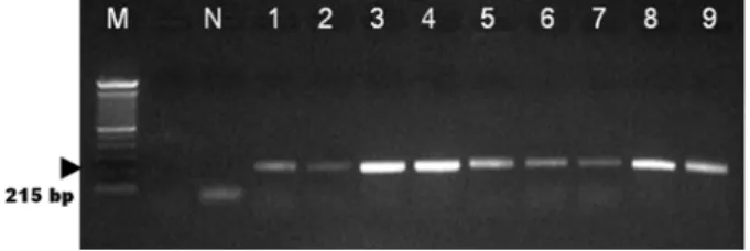
Fig. 2. Amplification of 16S rRNA gene from Mycoplasma species. Their PCR products were electrophoresed on a 2 % agarose gel. Lane: M: 1 kb marker, N: negative control, 1:
M. hyopneumoniae, 2: M. hyorhinis, 3: M. pneumonia, 4:
M. hominis, 5: M. pulmonis, 6: M. arthritiditis, 7: M. bovis, 8: M. fluccurare, 9: M. mycoides.
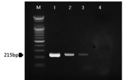
Fig. 3. Specificity of the developed consensus PCR targeted with 16S rRNA mycoplasma gene. Lane: M: 1 kb marker, 1: M.
hyopneumoniae, 2: M. hyorhinis, 3: Staphylococcus aureus, 4: Escherichia coli, 5: Helicobacter pylori, 6: Pasteurella multocida, 7: Salmonella pullorum, 8: Salmonella gallina- rum, 9: Actinobacillus pleuropneumoniae.
5 min followed by 30 cycles of 95
oC for 1 min, 55
oC for 1 min, and 72
oC for 1 min and a final extension step of 72
oC for 3 min, and samples were kept at 4
oC until analysis. Reactions were conducted using My Genie 32 Thermal Block PCR (Bioneer). After amplifi- cation, a 5 µl aliquot of each PCR was separated by electrophoresis on 2% agarose gels followed by ethi- dium bromide staining and UV transillumination.
Assay specificity and sensitivity
DNAs were extracted from the cultures of M. hy- opnenmoniae, M. hyorhinis, Staphylococcus aureus, Escherichia coli, Helicobacter pylori, Pasteurella multo- cida, Salmonella pullorum, Salmonella gallinarum and Actinobacillus pleuropneumoniae. Amplification of the 16S rRNA gene was performed with the same PCR procedure. In order to determine the sensitivity of the PCR assay, serial 10-fold dilutions of the purified ge- nomic DNA of M. hyopnenmoniae were tested.
Clinical samples
To evaluate the PCR system under field conditions, nasal were collected from conventional animal facility.
Sampling was performed from the nares with a dry, un- moistened swab. The tip of the collection swab was in- serted into the nares and rolled five times in each nostril. Collected specimens were transported and stored at room temperature. Specimens were processed for PCR analysis within 24 h of being collected. Each col- lection swab was put into 2 ml of 0.1 M PBS buffer
and vortexed and discarded, and the PBS was submitted to extract genomic DNAs for PCR assay.
RESULTS
Amplification of the consensus region of the 16S rRNA was performed with mycoplasma consensus primers. MycoF and MycoR was performed for primary amplification of target DNA. In this study, 215 bp am- plicons were amplified using the extracted DNAs from M. hyopneumoniae, M. hyorhinis, M. pneumonia, M.
hominis, M. pulmonis, M. arthritiditis, M. bovis, M.
fluccurare and M. mycoides (Fig. 2). The targeted 215 bp of 16S rRNA gene of Mycoplasma species were spe- cifically amplified by the optimized PCR system with the developed consensus primers, MycoF and MycoR.
The specificity of the developed consensus primers, MycoF and MycoR was confirmed by the study using other bacterial DNAs with high homology in their sequences. No positive signals were observed in the template DNA samples of Staphylococcus aureus, Escherichia coli, Helicobacter pylori, Pasteurella multo- cida, Salmonella pullorum, Salmonella gallinarum and Actinobacillus pleuropneumoniae (Fig. 3). However, the DNAs of M. hyopnenmoniae and M. hyorhinis were re- sulted in strong positive signal (Fig. 3).
Serial 10-fold dilutions of DNA from M. hyopnenmo-
niae were tested with our PCR assay. With our assay,
we could detect 10 pg of genomic DNA in 5 µl ali-
quots of PCR product by ethidium bromide staining
(Fig. 4). Nasal swabs were collected from conventional
Fig. 4. Sensitivity of genus-specific PCR targeted 16 SrRNA Mycoplasma gene. Lanes: M: 1 kb marker, 1: 10
3pg DNA of M. hyopnenmoniae, 2: 10
2pg DNA of M. hyopnenmo- niae, 3: 10 pg DNA of M. hyopnenmoniae, 4: 1 pg DNA of M. hyopnenmoniae.
Fig. 5. Direct PCR from M. hyopnenmoniae infected clinical samples. Lanes: M: 1 kb marker, 1: sample-1, 2: sample-2, 3: sample-3, 4: sample-4, 5: sample-5, 6: sample-6.
animal facility and submitted to PCR analysis (Fig. 5).
DISCUSSION
Mycoplasma species can induce a variety of problems in animals and human and in in vitro cell cultures.
Many species that can exist as commensal organisms are associated with certain diseases, but mycoplasma in- fections are often subtle or subclinical in nature. These non-apparent infections tend to be quite insidious, as they may affect a variety of biochemical and genetic as- pects of the infected cells, thereby resulting in unreliable experimental results and the possible transmission of diseases (Armstrong et al, 2010; Uphoff and Drexler, 2011; Degeling et al, 2012). Therefore, it is necessary to establish a routine diagnostic protocol for myco- plasma infection in order to ensure reliable research re- sults, as well as the safety of commercial biological products. However, the detection of Mycoplasma species in cell cultures remains a problem, despite the sub- stantial improvements that have been made in recent years in biochemical, immunological, and molecular bio- logical methods (Jung et al, 2003).
The diagnosis of Mycoplasma species is usually done by cultivation of the organism or by immuno- fluorescence tests performed on frozen thin lung sec- tions with polyclonal antibodies (Kobisch and Friis, 1996; Maes et al, 1996). However, due to the fastidious nature of Mycoplasma species, its culture and sero- logical identification may take up to 1 month.
Serological detection methods are further hampered by
cross-reactions which have been reported between M.
hyopneumoniae, M. hyorhinis, and M. flocculare (Armstrong et al, 1987; Freeman et al, 1984). With the advances made in molecular biology during the last few years, more is known about Mycoplasma species genes.
Hence, other methods can be used as diagnostic tools for this organism. Recently, PCR methods have been used to detect Mycoplasma species (Hong et al, 2011).
In order to circumvent those limitations, many nucleic acid technology-predicated procedures have been developed. PCR-based methods for the detection of cer- tain DNA regions of the mycoplasma genome have pro- ven both rapid and specific (Hu et al, 1995; Harasawa and Kanamoto, 1999; Kong et al, 2001; Loens et al, 2003; Khanna et al, 2005).
Classical diagnosis methods do not encompass a wide
range of mycoplasmas, and have some limitations, due
primarily to the fact that they require experience and
skill, as well as a series of time-consuming and labori-
ous steps (Chen, 1977). Despite its many limitations,
then, the time-consuming practice of microbiological
cultivation has traditionally constituted the method of
choice, in cases in which the other methods do not
yield conclusive results. However, certain mycoplasmas,
such as non-culturable M. hyorhinis strains, have proven
difficult to cultivate, and may elude such diagnostic
tests. In order to circumvent the problems posed by the
aforementioned classical methods, other methods have
been developed, some employing nucleic acid
technology. The results presented in this study indicate that 16S rRNA gene from 9 Mycoplasma species could be detected by the developed consensus PCR, using highly specific primers, MycoF and MycoR.
We suggested that this consensus PCR will be useful and effective for monitoring Mycoplasma species in var- ious kinds of animals and human and also cell cultures and could be used for detection of a new Mycoplasma species.
ACKNOWLEDGMENTS
This research was supported by Technology Develop- ment Program for Agriculture and Forestry (grant No.
610004-3), Ministry for Food, Agriculture, Forestry and Fisheries, Republic of Korea.
REFERENCES
Armstrong CH, Freeman MJ, Sands-Freeman L. 1987. Crossreac- tions between Mycoplasma hyopneumoniae and Myco-
plasma flocculare: practical implications for the sero-
diagnosis of mycoplasmal pneumonia of swine. Isr J Med Sci 23: 654-656.Armstrong SE, Mariano JA, Lundin DJ. 2010. The scope of my- coplasma contamination within the biopharmaceutical in- dustry. Biologicals 38: 211-213.
Ball HJ, Finlay D. 1998. Diagnostic application of monoclonal antibody (MAb)-based sandwich ELISAs. Methods Mol Biol 104: 127-132.
Chen TR. 1977. In situ detection of mycoplasma contamination in cell cultures by fluorescent Hoechst 33258 strain. Exp Cell Res 104: 255-262.
Degeling MH, Maguire CA, Bovenberg MS, Tannous BA. 2012.
Sensitive assay for mycoplasma detection in mammalian cell culture. Anal Chem 84: 4227-4232.
Freeman MJ, Armstrong CH, Sands-Freeman LL, Lopez-Osuna M. 1984. Serological cross-reactivity of porcine refer- ence antisera to Mycoplasma hyopneumoniae, M. floccu-
lare, M. hyorhinis and M. hyosynoviae indicated by the
enzyme-linked immunosorbent assay, complement fix- ation and indirect hemagglutination tests. Can J Comp Med 48: 202-207.Friis NF. 1973. The pathogenicity of Mycoplasma flocculare.
Acta Vet Scand 14: 344-346.
Harasawa R, Kanamoto Y. 1999. Differentiation of two biovars of Ureaplasma urealyticum based on the 16S-23S rRNA intergenic spacer region. J Clin Microbiol 37: 4135-
4138.
Hong S, Lee HA, Park SH, Kim O. 2011. Sensitive and specif- ic detection of Mycoplasma species by consensus poly- merase chain reaction and dot blot hybridization. Lab Anim Res 27: 141-145.
Hopert A, Uphoff CC, Wirth M, Hauser H, Drexler HG. 1993.
Specificity and sensitivity of polymerase chain reaction (PCR) in comparison with other methods for the de- tection of mycoplasma contamination in cell lines. J Immunol Methods 164: 91-100.
Hu M, Buck C, Jacobs D, Paulino G, Khouri H. 1995. Applica- tion of PCR for detection and identification of myco- plasma contamination in virus stocks. In Vitro Cell Dev Biol Anim 31: 710-715.
Ieven M, Ursi D, Van Bever H, Quint W, Niesters HGM, Goossens H. 1996. Detection of Mycoplasma pneminiae by two polymease chain reaction and role of M. pnemi-
niae in acute respiratory tract infection in pediatric
patients. J Infect Dis 173: 1445-1452.Jung H, Wang SY, Yang IW, Hsueh DW, Yang WJ, Wang TH, Wang HS. 2003. Detection and treatment of mycoplasma contamination in cultured cells. Chang Gung Med J 26:
250-258.
Khanna M, Fan J, Pehler-Harrington K, Waters C, Douglass P, Stallock J, Kehl S, Henrickson KJ. 2005. The pneumo- plex assays, a multiplex PCR-enzyme hybridization as- say that allows simultaneous detection of five organisms,
Mycoplasma pneumoniae, Chlamydia (Chlamydophila) pneumoniae, Legionella pneumophila, Legionella micda- dei, and Bordetella pertussis, and its real-time coun-
terpart. J Clin Microbiol 43: 565-571.Kobisch M, Friis NF. 1996. Swine mycoplasmoses. Rev Sci Tech 15: 1569-1606.
Kong F, James G, Gordon S, Zelynski A, Gilbert GL. 2001.
Species-specific PCR for identification of common con- taminant mollicutes in cell culture. Appl Environ Microbiol 67: 3195-3200.
Lindsey JR, Baker HJ, Overcash RG, Cassell GH, Hunt CE.
1971. Murine chronic respiratory disease. Significance as a research complication and experimental production with Mycoplasma pulmonis. Am J Pathol 64: 675-708.
Loens K, Ursi D, Goossens H, Ieven M. 2003. Molecular diag- nosis of Mycoplasma pneumoniae respiratory tract infections. J Clin Microbiol 41: 4915-4923.
Maes D, Verdonck M, Deluyker H, de Kruif A. 1996. Enzootic pneumonia in pigs. Vet Q 18: 104-109.
McAulifffe L, Ellis RJ, Ayling RD, Nicholas RA. 2003.
Differentiation of Mycoplasma species by 16S ribosomal DNA PCR and denaturing gradient gel electrophoresis fingerprinting. J Clin Microbiol 41: 4844-4847.
Muthomi EK, Rurangirwa FR. 1983. Passive haemagglutination and complement fixation as diagnostic tests for conta- gious caprine pleuropneumonia caused by the F-38 strain of mycoplasma. Res Vet Sci 35: 1-4.
Nicholas RA, Santini FG, Clark KM, Palmer NM, De Santis P,
Bashiruddin JB. 1996. A comparison of serological tests and gross lung pathology for detecting contagious bo- vine pleuropneumonia in two groups of Italian cattle.
Vet Rec 139: 89-93.
Razin S, Hayflick L. 2010. Highlights of mycoplasma research-an historical perspective. Biologicals 38: 183-190.
Taylor-Robinson D, Tully JG, Furr PM, Cole RM, Rose DL, Hanna NF. 1981. Urogenital mycoplasma infections of man: a review with observations on a recently dis-
covered mycoplasma. Isr J Med Sci 17: 524-530.
Uphoff CC, Drexler HG. 2011. Detecting my coplasma con- tamination in cell cultures by polymerase chain reaction. Methods Mol Biol 731: 93-103.
Volokhov DV, Graham LJ, Brorson KA, Chizhikov VE. 2011.
Mycoplasma testing of cell substrates and biologics:
review of alternative non-microbiological techniques.
Mol Cell Probes 25: 69-77.