ISSN 2288-1069 (Online)
http://dx.doi.org/10.12925/jkocs.2020.37.1.66
Effect of Polygonum multipolarum extract on the allergic reaction of NC/Nga mice causing atopic dermatitis
Ji-Sun Moon
*․Eun-Young Choi
✝Professor Dept. of Medical Beauty Care Jungwon University․Researcher RXAP KOREA (Received February 8, 2020; Revised February 19, 2020; Accepted February 21, 2020)
하수오 추출물(
Polygonum multiflorum)
이 아토피 피부염 유발 NC/Nga 생쥐의 알레르기 반응에 미치는 영향문지선*․최은영✝
중원대학교 의료뷰티케어학과 교수*, 알엑스에이피코리아 연구원✝
(2020년 2월 8일 접수: 2020년 2월 19일 수정: 2020년 2월 21일 채택)
Abstract : Atopic dermatitis (AD) usually develops in patients with an individual or family history of allergic diseases, and is characterized by chronic relapsing inflammation seen specially in childhood, association with IgE hyperproduction and precipitation by environmental factors. and wished to examine closely effect that
Polygonum multiflorum
isolated PM-E and PM-70M orally adminstration used to atopy dermatitis disease patient get in atopy eruption control experimentally. Atopic dermatitis is a chronically relapsing inflammatory skin disease. Animal models induced by relevant allergens play a very important role in the elucidation of the disease. This study was investigated the anti-allergic effect of PM-E and PM-70M on BMAC induced atopic dermatitis in NC/Nga mice. We summerized as the follow. PM-E and PM-70M significantly reduced the skin number of total cell number, CD4+ and CD11b+/Gr-1 cell compared with positive control and decreased the invasion of CD4+ cell in dorsal skin tissue compared with positive control group by using immunohistochemical staining and chemokine such as eotaxin and CCR3 compared with positive control group. PM-E and PM-70M markedly suppressed invasion and edema of leukocytes and mast cell in dorsal skin. Taken together, these findings suggested that PM-E and PM-70M has an anti-allergic activity and this might be useful for the clinical application to treat allergic diseases such as atopic dermatitis.Keywords : Polygonum multipolarum
extract,Atopic, Dermatitis, Allergy, Skin tissue
✝
Corresponding author
(E-mail: [email protected])
1. Introduction
Atopic dermatitis (AD) is a chronic inflammatory skin disease. It is also a chronic recurrent inflammatory skin disease with high incidence in infants and young children about 10-20% of the population. According to a survey conducted by the Korean Academy of Pediatric Allergy and Respiratory Diseases in 2000, 24.9% of elementary school students and 12.8% of middle school students have been diagnosed with atopic dermatitis[1]. The mechanisms of atopic dermatitis have not been fully established yet, but susceptibility to viral infections and several immunological abnormalities such as herpes simplex or scleroderma and reduced delayed immune responses to certain microbial antigens, increased production of IgE, decreased sensitivity to contact allergens and cleavage have been reported. In addition, the prevalence of this disease and the progression to refractory atopic dermatitis have been increasing[2]. There is a trend, which is evidence that genetic factors and environmental factors such as industrialization are important in the development of atopic dermatitis. These disease are reported to be characterized by pruritus, itching, lichen dermatitis, rash, tearing, and systemic dryness.
In addition, since histamine or inflammatory cytokines secreted from mast cells cause tissue damage and inflammatory response in conjunction with pruritus, drugs that effectively control their activity can be used as therapeutic agents for inflammatory diseases.
Recently, lots of anti-inflammatory drugs have been progressed through various studies, but there are many supplements in terms of fundamental treatment and side effects[3].
Therefore, Hasuo (
Polygoni Multiflori Radix
) is a tuber ofPolygonum multiflorum
Thunberg, and anthracuuinone derivative is a chrysophanol, emodin, physcion, questin, and stibene glucoside. As phenolic compounds such as 2, 3,5,4-tetrahydroxystilbene-2-O-β-D-gluco pyranoside, polygoacetophenoside have been studied.
Wilkinson et al. (2006) reported that anthracuuinone derivatives are effective in allergic contact dermatitis[4]. Th2-cell responses were obtained by administering
Rumex japonicus
Houtt herb containing Emodin, chrysophanol, and physcion to NC/Nga mice, atopy-induced mice and skin rashes were reported to be suppressed[5].Based on these basic data, the author would like to continue to study on the atopic dermatitis.
2. Experimental Method
2.1. Reagents and Instruments
2.1.1. Reagent
Reagents used in this experiment were diethyl pyrocarbonate (DEPC), chloroform, trichloroacetic acid, isopropanol, Tris-HCl, KCl, MgCl2, ACK lysis solution, DMEM culture, dulbecco's phosphate buffered saline (D-PBS), Sulforhodamin B (SRB), 2-isopropanol, Sodium dodecyl sulfate (SDS), PMA, Ionomycin, FK506, and antibiotics were manufactured by Sigma (USA). Fetal bovine serum (FBS) was used by Hyclone (Logan, USA) products, anti-CD4, anti-CD11b, anti-Gr-1 Pharmingen (Torreyana, USA), anti-mouse CD4 mAb DAKO (Glostrup, Denmark) was used, and other general reagents were used as express reagents.
2.1.2. Instruments
The equipment used in this experiment was a boiling water extractor (Daewoong, Korea), rotary vaccum evaporator (Büchi B-480, Switzerland), freeze dryer (EYELA FDU-540, Japan), CO2 incubator (Forma scientific Co., USA), clean bench (Vision scientific Co., Korea), autoclave (Sanyo, Japan), micro-pipet (Gilson, France), water bath (Vision scientific Co., Korea), vortex mixer (Vision scientific
Fig. 1 Experimental Design for the Induction of Atopic Dermatitis like NC/Nga Mouse.
Co., Korea), spectrophotometer (Shimazue, Japan), centrifuge (Sigma, USA), deep-freezer (Sanyo, Japan), Quantitative Real-Time RT-PCR (Applied Biosystems, USA), ice-maker (Vision scientific Co., Korea), homogenizer (OMNI , USA), plate shaker (Lab-Line, USA), Vario MACS (Bergisch Gladbach, Germany), FACS calibur (BD, USA) and ELISA leader (Molecular Devices, USA).
2.1.3. Animals
7-week-old male SPF (specific pathogen-free) NC/Nga mice (15-20 g) were supplied by Charles River Japan (Yokohama, Japan). Animals were used for the experiment after being given solid feed (no antibiotics, Samyang Feed Co.) and sufficient water until the day of the experiment and adapted to an environment of temperature 22 ± 2 °C, humidity 55 ± 15% for 12 hours (light-dark cycle).
2.1.4. Manufacturing of PM-E and PM-70 After grinding 200 g of Hasuo, PM-E and PM-70 were separated using Diaion HP-20 resin according to the fractionation method of Dong and Jeng (2004)[6]. Hasuo stock solution (PM-E) extracted with 70% ethanol was poured into adsorption chromatography (HP-20) containing resin. The width was left to be about 25-30 cm. Thereafter, 1000 mL of distilled water was poured to separate the Hasuo fraction passed through Diaion HP-20 resin (pass layer), 500 mL of 30% MeOH (PM-30M), 500 mL of 70% MeOH (PM-70% MFL), 30% 500 mL (PM-30A) of Acetone and 500 mL (PM-70A) of 70%
Acetone were passed through to separate the
separated fractions.
2.1.5. Animal Experiment
2.1.5.1. Dermatitis induction and sample processing
Seven-week-old NC/Nga mice were acclimated for one week, and 18-week-old NC/ Nga mice, which had already developed dermatitis, were co-cultured for two weeks in the same space for antigen reduction. Blood was drawn. After that, the mice were anesthetized with chloral hydrate (10%), anesthetized, and the dorsal neck area was depilated cleanly. Biostir Mite antigen cream (hereinafter referred to as BMAC:
Dermatophagoides farinae crude extract) provided by the central laboratory animal was evenly applied to the back and neck area twice a week for 3 weeks (10 to 13 weeks of age), and 4% SDS 2 hours before application.
The mice were used as a control by destroying the skin layer to induce dermatitis by spraying the solution (Fig. 1). Two weeks after the start of application (12 weeks age), the dermatitis was sufficiently induced and the scratching action was intensified. In the case of NC/Nga-Nr group (NC /Nga-normal group, Nr), 7-week-old NC/Nga mice were bred under SPF conditions until 15 weeks of age.
2.1.5.2. Drug treatment and evaluation The experiment was conducted in the NC/Nga-Nr group (Nr) and BMAC-coated control group in which 7-week-old NC/Nga mice were kept in SPF conditions up to 15 weeks of age. BMAC was applied and divided
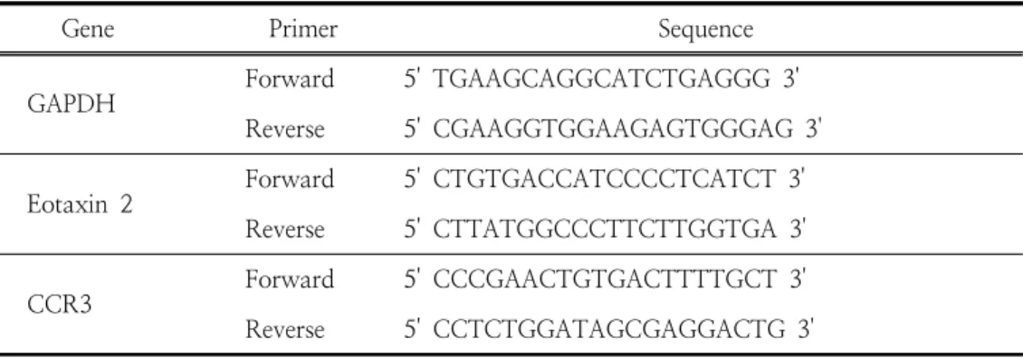
Gene Primer Sequence
GAPDH
Forward 5' TGAAGCAGGCATCTGAGGG 3' Reverse 5' CGAAGGTGGAAGAGTGGGAG 3'
Eotaxin 2
Forward 5' CTGTGACCATCCCCTCATCT 3' Reverse 5' CTTATGGCCCTTCTTGGTGA 3'
CCR3 Forward 5' CCCGAACTGTGACTTTTGCT 3'
Reverse 5' CCTCTGGATAGCGAGGACTG 3'
SYBR® Green PCR Master mix was used for cytokine gene expression, Taqman probe was used as GAPDH for internal standard, and the final concentration of primer was 200 nM. Eotaxin 2 and CCR3 mRNA expression was analyzed by synthesizing cDNA in the back skin tissue.
In the case of quantitative real-time PCR, pre-denaturation was performed at 2 min at 50 ℃, 10 min 94 ℃, and 40 cycles at 0.15 min at 95 ℃ and 1 min at 60 ℃. In the control group, the CSA group, the PM-E group, and the PM-70% MFL group, GAPDH was used as an internal standard. In the case of quantitative PCR in a target group, a relative quantitative (RQ) value was measured by calculating y = x (1 + e) n x = starting quantity, y = yield, n = number of cycles, and e = efficiency.
Table 1. Primer Sequence
into experimental groups administered PM-E and PM-70% MFL.
BMAC was injected with 4% SDS solution twice a week for a total of three weeks (10 to 13 weeks of age), and 200 μl each was applied after 2 hours. Dermatitis rash was confirmed 2 weeks after the start of BMAC application (12 weeks of age), and once every 3 weeks from 12 weeks to 15 weeks of age, CsA (10 mg / kg), PM-E (200 mg / kg) and PM, respectively. -70% MFL (75 mg / kg) was administered.
2.1.5.3. Leukocyte Analysis
After the final clinical visual evaluation, the blood was collected and Biotoxtec measured the number of neutrophils, eosinophils, basophils and lymphocytes, and monocytes.
The measurement was performed with an automatic hemocytometer (MS9-5, MELET SCHLOESING, France) using Minos-ST according to the Fonio method[7].
2.1.5.4. Fluorescence Flow Cytometry at Back Skin Tissue
After drug administration (15 weeks old), fine chopping of the dorsal skin tissue of NC/Nga mice was performed, followed by collagenase 1 mg / ml (in 2% FBS + RPMI 1640), in a 37 ℃ shaker (180 rpm, 20 min.) Incubator. After incubation, the supernatant was recovered. This method was repeated four times.
After measuring the total cell number of the isolated skin tissue infiltrating cells, after adjusting the dorsal cells of all tissues to 5 × 10⁵ cells, immunofluorescence staining was performed at 4 ℃. Anti-CD4-, anti-CD11b and anti-Gr-1 were added to each tissue and allowed to react on ice for 30 minutes. After the reaction, the cells were washed with PBS three times or more, and then the number of CD4+ and CD11b/Gr-1+ cells was analyzed by percentage (%) using the Cell Quest program of flow cytometry. Was calculated.
2.1.5.5. Quantitative real-time PCR in dorsal skin
Atopic dermatitis-like skin The back skin tissue of NC/Nga mice was extracted, and 500 μl of RNAzolB was added to each tissue and ground by homogenizer until dissolved. 50 μl of chloroform (CHCl3) was added to the tissue grinding mixed suspension, and then mixed again for 15 seconds. After standing on ice for 15 minutes, centrifuged at 13,000 rpm, about 200 μl of the supernatant was collected, mixed with the same amount as 200 μl of 2-propanol, and slowly shaken and left for 15 minutes on ice. This was again centrifuged at 13,000 rpm, washed with 80%
EtOH and dried for 3 minutes by vacuum pump to extract RNA. The extracted RNA was dissolved in 20 μl of distilled water treated with DEPC, inactivated at 75 ℃. of heating block, and then used for first-strand cDNA synthesis. Quantitative real-time PCR was performed using a 7500 Real-Time PCR system[8].
2.2. Statistical processing
Results obtained from the experiments were recorded as mean ± standard error, and significance test was performed using Student's t-test analysis method[9].
3. Results and Discussion
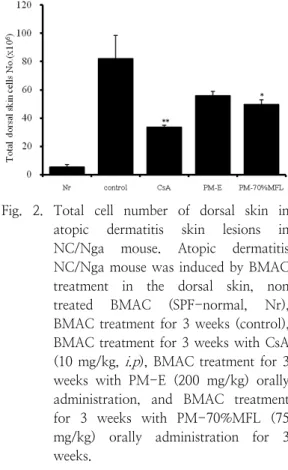
3.1. Effect of Skin Tissue on Total Cell Number
Atopic dermatitis causes inflammatory cells to be recruited into the skin tissue, increasing the total number of cells. As a result of measuring the total cell number of the skin, the control was significantly increased compared to the NC/Nga-Nr group. In addition, inflammatory cells were significantly decreased in the CsA group and the PM-70%
MFL group (p <0.05) compared to the control group (Fig. 2).
Fig. 2. Total cell number of dorsal skin in atopic dermatitis skin lesions in NC/Nga mouse. Atopic dermatitis NC/Nga mouse was induced by BMAC treatment in the dorsal skin, non treated BMAC (SPF-normal, Nr), BMAC treatment for 3 weeks (control), BMAC treatment for 3 weeks with CsA (10 mg/kg,
i.p
), BMAC treatment for 3 weeks with PM-E (200 mg/kg) orally administration, and BMAC treatment for 3 weeks with PM-70%MFL (75 mg/kg) orally administration for 3 weeks.3.2. Effect of Fluorescent Flow Cytometry in Back Skin Tissue on the Immune Cells
As atopic skin rashes intensify, immune inflammatory cells, such as TH2 cells, eosinophils, monocytes, and mast cells, increase, which continues to exacerbate skin rashes in animals with chronic skin diseases.
The role of myeloid cells in immune regulation is known to play an immunosuppressive effect in various disease states and plays an important role in cell growth and differentiation. The characteristics of myeloid suppressor cells (MSC) express both surface molecules in macrophage (Mac-1/CD11b) and granulocyte (Gr-1) lineages. This CD11b+Gr-1+ MSC migrates from spleen and lymph nodes to inflamed skin and tissues[10].
These cells inhibit antibody production,
cytotoxic T lymphocyte (CTL) generation, and lymphocyte proliferative responses, and in the case of atopy, continue to exacerbate chronic skin diseases[11].
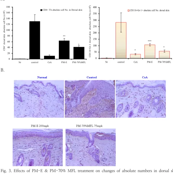
As a result of measuring the absolute cell number of CD4+ cells in the dorsal skin tissue, the control group increased significantly compared to NC/Nga-Nr group, and the CsA-administered group significantly decreased more than 6 times compared to the control group (p<0.001). PM-E group, and PM-70%
MFL group also showed a significant decrease compared to the control group (p<0.01, p<0.05) (Fig. 3A). In the case of CD11b/Gr-1, the PM-E and PM-70% MFL groups also showed a significant decrease compared to the control group (p<0.001, p<0.05) (Fig. 3A).
As a result of these studies, CD4+ Th cells (brown color, arrow) inactivated by CsA, PM-70% MFL-treated group and ALN and back skin tissue after immunochemical tissue staining were observed. Compared to the Nga-Nr group (Fig. 3B), CD4+ Th cells in the control group increased significantly with clusters at the bottom of the epidermis. In the CsA-administered group and PM-E- administered group, all CD4+ Th cells were significantly decreased compared to the control group, especially in the PM-70% MFL group (Fig. 3B).
3.3. Effect on skin inflammation
3.3.1. Effect on white blood cells
White blood cells are divided into granulocytes, lymphocytes, and monocytes.
Peripheral blood generally has an immune function against external antigens. Recent studies have shown that the number of white blood cells increases as the number of granulocytes increases in peripheral blood of chronic asthma and atopic patients. The characteristics of these granulocytes are known as neutrophils, eosinophils, basophils having granules characteristic of the cytoplasm. These cells are made in the bone marrow and
distribute through the blood to systemic tissue.
From the promyelocytic stage, specific granules of each cell appear and classify the cells into neutrophils, eosinophilic, basophils, etc[12].
As these granulocytes mature, peroxidase- positive azurophilic granules decrease, peroxidase-negative specific granules increase, and cell surface Fc receptors and receptors for complement such as CRI and CR3 appear.
Neutrophil is 2 to 6%, the number of bacteria increases dramatically as soon as it enters the human body, and it recognizes and binds common components on the surface of various bacteria with macrophages to act as phagocytosis and secrete biologically active molecules called cytokines. These active molecules dilate nearby blood vessels, increase local blood flow, increase fluid leakage, and mobilize nearby white blood cells[13].
Monocytes differentiate into macrophages, which are the largest and most mobilized among white blood cells, and when pathogens or toxins invade, they exit the blood vessels with neutrophils and treat the invading pathogens by phagocytosis. Eosiniphils play an important role in detoxifying toxic substances entering the body from the outside by mass gathering in the external contact area and are increased in atopic diseases. Basophil is known to play an important role in the production of inflammation-related substances, asthma, allergies and hypersensitivity[14].
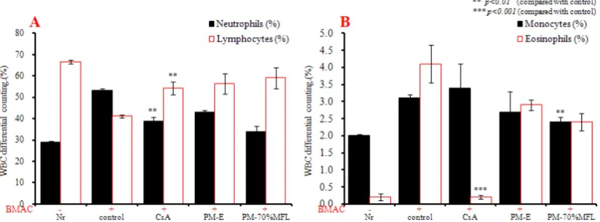
At the end of the experiment (15 weeks), WBC neutrophil and eosinophils ratios were significantly higher in the control group than in the NC/Nga-Nr group. The CsA group was significantly decreased (p<0.01, p<0.001) compared to the NC/Nga-Nr group compared to the control group, and the neutrophil and eosinophils ratios in the PM-E group and the PM-70% MFL group were decreased compared to the control group. However, there was no significance. The percentage of monocytes was significantly decreased in the PM-70% MFL group compared to the control group (p<0.01) (Fig. 4). This result was similar
A.
**
*
0 20 40 60 80 100 120 140 160 180
Nr control CsA PM-E PM-70%MFL
CD4+ dorsal skin absolute cell No.(x105) CD4+ T h absolute cell No. in Dorsal skin
*
***
*
0 50 100 150 200 250 300 350 400
Nr control CsA PM-E PM-70%MFL
CD11b+Gr-1 dorsal skin absolute cell No.(x105)
CD11b+Gr-1+ absolute cell No. dorsal skin
B.
Fig. 3. Effects of PM-E & PM-70% MFL treatment on changes of absolute numbers in dorsal skin cells in NC/Nga mouse(A). Immunohistochemical staining of the skin stained with CD4+ Th cells of dorsal skin in atopic dermatitis skin lesions in NC/Nga mouse(B).
Atopic dermatitis NC/Nga mouse was induced by BMAC treatment in the dorsal skin, non treated BMAC (SPF-normal, Nr), BMAC treatment for 3 weeks (control), BMAC treatment for 3 weeks with CsA (10 mg/kg,
i.p
), BMAC treatment for 3 weeks with PM-E (200 mg/kg) orally administration, and BMAC treatment for 3 weeks with PM-70%MFL (75 mg/kg) orally administration for 3 weeks. NC/Nga mouse dorsal skin cells(2×10⁵ cells/㎖) were isolated from dorsal skin, and the dorsal skin cells were washed twice and analyzed by flow cytometry.Absolute numbers of CD4+ Th ,& CD11b+Gr-1+ in NC/Nga mouse(Fig. 3.A). Following 3 weeks, mouse dorsal skin biopsy were stained with anti-mouse CD4mAb respectively. Dorsal skin biopsy were stained with anti-mouse CD4mAb, used LSAB2 HRP. Rabbit/mouse(DAB) kit and shows the CD4+ T cells in the dermis(red arrow) by bright microscope(×400)(Fig.
3.B).
Fig. 4. WBC differential counting in atopic dermatitis skin lesions in NC/Nga mouse.
Atopic dermatitis NC/Nga mouse was induced by BMAC treatment in the dorsal skin, non treated BMAC (SPF-normal, Nr), BMAC treatment for 3 weeks (control), BMAC treatment for 3 weeks with CsA (10 mg/kg,
i.p
), BMAC treatment for 3 weeks with PM-E (200 mg/kg) orally administration, and BMAC treatment for 3 weeks with PM-70%MFL (75 mg/kg) orally administration for 3 weeks. Blood was collected from the retro-orbital plexus under ether anesthesia and heparinized immediately thereafter. Cell contents were measured by hematology(BD, USA).**
* *
0.0 0.2 0.4 0.6 0.8 1.0 1.2 1.4
Nr control CsA PM-E PM-70%MFL
Eotaxin2 mRNA RQ of Mite-CT in dorsal skin
Eotaxin2 mRNA RQ level in dorsal skin
***
*
***
0.0 0.2 0.4 0.6 0.8 1.0 1.2 1.4
Nr control CsA PM-E PM-70%MFL
CCR3 mRNA RQ of Mite-CT in dorsal skin
CCR3 mRNA RQ level in dorsal skin
Fig. 5. Effects of PM-E & PM-70%MFL treatment on Eotaxin2, and CCR3, mRNA expression in NC/Nga mouse. Atopic dermatitis NC/Nga mouse was induced by BMAC treatment in the dorsal skin, non treated BMAC (SPF-normal, Nr), BMAC treatment for 3 weeks (control), BMAC treatment for 3 weeks with CsA (10 mg/kg,
i.p
), BMAC treatment for 3 weeks with PM-E (200 mg/kg) orally administration, and BMAC treatment for 3 weeks with PM-70%MFL (75 mg/kg) orally administration for 3 weeks. Total RNAs were extracted in dorsal skin tissue, and eotaxin 2, and CCR3, mRNA synthesized by real-time PCR was analyzed. The amount of SYBR Green was measured at the end of each cycle. The cycle number at which the emission intensity of the sample rises above the baseline is referred as to the RQ(relative quantitative) and is proportional to the target concentration. Real-time PCR was performed in duplicate and analyzed by a Applied Biosystems 7500 Real-Time PCR system.to that of human atopic disease in atopic animal models with increased IgE and skin rashes in NC/Nga mice, and is consistent with the increase in the proportion of neutrophils, eosinophils and basophils in the blood[15].
3.3.2. Inflammatory gene expression analysis in back skin tissue
In order to determine whether eotaxin expression could be regulated in NC/Nga mice that induced atopic dermatitis, the RNA amount of eotaxin 2 expressed in skin tissues of mice was analyzed using real time-PCR.
Based on the amount of mRNA present in the dermal tissue of mice that induced dermatitis, the administration of PM-E and PM-70%
MFL groups resulted in significant reduction of eotaxin 2 expression compared to the control group. (p<0.05, p<0.001). In addition, chemokine CCR3 on the surface of chemokine eotaxin and T lymphocytes[16], eosinophils, basophils, etc., which collect eosinophils into the site of inflammation, also showed a significant decrease in mRNA gene expression compared to controls(Fig. 5).
4. Conclusion
Using the Hasuo sample (PM-E, PM) as a sample, the author investigated the effects of changes in immune cell number, chemokine expression, and inflammation of skin tissues using atopic dermatitis animal model induced by BMAC, and came to the same conclusion.
1. The PM-70% MFL administration group showed a decrease in skin total cell compared to the control group.
2. The effect of PM-70% MFL on the immune cells through fluorescence flow cytometry in the back skin tissue showed that the number of CD4+, CD11b+/GR-r1+ cells in the skin was significantly reduced compared to the
control group. Histochemical staining inhibited the infiltration of CD4+ cells compared to the control.
3. In the PM-70% MFL group, the cell number was significantly decreased due to the decrease of neutrophil, eosinophils, monocytes and basophils even in the effect of leukocyte.
As a result, PM-70% MFL showed significant clinical symptom improvement effect through skin inflammation inhibitory effect in BMAC-induced atopic dermatitis animal model, and it was recognized as efficacy for the treatment of atopic dermatitis.
References
1. B. Andrew, T. H. Sam, C. U. Mark,
“Allergic and immunologic disease of the skin",
Journal of Allergy and Clinical Immunology.
Vol.111, pp.560-570, (2003).2. J. A. Bellanti, “Cytokines and allergic diseases: clinical aspects",
Allergy and Asthma Proceedings,
Vol.19 No.6, pp.337-41, (1998).
3. B. Huang, P. Y. Pan, L. I. Qingsheng, I.
S. Alice, E. David, L. Jonathan Bromberg, C. M. Divino, S. H. Chen. “Gr-1+CD115+ Immature Myeloid Suppressor Cells Mediate the Development of Tumor- Induced T Regulatory Cells and T-Cell Anergy in Tumor-Bearing Host",
Cancer Research,
Vol.15, No.66, pp. 1123-1131, (2006).4. M. K, Church, J. Hiroi, “Inhibition of IgE-dependent histamine release from human dispersed lung mast cells by anti-allergic drugs and salbutamol",
British Journal of Pharmacology,
Vol. 90, No.2, pp. 421-429, (1987).5. W. W. Danniel, “A foundation for analysis in the health science",
Biostics,
pp.136-146, (1983).
6. S. Dong, S. H. Jung, J. S. Moon, S. K.
Rhee, J. Y. Son “Antioxidant activities of clove by extraction solvent",
Journal of the Korean Society of Food Science and Nutrition,
Vol. 33, No.4, pp. 609-613, (2004).7. M. Ebtekar, “Effects of Persistent Organic Pollutants on the Immune System: The Case of Dioxins",
Journal of Environmental Health Science &
Engineering,
Vol.1, No.2, pp.1-7, (2004).8. E. Gonzalez-Rey, A. Chorny, M. Delgado,
“Regulation of immune tolerance by anti-inflammatory neuropeptides",
Nature Reviews Immunology,
Vol.7, No.1, 52-63, (2007).9. J. M. Hanifin, “Atopic dermatitis:
broadening the perspective",
Journal of the American Academy of Dermatology,
Vol.51, No.1, pp. 23-24, (2004).10. U. Hoffler, K. Oette, “Comparative studies on four thrombocyte counting methods:
counting in the smear preparation after Fonio, chamber counting in the phase contrast microscope, counting with the Coulter Thrombocounter, and the Technicon Auto-Counter",
Zeitschrift fur klinische Chemie und klinische Biochemie,
Vol.12, No.5, pp. 198-206, (1974).11. H. Matsuda, N. Watanabe, G. P. Geba, J.
Sperl, M. Tsudzuki, J. Hiroi, M.
Matsumoto, H. Ushio, S. Saito, P. W.
Askenase and C. Ra. “Development of atopic dermatitis-like skin lesion with IgE hyperproduction in NC/Nga mice",
International Immunology,
Vol.9, No.3, pp. 461-466, (1997).12. S. M. Wilkinson, J. Brittain, M. H. Beck,
“Allergic contact dermatitis from an anthraquinone derivative in a chemical plant",
Contact Dermatitis,
Vol.30, No.4, pp. 241-242, (2006).13. K. Jo¨hrens, I. Anagnostopoulos, H. Du¨
rkop, H. Stein, “Different T-bet expression patteerns characterize particular reactive lymphoid tissue lesions",
Histopathology,
Vol.48, No.4, pp. 343-352, (2006).14. H. S. Lee, S. K. Kim, J. B. Han, H. M.
Choi, J. H. Park, E. C. Kim, M. S. Choi, H. J. An, J. Y. Um, H. M. Kim, B. I.
Min. “Inhibitory effects of Rumex japonicus Houtt. on the development of atopic dermatitis-like skin lesions in NC/Nga mice",
British Journal of Dermatology,
Vol.155, No.1, pp. 33-38 (2006).15. W. Ma, P. J. Bryce, A. A. Humbles, D.
Laouini, A. Yalcindag, H. Alenius, D. S.
Friend, H. C. Oettgen, C, Gerard, R. S.
Geha, “CCR3 is essential for skin eosinophilia and airway hyperresponsiveness in a murine model of allergic skin inflammation",
The Journal of clinical investigation,
Vol.109, No.5, pp. 621-8, (2002).16. Y. Hashimoto, I. Arai, Y. Nakanishi, T.
Sakurai, “Atsushi Nakamura. : Scratching of their skin by NC/Nga mouse leads to development of dermatitis",