Effects and Molecular Mechanisms of Eupatorium chinensis var. simplicifolium Extract on Abnormal Proliferation of Vascular Smooth Muscle Cells
Min-Jeong Kim1, Jihee Kim1,2, Jin-Ho Lee1, Minah Kim1, Keunjung Woo1, Han Sung Kim3 and Tack-Joong Kim1,4*
1Division of Biological Science and Technology, Yonsei University, Wonju 26493, Korea
2Division of Translational Science, Research Institute, National Cancer Center, Goyang 10408, Korea
3Division of Biomedical Engineering, Yonsei University, Wonju 26493, Korea
4Research & Development Center, Doctor TJ Co. Ltd., Wonju 26493, Korea Received May 23, 2021 /Revised July 15, 2021 /Accepted August 26, 2021
Eupatorium chinensis var. simplicifolium (EUC) has anti-inflammatory and antioxidant effects. Young sprouts of EUC have been used as food for a long time, and the whole EUC plant has been used as an herbal remedy in oriental medicine. Arteriosclerosis, or chronic inflammation in arterial vessels, is a cardiovascular disease and is involved in various disorders. Cardiovascular diseases such as reste- nosis and neuropathic hyperplasia are mainly caused by abnormal growth and movement due to mul- tiple growth factors in vascular smooth muscle cells (VSMCs). Platelet-derived growth factor (PDGF) is a mitogen released from damaged vessel walls and is involved in the proliferation and migration of VSMCs. To determine the effects of EUC on the abnormal proliferation and migration of VSMCs, the present study investigated intracellular signaling pathways in PDGF-BB-induced VSMCs treated with and without EUC. Pretreating PDGF-BB-induced VSMCs with EUC tended to effectively decrease cell proliferation and migration. Subsequently, the intracellular growth-related signaling pathways of AKT, phospholipase C gamma (PLC-γ), and mitogen-activated protein kinase (MAPK) were inves- tigated using western blotting to confirm inhibited phosphorylation. Furthermore, flow cytometry data showed that EUC blocked the cell cycle of VSMCs. These results suggest that EUC can inhibit the proliferation and migration of VSMCs by controlling the cell cycle and growth factor receptors.
Furthermore, this indicates that EUC can be used as a preventative against cardiovascular disease re- sulting from abnormal proliferation and migration of VSMCs.
Key words : Cardiovascular disease, Eupatorium chinensis var. simplicifolium, platelet-derived growth factor, vascular smooth muscle cell
*Corresponding author
*Tel : +82-33-760-2242, Fax : +82-33-760-2183
*E-mail : [email protected]
This is an Open-Access article distributed under the terms of the Creative Commons Attribution Non-Commercial License (http://creativecommons.org/licenses/by-nc/3.0) which permits unrestricted non-commercial use, distribution, and reproduction in any medium, provided the original work is properly cited.
Introduction
Cardiovascular disease is the most common cause of death worldwide. In the 2013 Global Burden of Disease study, 17.3 million deaths were reportedly caused by car- diovascular disease, which can cause several vascular system disorders [14]. Atherosclerosis is a type of cardiovascular disease causing peripheral blood flow disorders and results from excessive inflammatory fibroproliferative reactions in the arterial wall [12]. Percutaneous coronary intervention with stents or balloon catheter commonly is used for treat-
ment of patients with atherosclerosis [15]. However, issues such as restenosis and neointimal hyperplasia can occur [4, 10]. These problems are usually caused by abnormal growth of vascular smooth muscle cells (VSMCs) [5].
Various growth factors are involved in VSMC pro- liferation and migration. Inflammatory cells, platelets, and VSMCs are activated in response to vascular damage and release growth factors, particularly, members of the plate- let-derived growth factor (PDGF) group [9]. PDGF is a mi- togen released from damaged vessel walls and is involved in proliferation and movement of VSMCs [1]. The PDGF group consists of five entities: PDGF-AA, PDGF-AB, PDGF- BB, PDGF-CC, and PDGF-DD. PDGF-BB is one of the stron- gest stimulants for proliferation of VSMCs compared with other members of the PDGF group, all of which can be com- bined with all types of PDGF receptors (PDGF-Rs) [2, 3].
After PDGF-BB and PDGF-R binding, they induce phosphor- ylation of the PDGF-R beta (PDGF-Rβ) tyrosine residue.
Signal molecules such as phospholipase C (PLC) γ1 and phosphatylinositol 3-kinase (PI3-K) are initiated by PDGF- BB to activate mitogen-activated protein kinase (MAPK) pathways [6]. Furthermore, cell signaling pathways deter- mine growth and arrest of cells [7].
Eupatorium chinensis var. simplicifolium (Euc) is a per- ennial herb belonging to the chrysanthemum family and grows in the mountain meadows and grasslands of East Asia. In oriental medicine, this plant is used in Korean folk- lore medicine to treat various diseases such as influenza, acute bronchitis, rheumatoid disease, circulatory disease, skin disease,and constipation [8]. Euc is known to exert an- ti-inflammatory and antioxidant effects [8]. However, how Euc affects the proliferation and migration of VSMCs has yet to be determined. In the present study, the effects of Euc on proliferation and migration of VSMCs and its molec- ular mechanisms were investigated. In addition, the possi- bility of Euc to prevent restenosis and neointimal hyper- plasia after stent insertion was evaluated.
Materials and Methods
Preparation of Eupatorium chinensis var. simplici- folium extract
Eupatorium chinensis var. simplicifolium was cultivated by the Good Agricultural Practice (GAP) of the Rural Develop- ment Administration (RDA) and was harvested in Eum- seong, North Chungcheong Province (GPS: E 128°62´ N36°
56´). Samples were extracted by reacting and filtering at 150 rpm for 48 hr with 99% ethanol added to 50 g of Euc stems to produce a concentrated freeze-dried powder for use in experiments.
Chemicals and reagents
The Quanti-Max WST-8 cell viability kit was purchased from Biomax (Seoul, Korea). Fetal bovine serum (FBS) was obtained from Access Biological (Vista, CA, USA). Penicillin- streptomycin, trypsin-EDTA solution, and propidium iodide (PI) were purchased from Sigma-Aldrich (St. Louis, MO, USA). Phosphate-buffered saline (PBS) was purchased from Gibco Life Technologies, Inc. (Rockville, MD, USA). Anti- bodies against AKT, p-AKT (Ser473), ERK1/2, p-ERK1/2 (Thr202/Tyr204), PLC-γ, p-PLC-γ (Tyr783) and anti-rabbit IgG were purchased from Cell Signaling Technology, Inc.
(Danvers, MA, USA). RNase A was purchased from Invitro- gen (Carlsbad, CA, USA). Ethanol was purchased from
Merck KGaA (Darmstadt, Germany). PDGF-BB was obtained from Komabiotech (Seoul, Korea).
Cell culture
Rat aortic VSMCs were purchased from BioBud Co. Ltd.
(Seoul, Korea) and were cultured in high-glucose Dulbecco’s Modified Eagle Medium (DMEM; Sigma-Aldrich) supple- mented with 10% FBS, 100 U/ml penicillin, and 100 U/ml streptomycin at 37℃ in a humidified atmosphere of 95% air and 5% CO2. VSMCs between passage 5 and 14 were used for all experiments.
Cytotoxicity
Cytotoxicity was measured using a Quanti-Max WST-8 cell viability kit following the manufacturer’s instructions.
VSMCs were seeded at 1×10⁵ cells/ml in 96-well plates for 48 hr. After removing the supernatant, serum-free DMEM was added to every well for 22 hr. After removing the super- natant, the cells were incubated with various concentrations of Euc (0, 25, or 50 μg/ml) in serum-free DMEM for 2 hr.
After removing the supernatant, DMEM with 10% Quanti- Max kit reagent was added to the cells for 1 hr at 37℃.
Cytotoxicity was measured using the FLx800 microplate reader (BioTek Instruments Inc., Winooski, VT, USA).
Cell proliferation assay
VSMCs were seeded at 1×10⁵ cells/mL in 96-well plates.
When VSMCs reached 70% confluency, the medium was re- moved, and the cells were incubated with serum-free DMEM for 22 hr and various concentrations of Euc (0, 25, or 50 μg/
ml) for 2 hr. Then, 50 ng/ml of PDGF-BB was added for 48 hr. After removing the supernatant, DMEM with 10%
Quanti-Max kit reagent was added to the cells for 1 hr at 37℃. The cell viability was measured at 450 nm with a FLx800 Microplate reader (BioTek Instruments, Inc.).
Cell migration assay
VSMCs were seeded at 1×10⁵ cells/ml in 6-well plates.
After VSMCs achieved confluency, they were incubated with serum-free DMEM for 22 hr after removing supernatant.
Various concentrations of Euc (0, 25, or 50 μg/ml) were add- ed for 2 hr. Then, the middle of the wells was scraped using an SPLScar Scratcher (SPL, Korea) to remove cells. These cells were washed twice with PBS and were treated for 0 hr and 48 hr with 50 ng/ml of PDGF-BB and serum-free DMEM after removing the supernatant. After 48 hr, crystal
A B
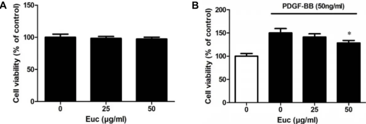
Fig. 1. Cytotoxicity of Euc on viability of VSMCs (A) and PDGF-BB-induced proliferation of VSMCs (B). To investigate the cytotoxic effect of Euc on VSMCs, the cells were cultured in 96-well plates (1×10⁵ cells/ml) for 48 hr, and the medium was replaced with serum-free medium for 22 hr. This medium was replaced with serum-free medium with or without Euc (0, 25, or 50 μg/ml) for 2 hr. The Quanti-Max kit reagent was added to serum-free medium, and the cells were incubated for 1 hr.
The optical density was determined at 450 nm using a microplate reader. Results are shown as mean (± SEM) of four in- dependent experiments, each performed in quadruplicate. To investigate the inhibitory of Euc on PDGF-BB-induced VSMC proliferation, VSMCs were cultured in 96-well plates at a density of 1×10⁵ cells/ml until they reached 70% confluence, and then the medium was replaced with serum-free medium and further cultured for 22 hr. Next, the medium was replaced with serum-free medium with or without Euc (0, 25, or 50 μg/ml) for 2 hr. The cells were incubated with in serum-free medium containing PDGF-BB (50 ng/ml) for 48 hr. The Quanti-Max kit reagent and serum-free medium were added to the cells, followed by incubation for 1 hr. The optical density was determined at 450 nm using a microplate reader. Results are shown as mean (± SEM) of four independent experiments, each performed in quadruplicate.
violet was added, and cells were washed with distilled water and imaged with a microscope (Nikon, Japan).
Western blotting
VSMCs were seeded at 1×10⁵ cells/ml in 6-well plates.
When VSMCs reached 80% confluency, the supernatant was removed, and the cells were incubated with serum-free DMEM for 22 hr and various concentrations of Euc (0, 25, or 50 μg/ml) for 2 hr. Then, PDGF-BB was added for approx- imately 5-30 min. After removal of medium, lysis buffer was added to the cells. Cell lysates were collected and analyzed using sodium dodecyl sulfate polyacrylamide gel electro- phoresis (SDS-PAGE) on 10% polyacrylamide gels, and pro- teins were transferred to PVDF membranes. The membranes were blocked for 30 min at room temperature and incubated with primary antibodies (1:2,500 dilution) overnight at 4℃
on a rocker. After incubation, membranes were incubated for 2 hr with secondary antibodies (1:5,000 dilution) at room temperature. The blots were detected using chem- iluminescent plus detect reagent for Western blots with the ImageQuant LAS 4000 system (GE Healthcare, Buckin- ghamshire, UK).
Flow cytometric analysis
VSMCs were seeded at 1×10⁵ cells/ml in 6-well plates.
When VSMCs reached 70% confluency, the supernatant was removed, and the cells were incubated with serum-free DMEM for 22 hr and various concentrations of Euc (0, 25, or 50 μg/ml) for 2 hr. Then, PDGF-BB was added for 48 hr. After removing supernatant, the cells were washed twice with PBS and removed from the culture surface using tryp- sin-EDTA. The cells were centrifuged at 1,000×g for 10 min, and the supernatant was removed. Then, cells were washed twice with PBS/EDTA and fixed in 70% ethanol with PBS/
EDTA at 4℃ overnight. After vortexing, the cells were cen- trifuged at 1,000×g for 10 min, the ethanol was removed, and cells were treated with RNase A (50 μg/ml) for 1 hr at room temperature. Next, the cells were stained with 20 μg/ml PI solution for 10 min. DNA-PI complexes were esti- mated using a flow cytometry calibur system (Becton Dickin- son, CA, USA).
Statistical analysis
Experimental results are expressed as mean (± standard error of the mean, SEM). Comparison between experimental treatments was performed using two-tailed independent t-test. At least three samples or independent experiments, each in triplicate or quadruplicate, were analyzed for each experimental treatment. A P-value < 0.05 was considered statistically significant.
Fig. 3. Effects of Euc on AKT phosphorylation in PDGF-BB-in- duced VSMCs. Cells were cultured in 6-well plates (1×
105 cells/ml). After VSMCs reached 80% confluency, the medium was replaced with serum-free medium. After 22 hr, cells in serum-free medium were treated with or without Euc (0, 25, or 50 μg/ml) for 2 hr. Next, the cells were incubated with PDGF-BB in serum-free medium for 30 min. Cell lysates were subjected to SDS-PAGE and analyzed using western blotting with anti-phospho-AKT and anti-AKT antibodies. The results are an average of three similar experiments and are expressed as mean ± SEM.
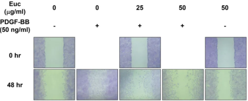
Fig. 2. Inhibitory effects of Euc on PDGF-BB-induced VSMC migration. Cells were cultured in 6-well plates (1×10⁵ cells/ml). When VSMCs reached confluency, the medium was replaced with serum-free medium for 22 hr. After 22 hr, the medium was replaced with serum-free medium with or without Euc (0, 25, or 50 μg/ml) for 2 hr. Next, cells were incubated with PDGF-BB dissolved in serum-free medium for 0 or 48 hr. Cells were treated with crystal violet after removal of serum-free medium with PDGF-BB, and the stained cells were captured using microscopy.
Results
Effects of Euc on proliferation of PDGF-BB-induced VSMCs
Cell cytotoxicity assays were conducted to identify wheth- er Euc affected the number of VSMCs. Cells were treated with serum-free medium for 22 hr, followed by treatment with various concentrations of Euc (0, 25, or 50 μg/ml) for 2 hr, and cell survival rates measured. Cell viability was measured using a Quanti-Max WST-8 cell viability kit. The survival rates of VSMCs treated with 25 μg/ml and 50 μg/
ml of Euc were 98.34±2.96 and 97.36±2.83%, respectively.
The number of cells did not decrease or increase when treat- ed with Euc, indicating that Euc does not have a direct toxic effect on cells (Fig. 1A). PDGF-BB is a substance that induces proliferation of VSMCs. Cell viability assays were conducted to determine if Euc inhibited VSMC proliferation via PDGF- BB. Proliferation of VSMCs occurred with PDGF-BB treat- ment; after treatment with Euc, he proliferation decreased in a dose-dependent manner. The cell survival rate in the PDGF-BB-treated cells without Euc increased to 150.17±9.49
% and tended to decrease concentration-dependently with Euc treatment. The survival rates of cells were 141.50±6.71%
and 128.60±5.37% at 25 μg/ml and 50 μg/ml of Euc, respec- tively (Fig. 1B).
Effects of Euc on migration of PDGF-BB-induced VSMCs
Crystal violet assays were conducted to identify the in- hibition of migration of VSMCs treated with PDGF-BB and Euc. Consequently, cell migration increased in the PDGF-BB-
induced cells, and Euc inhibited migration of PDGF-BB-in- duced VSMCs in a concentration-dependent manner (Fig. 2).
Effects of Euc on AKT phosphorylation in PDGF- BB-induced VSMCs
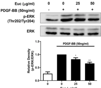
Fig. 4. Effects of Euc on ERK phosphorylation in PDGF-BB-in- duced VSMCs. Cells were cultured in 6-well plates at a density of 1×10⁵ cells/ml. After VSMCs reached 80%
confluency, the medium was replaced with serum-free medium. After 22 hr, the medium was replaced with serum-free medium with or without Euc (0, 25, or 50 μg/ml) for 2 hr. Next, the cells were incubated with PDGF-BB dissolved in serum-free medium for 5 min.
Cell lysates were subjected to SDS-PAGE and analyzed using Western blotting with anti-phospho-ERK1/2 and anti-ERK1/2 antibodies. The results are an average of four similar experiments and expressed as mean ± SEM.
Statistical differences from PDGF control (PDGF-stimu- lated but without Euc) are indicated with *p<0.05.
Fig. 5. Effects of Euc on PLC-γ phosphorylation in PDGF-BB-in- duced VSMCs. Cells were cultured in 6-well plates (1×10⁵ cells/ml). After VSMCs reached 80% confluency, the medium was replaced with serum-free medium for 22 hr. The medium was replaced with serum-free me- dium with or without Euc (0, 25, or 50 μg/ml) for 2 hr.
Next, the cells were incubated with PDGF-BB dissolved in serum-free medium for 5 min. Cell lysates were sub- jected to SDS-PAGE and analyzed using western blotting with anti-phospho-PLC-γ and anti-PLC-γ antibodies. The results are an average of three similar experiments and are expressed as mean ± SEM.
The PI3K-Akt signaling pathway is essential for VSMC proliferation and migration [13]. The PI3K-Akt signaling pathway inhibits apoptosis in cells, affecting survival [7].
Therefore, in the present study, western blotting was per- formed to identify the molecular mechanism of Euc in cell proliferation. Compared with PDGF-BB untreated controls (0.01±0.01), AKT phosphorylation was increased in controls treated with PDGF-BB (1.00±0.00). When cells were treated with various concentrations of Euc and compared with PDGF-BB-induced controls, AKT phosphorylation tended to decrease weakly when treated with 50 μg/ml of Euc (0.81±
0.11; Fig. 3).
Effects of Euc on ERK phosphorylation in PDGF- BB-induced VSMCs
The MAPK/ERK cell signaling pathway affects cell pro- liferation and migration and is an important signaling path- way associated with cell survival [13]. ERK is part of the MAPK/ERK cell signaling pathway and mediates intra-
cellular signaling through phosphorylation. PDGF enhances ERK phosphorylation, increasing proliferation and move- ment of VSMCs [13]. Western blotting was performed to identify the molecular mechanisms involved in the effects of ERK phosphorylation on VSMC cell proliferation and migration. Compared with controls (0.26±0.06), ERK phos- phorylation was increased in the PDGF-BB-treated cells (1.00±0.00). In addition, ERK phosphorylation tended to de- crease in the cells treated with 25 μg/ml (0.81±0.05) or 50 μg/ml (0.65±0.04) of Euc compared with PDGF-BB only- treated cells (Fig. 4).
Effects of Euc on PLC-γ phosphorylation in PDGF- BB-induced VSMCs
The PLC-γ signaling pathway is important for activating proliferation and migration of VSMCs. PLC-γ is an isozyme of phospholipase C activated through phosphorylation and involved in VSMC proliferation [13]. Western blotting con- firmed PLC-γ phosphorylation in PDGF-BB-treated cells, and PLC-γ phosphorylation tended to decrease in cells treated with PDGF-BB and Euc. PLC-γ phosphorylation increased
Fig. 6. Effects of Euc on PDGF-BB-induced VSMC cell cycle progression. Cells were cultured in 96-well plates (1×10⁵ cells/ml).
When VSMCs reached 70% confluency, the medium was replaced with serum-free medium for 22 hr. Next, the medium was replaced with serum-free medium with or without Euc (0, 25, or 50 μg/ml) for 2 hr. Then, cells were incubated with PDGF-BB dissolved in serum-free medium for 48 hr. Cells were fixed in 70% ethanol and incubated with RNase A for 1 hr. The cells were stained with PI, and cell cycle progression was analyzed using a flow cytometer.
Fig. 7. Euc inhibits proliferation and migration of PDGF-BB-induced VSMCs.
from 0.06±0.06 to 1.00±0.00 after PDGF-BB treatment, and decreased with 25 μg/ml (0.75±0.29) and 50 μg/ml (0.40±
0.20) of Euc treatment (Fig. 5).
Effects of Euc on PDGF-BB-induced cell cycle pro- gression
Cell cycle progression was investigated using flow cy- tometry to determine the cellular mechanisms associated with the proliferation and migration inhibition caused by Euc in PDGF-BB-induced VSMCs. The serum-deprivation of VSMCs in primary culture for 24 hr resulted in an approx- imately 83.62±0.22% synchronization of the cell cycle in the G0/G1 phase. And the percentage of cells in G0/G1 phase was decreased to 57.81±0.21% for 24 hr after PDGF-BB. The percentage of cells in G2/M phase was increased from 11.86
±0.32 to 37.14±0.21% for 24 hr after PDGF-BB was added.
In contrast, Euc-treated cells showed a significant blocking of cell cycle progression. Euc reduced the percentage of cells in G2/M phase to 14.81±0.45% at a concentration of 50 μg/
ml. This finding indicates that Euc may act at the events of the cell cycle to be effective against DNA synthesis (Fig. 6).
Discussion
Cardiovascular disease is the most common cause of death worldwide [14]. The commonly used surgical treat- ment for patients with atherosclerosis is percutaneous coro- nary intervention with stent or balloon catheter [15]. Howev- er, problems can occur after these surgeries, including abnor- mal growth of VSMCs caused by various growth factors, resulting in arteriosclerosis, vascular stenosis, or neointimal hyperplasia [5]. PDGF-BB is an important factor in migration and proliferation of VSMCs [11]. After PDGF-BB and PDGF- R binding, signal molecules such as PLC-γ1 and PI3-K/AKT are initiated by PDGF-BB, followed by activation of MAPK pathways [6]. Thus, in the present study, the effects of Euc on proliferation and migration of VSMCs and the molecular mechanisms involved were investigated as a possibility for inhibition of abnormal growth of VSMCs.
The cytotoxicity experiments showed that Euc was not toxic to and did not have a direct effect on VSMCs (Fig.
1A). Therefore, cells were treated with various Euc concen- trations, and the cell viability of PDGF-BB-induced VSMCs was confirmed. Proliferation of PDGF-BB-induced VSMCs decreased dose-dependently with 0-50 μg/ml of Euc (Fig.
1B). Furthermore, Euc inhibited the migration and pro- liferation of VSMCs treated with PDGF-BB (Fig. 2). Prolifera- tion and migration of VSMCs are induced by cell signaling pathways such as PI3K-AKT, MAPK/ERK, and PLC-γ; thus, PDGF-BB-induced VSMCs were investigated using western blotting. The PDGF-BB and PDGF-R binding induces phos- phorylation of AKT, ERK, and PLC-γ. AKT is a protein kin- ase B involved in cell survival pathways by inhibiting apop- tosis. Euc inhibited AKT phosphorylation in PDGF-BB-in- duced VSMCs (Fig. 3). ERK, a component of the MAPK/ERK signaling pathway, is a kinase activated by growth factors to induce cell processes involving proliferation and differ- entiation. ERK phosphorylation was increased in cells treat- ed with only PDGF-BB but was decreased dose-dependently in cells treated with Euc (Fig. 4). PLC-γ is an isozyme of phospholipase C, activated by phosphorylation and involved in proliferation of VSMCs. Euc inhibited PLC-γ phosphor- ylation in cells treated with PDGF-BB (Fig. 5). This result indicates that Euc inhibits the proliferation and migration of cells treated with PDGF-BB by inhibiting phosphorylation of AKT, ERK, and PLC-γ. Previous results have shown that Euc inhibits the cell signaling processes. Results of Flow cy- tometric analysis results confirmed that inhibition of these cell signaling pathways inhibited cell proliferation and mi- gration by affecting cell cycles, and the transition from G0/
G1 to G2/M phase was suppressed by Euc in PDGF-BB- treated VSMCs (Fig. 6).
In conclusion, Euc inhibited proliferation and migration by inhibiting the cell cycle through the PI3K-AKT, MAPK/
ERK, and PLC-γ signaling pathways. Euc inhibited phos- phorylation of AKT, ERK, and PLC-γ. These results indicate that Euc can inhibit abnormal proliferation and migration of VSMCs (Fig. 7). Therefore, Euc could potentially be used for treatment of cardiovascular disease mediated by abnor- mal proliferation and migration of VSMCs.
Acknowledgements
This work was supported by the National Research Foundation of Korea (NRF) Grant funded by the Korean Government (NRF-2016S1A5B8925203). We are grateful to Dr. Yusu Shin (National Institute Horticultural & Herbal Science, RDF, KOREA) for providing us with Eupatorium chi- nensis var. simplicifolium (Euc) extrect.
The Conflict of Interest Statement
The authors declare that they have no conflicts of interest with the contents of this article.
References
1. Fasciano, S., Patel, R. C., Handy, I. and Patel, C. V. 2005.
Regulation of vascular smooth muscle proliferation by hep- arin: inhibition of cyclin-dependent kinase 2 activity by p27(kip1). J. Biol. Chem. 280, 15682-15689.
2. Ha, J. M., Yun, S. J., Kim, Y. W., Jin, S. Y., Lee, H. S., Song, S. H., Shin, H. K. and Bae, S. S. 2015. Platelet-derived growth factor regulates vascular smooth muscle phenotype via mammalian target of rapamycin complex 1. Biochem. Biophys.
Res. Commun. 464, 57-62.
3. Hannink, M. and Donoghue, D. J. 1989. Structure and func- tion of platelet-derived growth factor (PDGF) and related proteins. Biochim. Biophys. Acta 989, 1-10.
4. Kanzaki, T., Shinomiya, M., Ueda, S., Morisaki, N., Saito, Y. and Yoshida, S. 1994. Enhanced arterial intimal thicken- ing after balloon catheter injury in diabetic animals accom- panied by PDGF β-receptor overexpression of aortic media.
Eur. J. Clin. Invest. 24, 377-381.
5. Kim, T. J., Han, H. J., Lim, Y., Song, M. C., Kim, J., Hong, J. T., Yoo, H. S., Pyo, M. Y., Hwang, B. Y., Lee, M. K. and Yun, Y. P. 2009. Antiproliferative action of cudraflavone B, isolated from Cudrania tricuspidata, through the down- regulation of pRb phosphorylation in aortic smooth muscle cell proliferation signaling. J. Cardiovasc. Pharmacol. 53, 341- 348.
6. Kim, T. J., Lee, J. H., Lee, J. J., Yu, J. Y., Hwang, B. Y., Ye, S. K., Shujuan, L., Gao, L., Pyo, M. Y. and Yun, Y. P. 2008.
Corynoxeine isolated from the hook of Uncaria rhyncho- phylla inhibits rat aortic vascular smooth muscle cell pro- liferation through the blocking of extracellular signal regu- lated kinase 1/2 phosphorylation. Biol. Pharm. Bull. 31, 2073-
2078.
7. Kim, T. J., Lim, Y., Kim, D. W., Kwon, J. S., Son, J. H., Jin, Y. R., Son, D. J., Jung, J. C., Avery, M. A., Hong, J. T. and Yun, Y. P. 2007. Epothilone D, a microtubule-stabilizing compound, inhibits neointimal hyperplasia after rat carotid artery injury by cell cycle arrest via regulation of G1-check- point proteins. Vascul. Pharmacol. 47, 229-237.
8. Lee, J. H., Kim, D. H., Shin, J. W., Park, S. J., Kim, Y. S., Shin, Y. S., Yu, J. Y. and Kim, T. J. 2012. Eupatorium chi- nensis var. simplicifolium root extract inhibits the lip- opolysaccharide-induced inflammatory response in Raw 264.7 macrophages by inhibiting iNOS and COX-2 expression. J. Life Sci. 22, 1137-1144.
9. Lu, Q. B., Wan, M. Y., Wang, P. Y., Zhang, C. X., Xu, D.
Y., Liao, X. and Sun, H. J. 2018. Chicoric acid prevents PDGF-BB-induced VSMC dedifferentiation, proliferation and migration by suppressing ROS/NFκB/mTOR/P70S6K sig- naling cascade. Redox Biol. 14, 656-668.
10. Muto, A., Fitzgerald, T. N., Pimiento, J. M., Maloney, S. P., Teso, D., Paszkowiak, J. J., Westvik, T. S., Kudo, F. A., Nishibe, T. and Dardik, A. 2007. Smooth muscle cell signal transduction: implications of vascular biology for vascular surgeons. J. Vasc. Surg. 45 Suppl A, A15-24.
11. Raines, E. W. 2004. PDGF and cardiovascular disease.
Cytokine Growth Factor Rev. 15, 237-254.
12. Ross, R. 1993. The pathogenesis of atherosclerosis: a per- spective for the 1990s. Nature 362, 801-809.
13. Schwartz, S. M. and Reidy, M. A. 1987. Common mecha- nisms of proliferation of smooth muscle in atherosclerosis and hypertension. Hum. Pathol. 18, 240-247.
14. Townsend, N., Wilson, L., Bhatnagar, P., Wickramasinghe, K., Rayner, M. and Nichols, M. 2016. Cardiovascular disease in Europe: epidemiological update 2016. Eur. Heart J. 37, 3232-3245.
15. Yahagi, K., Kolodgie, F. D., Otsuka, F., Finn, A. V., Davis, H. R., Joner, M. and Virmani, R. 2016. Pathophysiology of native coronary, vein graft, and in-stent atherosclerosis. Nat.
Rev. Cardiol. 13, 79-98.
초록:등골나물추출물의 혈관 평활근 세포의 비정상 증식에 대한 억제 효과 및 분자기작
김민정1․김지희1,2․이진호1․김민아1․우근정1․김한성3․김택중1,4*
(1연세대학교 생명과학기술학부, 2국립암센터 이행성연구부, 3연세대학교 의공학부, 4(주)닥터티제이 R&D 연구소)
등골나물은 오랫동안 음식으로 사용되어 왔으며 한방에서 한약재로 사용되어왔으며, 특히 등골나물 추출물은 항염 및 항산화 효과가 보고되었다. 심혈관 질환인 동맥경화증은 동맥 혈관에서 발생하는 만성 염증이며 다양한 질환에 관여하고 있다. 재협착 및 신경병증 증식과 같은 심혈관 질환은 주로 혈관 평활근 세포의 다중 성장인자로 인한 비정상적인 성장과 이동으로 인해 발생하며, 혈소판유래 성장인자는 손상된 혈관벽에서 방출되며 혈관 평활 근 세포의 증식 및 이동에 관여한다. 본 연구에서는 혈관 평활근 세포의 비정상적인 증식 및 이동에 대한 등골나 물 추출물의 효과를 확인하였고, 혈소판유래 성장인자로 유도된 혈관 평활근 세포내에서 신호전달 경로를 조사하 였다. 혈소판유래 성장인자로 유도된 혈관 평활근 세포에서 등골나물 추출물의 전처리는 세포 증식 및 이동을 효과적으로 감소시켰다. 또한, 세포 내 신호전달 경로 AKT, phospholipase C gamma (PLC-γ) 및 mitogen-acti- vated protein kinase (MAPK)의 웨스턴 블롯 실험 결과, 이들 경로의 인산화 억제를 확인하였다. 유세포분석 데이 터는 혈관 평활근 세포의 세포주기가 등골나물 추출물에 의해 차단되었음을 보여주었다. 이 결과는 혈소판유래 성장인자가 세포주기와 세포 내 신호전달 인자를 조절하여 혈관 평활근 세포의 증식과 이동을 억제 할 수 있음을 시사한다. 또한, 이는 등골나물 추출물이 혈관 평활근 세포의 비정상적인 증식 및 이동을 차단하여 심혈관 질환 예방에 하나의 소재로 사용될 수 있음을 시사한다.