Copyright ⓒ 2009, The Korean Academy of Oral Biology
43
Journal of Oral Biology
Effect of Antibiotics upon the Antibacterial Activity of Platelet Microbicidal Protein against Streptococcus rattus BHT
Jae-Wook Kim
1, Son-Jin Choe
1, and Si Young Lee
2*
1
Department of Oral Microbiology, College of Dentistry, Seoul National University, Seoul, Korea and
2Department of Oral Microbiology, College of Dentistry, Research Institute of Oral Science, Kangnung National University, Kangnung, Korea (received March 4, 2009 ; revised March 16, 2009 ; accepted March 20, 2009)
Thrombin-induced platelet microbicidal protein (tPMP) is a small cationic peptide that exerts potent in vitro microbicidal activity against a broad spectrum of human pathogens, including Staphylococcus aureus and Streptococcus rattus BHT. Earlier evidence has suggested that tPMP targets and disrupts the bacterial membrane.
However, it is not yet clear whether membrane disruption itself is sufficient to kill the bacteria or whether subsequent, presumably intracellular, events are also involved in this process. In this study, we investigated the microbicidal activity of rabbit tPMP toward S. rattus BHT cells in the presence or absence of a pretreatment with antibiotics that differ in their mechanisms of action. The streptocidal effects of tPMP on control cells (no antibiotic pretreatment) were rapid and concentration-dependent. Pretreatment of S. rattus BHT cells with either penicillin or amoxicillin (inhibitors of bacterial cell wall synthesis) significantly enhanced the anti-S. rattus BHT effects of tPMP compared with the effects against the respective control cells over most tPMP concentration ranges tested. On the other hand, pretreatment of S. rattus BHT cells with tetracycline or doxycycline (30S ribosomal subunit inhibitors) significantly decreased the streptocidal effects of tPMP over a wide peptide concentration range. Furthermore, pretreatment with rifampin (an inhibitor of DNA-dependent RNA polymerase) essentially blocked the killing of S. rattus BHT by tPMP at most concentrations compared with the respective control cells. These results suggest that tPMP
exerts anti-S. rattus BHT activity through mechanisms involving both the cell membrane and intracellular targets.
Key words: platelet microbicidal protein, antibiotics, streptococcus
서 론
혈소판은 외상이 생긴 혈관부위나 맥관내피에 감염이 생기는 경우 조기에 나타나는 수적으로 가장 많은 세포 이다(Durack, 1975; Durack et al., 1973; Ferguson et al., 1986; Sullam et al., 1985). 맥관내피 감염의 하나는 감 염성 심내막염이다. 전통적으로, 판막 표면에 혈소판이 침 적하여 큰 응괴가 형성되면 이 응괴내의 세균들은 숙주 방어계에 의하여 쉽게 제거되지 않는 것으로 생각되어 왔다. In vitro 연구에 의하면, 혈소판들이 응괴할 때 세 균은 이 응괴 속에 매몰되지만 혈소판에 의하여 포식되 거나 치사되지 않는다 (Clawson과 White, 1971). 이런 이유 때문에, 혈소판은 숙주의 손상과 병의 진전에 기여 하는 것으로 간주되어 왔다.
그러나 근래의 연구들에 의하여, 혈소판이 심내막 감염 을 조장하는 역할뿐 아니라, 반대로 감염을 제한할 수 있 음도 제시되었다. 혈소판에 의한 감염의 제한은 부착하는 세균을 저해하는 혈소판-유래 항미생물 인자들에 기인할 수 있다. 예로서, 소분자량의 양이온성 항미생물 펩타이 드들이 사람과 토끼의 혈소판으로부터 분리되었고 이것 들은 혈소판 항미생물 단백질(Platelet Microbicidal Protein (PMP))이라고 지칭된다(Yeaman et al., 1992b).
트롬빈으로 자극한 토끼 혈소판으로부터 분비되는 주된 PMP인 tPMP-1은 가장 잘 연구된 PMP이다(Yeaman et
*Corresponding author: Si Young Lee, Department of Oral
Microbiology, College of Dentistry, Research Institute of
Oral Science, Kangnung National University, Kangnung,
210-702, Korea. Tel.: +82-33-640-2455, Fax.: +82-33-642-
6410, E-mail: [email protected]
al., 1997). tPMP는 Staphylococcus aureus, Streptococcus rattus BHT를 포함하여 넓은 스펙트럼의 병원균에 대하 여 강한 살미생물 활성을 나타낸다(Lee et al., 2001;
Yeaman, 1997). tPMP의 주된 활성 메커니즘의 하나는 미생물의 세포질막의 투과성화 (permeabilization)에 있는 것으로 판명되고 있다. 최근 Choi등의 연구에 따르면(Choi et al., 2005) 구강연쇄구균에서도 tPMP에 의한 세포막 의 depolarization과 permeabilization이 tPMP에 의한 이 들 세균의 치사와 상호연관이 있음을 보였다. S. aureus 를 tPMP에 노출시키면, 세포질막의 파괴는 1분내에 일 어나지만 세포 자체의 치사는 1-2 시간 이후에 이루어진 다(Yeaman et al., 1992b; Yeaman et al., 1998; Yeaman et al., 1997). 이런 사실은 세포내의 어떤 표적이 포도 구균의 치사 메커니즘에 관련되는 것으로 해석된다. Xiong 등(1999)은 tPMP-1의 잠재적 세포내 표적의 존재여부를 탐색하기 위하여 작용 메커니즘이 서로 상이한 항생제들 로 S. aureus를 전처리한 뒤에 이 펩타이드의 효과를 연 구한 결과, 항생제들이 tPMP로써 유도되는 항 포도구균 효과를 간섭하는 사실을 보고하였다. 이런 관찰에 근거하 여 Xiong 등(1999)은 tPMP-1의 항 S. aureus 활성이 세포막과 세포내 표적을 포함하는 메커니즘을 통해서 일 어난다고 제안하였다.
이 연구는 tPMP의 구강연쇄구균 치사 메커니즘에 제 2의 치사과정이 S. aureus에서 처럼 관련되는가를 구명 하기 위하여 고안되었으며, 서로 다른 작용 메커니즘을 소유한 항생제들로써 S. rattus BHT를 전처리함으로써 tPMP의 잠재적 세포내 표적을 탐색함을 목적으로 하였다.
재료와 방법
세균과 배양
tPMP의 살균활성 결정에 이용한 Bacillus subtilis ATCC 6633 균주는 American Type Culture Collection (ATCC; Manassas, VA, USA)에서 구입하였다. 실험에 사용한 구강 연쇄구균인 Streptococcus rattus BHT는 서 울대학교 치과대학 구강 미생물학교실에 보관중인 실험 실 균주이다. 구강 연쇄구균의 배양은 Todd-Hewitt broth (THB) (Difco, Detroit, MI, USA)를 사용하여 37oC에 서CO2가5 % 추가된 상태에서 배양하였으며 B. subtilis 는Brain Heart Infusion (BHI) broth (Difco)로 37oC에 서 배양하였다.
tPMP의 준비
트롬빈으로 자극된 PMP는 Yeaman 등(1992b; 1993)의 방법으로 준비하였다. 완충된 citrate 항응고 약제 (0.11 M sodium citrate2H2O and 0.002 M citric acidH2O, pH 5.5)가 든 실리콘 처리된 유리튜브 (Vacutainer Systems,
Becton Dickinson and Company, Franklin Lakes, NJ, USA)에 토끼로부터 혈액을 채취한 뒤 (항응고 약제: 혈 액의 비율, 1:10), 20oC에서 원심 분리하여 (150 × g, 15 분) 튜브 상부에 혈소판이 풍부한 플라즈마 (platelet rich plasma, PRP) 현탁을 얻었다. 이 PRP 분획의 위쪽 2/3 를 다른 튜브에 수집한 뒤 원심 분리하여 (2,000 × g, 10 분) 혈소판 침전을 얻고, 혈소판을 Tyrode 용액 (pH 6.8) 으로 2회 세척하였다. 세척한 혈소판을 37oC로 미리 가 온된 Eagle의 Minimum Essential Medium (MEM) (GibcoBRL, Gaithersburg, MD, USA)으로 현탁하고, Hausser 혈구 측정기로 수를 산정하여 1 × 10⁹ 혈소판/ml 로 되게 하였다. 혈소판 현탁액 1 ml 당 1 unit의 bovine thrombin (Sigma Chemical Co. St. Louis, MO, USA) (12.5µl의 0.2 M CaCl2포함)으로 37oC에서 30 분간 자 극하여 tPMP가 혈소판으로부터 분비되게 하고, 원심분리 (3,000× g, 15분)하여 tPMP가 풍부한 상층액을 얻었다.
tPMP는 실리콘 처리된 유리 튜브에 분주한 후 -70oC에 보관하며 실험에 사용하였다.
tPMP의 살균활성의 결정 및 표준화
트롬빈으로 유도된 tPMP의 살균활성은 Lee 등(2001) 의 방법으로 결정하였다. tPMP의 생물활성의 측정은 tPMP 의 살균작용에 민감한 표지 (indicator) 세균인 B. subtilis ATCC 6633으로 시행하였다. tPMP를 넣은, 단백질이 적 게 부착하는 microtiter 접시 (Corning Costar, Cambridge, MA, USA)에 B. subtilis (105CFU/ml)를 식균하여 세균의 농도는 104CFU/ml, tPMP의 희석범위는 1:1 (비희석)에 서 1:1,024 (최종 웰 용량=200 µl)되게 하였다. 한 웰에 는 MEM에 현탁한 B. subtilis만 넣어 양성 성장 기준군 으로 삼았다. Microtiter 접시를 37oC에서 1시간 배양한 후, 각 웰에서 20 µl 씩 채취하여 0.01 %의 sodium polyanetholsulfonate (SPS) (PMP 유도에 의한 살균을 저해하는 제제)를 함유한 PBS (PBS-SPS)로 희석하여 BHI 고체 배지에 식균, 배양 (37oC에서 18시간)한 뒤 CFU를 산정하였다. PMP의 생물활성은 B. subtilis에 대 하여 ≥95%의 치사율을 보이는 가장 높은 PMP 희석의 역수 (Unit/ml)로서 표시하였다.
구강 연쇄구균에 대한 tPMP의 살균성 측정
배양된 세균을 수집하여 PBS로 두 번 씻은 후 660 nm 에서의 흡광도를 측정하고 미리 준비된 표준 곡선 (세균 수:흡광도)을 이용하여 세균수를 산정 하였다. 최종 세균 농도가 104-105CFU/ml 되게 PMP (최종 농도 200 Unit/
ml)가 든 MEM 이나 PMP가 안든 MEM (기준군)에 넣 고 1 시간 배양한 후 20 µl 씩 채취하여 PBS-SPS로 희 석하고 경우에 따라서는 생리식염수로 추가적으로 더 희 석한 후 THB 한천배지에 식균, 배양하였다. CO2가 추 가 (5 %)된 상태로 37oC에서 3일간 배양 후 CFU를 세
어 기준군에 대한 실험군의 백분율로서 PMP의 구강 연 쇄구균에 대한 살균활성을 표시하였다. 모든 실험에서 tPMP 생물활성의 양성 기준으로 B. subtilis를 포함시켰다.
항생제 최소 생육저해농도 (minimum inhibitory concentration; MIC)의 결정
실험에 사용한 구강 연쇄구균들의 페니실린, 아목시실 린, 테트라사이클린, 독시사이클린, 그리고 리팸핀(Sigma Chemical Co.)의 항생제 감수성 검사는 MHB (Muller- Hinton broth) 배지를 사용하여National Committee for Clinical Laboratory Standards의 지침(1993)에 근거하여 시행하였다. 항생제 저장 용액들은 분말상태의 항생제들 을 증류수에 용해시킨 다음 pore size가 0.22 µm인 여과 지를 통과시켜 준비하였다. 사용한 세균들의 최종농도는 105CFU/ml되게 식균하였다. MIC는 세균을 37oC에서 18시간 배양후에 판독하였으며 MIC는 세균의 성장을 보 이지 않은 최소의 항생제 농도로 하였다. 모든 MIC는 서로 다른 날에 최소한 두 번 이상 재검사하였다.
항생제로써 전처리한 S. rattus BHT에 대한 tPMP의 살균효과
tPMP의 살연쇄구균 효과에 미치는 항생제 전처리의 영 향을 평가하기 위하여 배지 THB에서 배양한 S. rattus BHT 세포를 tPMP에 노출시키기 이전에, 작용 메커니즘 측면에서 서로 상이한, 아래의 항생제들 가운데 한 종류 에 노출시켰다. 1) 세균의 세포벽합성 저해제 (페니실린 또는 아목시실린), 2) 30S 라이보좀 단위체 단백질합성 저해제 (테트라사이클린 또는 독시사이클린), 3) DNA- dependent RNA 중합효소에 부착하여 RNA합성 개시단 계를 저해하는 제제 (리팸핀). 항생제 전처리는 MHB 배 지에 세균을 107CFU/ml 의 수로 식균하여 37oC에서 1 시간의 진탕배양으로 수행하였다. 항생제 전처리에 사용 한 모든 항생제는 각 항생제 MIC 값의 5 배 농도를 사 용하였다. MHB 배지에서의 배양 뒤에, 항생제로 전처리 한 S. rattus BHT 세포와 기준 세포(항생제로 전처리 안 한 것)를 원침(1,000 × g에서 10 분간)하고, 잔존하는 배 지 및 항생제의 제거를 위하여 PBS (pH 7.2)로 두 번 씻고 PBS에 다시 현탁하여, tPMP로 처리키 위하여 필 요한 농도로 희석하였다. S. rattus BHT는 최종 접종균 수가 104-105CFU/ml 되게, 그리고 tPMP는 0-50 Unit/
ml의 농도범위가 되도록 첨가하였다. 접종에 사용한 세 균농도는 tPMP의 살균성을 연구한 이전의 보고(Lee et al., 2001)에서 사용된 것이다. 위의 검사는 모두 37oC 에서 1시간에 걸쳐 행하였다. 배양 종료 뒤에 개개의 튜 브에서 20 µl씩 채취하여 PBS-SDS로 희석한 다음 THB 한천배지에 식균하였다. 생존하는 세균세포는 3 일간의 배양 뒤에 산정하여 데이터를 ∆log10CFU/ml/h (살균속도) 로 표시하였다 (∆log10CFU/ml/h = 실험군의 log10CFU/
ml/h - 기준군의 log10CFU/ml/h (Xiong et al., 1999)).
실험은 모두 2 개 한벌로써 2 번 이상 실시하였으며 최 종 데이터는 평균 ± 표준편차로 표시하였다.
통계처리
항생제로써 전처리하거나 하지않은 S. rattus BHT 세 포를 각 농도의 tPMP에 노출시킬 때의 살균속도 (∆log10CFU/ml/h)에서의 차이는 Student t 검사로써 비교 하였고, p<0.05의 값을 유의한 차이를 보이는 것으로 간 주하였다.
실험 결과
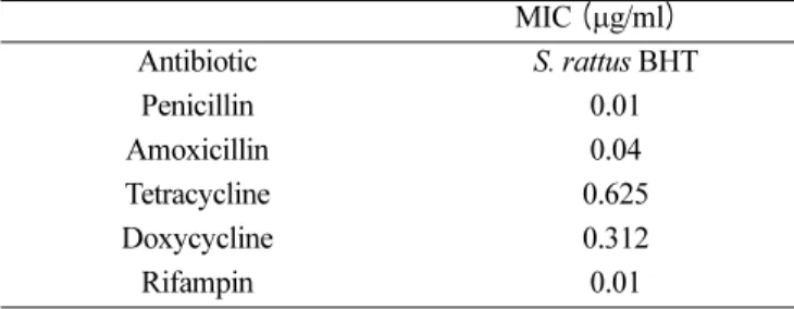
S. rattus BHT의 항생제 감수성
이 검사에 사용된 세균세포 농도인 105CFU/ml에서 조 사한 모든 항생제에 감수성을 보였다 (Table 1).
작용 메커니즘들이 상이한 항생제로써 전처리한 세 균에 대한 tPMP의 살균효과
1) 세균 세포벽합성 저해제들의 tPMP 살균활성에 대한 영향
항생제로써의 전처리 없이, tPMP 자체로서는 농도 의 존적 연쇄구균 치사활성을 보였다. 그러나, tPMP 처리 이전에 S. rattus BHT 세포들을 페니실린이나 아목시실 린에 노출시키면, tPMP 단독으로써 성취하는 치사에 비 교하여 S. rattus BHT의 치사가 증가되었다 (Fig. 1). 이 항진된 효과는 페니실린 혹은 아목시실린 전처리의 경우 비슷한 수준이었으나 통계학적 유의성에 있어서, 페니실 린의 경우 1.56, 25, 그리고 50 Unit/ml에서 그차이에 유 의성이 있었고 아목시실린의 경우에는 1.56, 3.13, 6.25, 그리고 50 Unit/ml에서 유의한 차이를 보였다. 예로서, tPMP (50 Unit/ml) 단독으로는 평균 살균속도 -1.22
∆log10CFU/ml/h을 보였다. 그러나 동일 tPMP 농도에 노 출되기 이전에 페니실린이나 아목시실린으로 전처리했을 때는 살연쇄구균 효과가 증가하였다 (각각 -1.54와 -1.56 log10∆CFU/ml/h; 기준세포에 대비하여 페니실린 및 아목 시실린 전처리 세포의 경우 p<0.05).
Table 1. In vitro susceptibility of S. rattus BHT to antibiotics.
MIC (µg/ml)
Antibiotic S. rattus BHT
Penicillin 0.01
Amoxicillin 0.04
Tetracycline 0.625
Doxycycline 0.312
Rifampin 0.01
2) 세균 단백질합성 저해제의 tPMP 살균활성에 대한 영향 세균의 30S 라이보좀 단위체에 부착하여 단백질 합성 을 저해하는 항생제들인 테트라사이클린이나 독시사이클 린으로의 전처리는 tPMP의 살연쇄구균 효과를 펩타이드 의 농도 6.26-50 Unit/ml 범위에서 유의하게 (p<0.05) 비 슷한 수준들로 감소시켰다 (Fig. 2).
3) 세균의 RNA 합성저해 항생제의 tPMP 살균활성에 대한 영향
세균의 DNA-dependent RNA 중합효소에 부착하여 RNA 합성 개시단계를 저해하는 항생제인 리팸핀으로써 의 전처리가 tPMP의 살균활성에 미치는 영향은 Fig. 3 에 보였다. 리팸핀으로 전처리한 S. rattus BHT 세포는 뒤따라서 tPMP에 노출될 때 그 살미생물 효과에, 기준 세포에 비하여, 유의하게 덜 민감하였다 (펩타이드의 농 도 6.25에서부터 50 Unit/ml의 범위에서 p<0.05). 예로서,
Fig. 1. Susceptibility of S. rattus BHT pretreated with a bacterial cell wall synthesis inhibitor (penicillin or amoxicillin) to tPMP. S.
rattus BHT cells were pretreated with either 5 × the MIC of peni- cellin, 5 × the MIC of amoxicillin, or THB only at 37
oC for 1 h prior to exposure to tPMP for 1 h in MEM buffer (pH 7.2). Survi- vors were enumerated on solid medium. Values indicate means of duplicate determinations and error bars represent standard errors.
The data are a representation of at least two experiments. *, statisti- cally significant increase (p<0.05).
Fig. 2. Susceptibility of S. rattus BHT pretreated with a bacterial protein synthesis inhibitor (tetracycline or doxycycline) to tPMP. S.
rattus BHT cells were pretreated with either 5 × the MIC of tetra- cycline, 5 × the MIC of doxycycline, or THB only at 37
oC for 1 h prior to exposure to tPMP for 1 h in MEM buffer (pH 7.2). Survi- vors were enumerated on solid medium. Values indicate means of duplicate determinations and error bars represent standard errors.
The data are a representation of at least two experiments. *, statisti- cally significant increase (p<0.05).
Fig. 3. Susceptibility of S. rattus BHT pretreated with a RNA syn- thesis inhibitor (rifampin) to tPMP. S. rattus BHT cells were pre- treated with 5 × the MIC of rifampin or THB only at 37
oC for 1 h prior to exposure to tPMP for 1 h in MEM buffer (pH 7.2). Survi- vors were enumerated on solid medium. Values indicate means of duplicate determinations and error bars represent standard errors.
The data are a representation of at least two experiments. *, statisti-
cally significant increase (p<0.05).
최고의 tPMP 농도인 50 Unit/ml에서 S. rattus BHT 기 준세포에 대한 살연쇄구균 속도는 -1.22 ∆log10CFU/ml/
h이었다. 그러나 동일한 펩타이드 농도에서, 리팸핀으로 전처리한 세균은 단지 -0.06 ∆log10CFU/ml/h의 치사속 도를 보였다 (p<0.05).
고 찰
tPMP의 살균 메커니즘을 연구하기 위하여 연쇄구균 S.
rattus BHT 균주를 사용하여 수행한 우리의 연구성적의 일부는 포도구균인 S. aureus를 이용한 Xiong 등(1999) 의 것과 일치하였다. Xiong 등은 DNA 및 단백질 합성 에 대한 영향이 tPMP-1의 살미생물 메커니즘의 하나가 될 수 있음을 보였는데 S. rattus BHT에서도 단백질 합 성의 저해가 tPMP의 살균 메커니즘이 될 수 있다는 결 과를 얻었다. 여기에 더하여 이 연구에서는 리팸핀으로써, DNA- dependent RNA 중합효소에 의한 RNA 합성을 차단하면, 세균을 tPMP에 노출시킴으로써 초래되는 살미 생물 효과가 크게 제거될 수 있었다. 이 관찰은 RNA 합 성저해도 tPMP의 살미생물 메커니즘의 일환이 될 수 있 음을 보이는 것이고 이 연구의 전체적 결과는 구강 연쇄 구균인 S. rattus BHT에서도 tPMP가 세포내 표적에 작 용한다는 것을 제시하는 것이다. 그리고 우리의 연구성적 과 Xiong 등(1999)의 것을 종합하면, tPMP의 살미생물 메커니즘에는, 미생물의 유전자의 증식을 포함하여 유전 자의 전사 및 번역 과정이 관련된다는 것이다. S. gordonii DL1을 포함하여(Lee et al., 2001) 구강 연쇄구균들이 보 이는 tPMP 저항성의 메커니즘은 아직 정확하게 알려져 있지 않지만 S. aureus에서는 tPMP 저항성의 원인이 이 세균에 들어있는 플라즈미드에 기인한다는 보고가 있다 (Kupferwasser et al., 1999).
선천성 (innate) 숙주방어에서 작용하는 양이온성 항미 생물 펩타이드들 가운데에서 tPMP와 더불어 비교적 많 이 연구된 것은 Human Neutrophil Defensin-1 (HNP- 1)이다. HNP-1은 중성구의 defensin으로서 분자량이 적 고 중성구의 azurophilic granule 속에 들어 있다(Ganz et al., 1990). HNP-1도 tPMP처럼 여러 종류의 미생물 을 죽일 수 있고(Lehrer et al., 1989), 표적 생물의 세 포막을 파괴하며(Shimoda et al., 1995), 세포막의 전압- 의존성 투과성화를 일으키고, 막의 기능을 교란시킨다 (Yeaman et al.,1998). 또 E. coli를 모델 시스템으로 사 용한 Lehrer 등(Lehrer et al., 1989)의 연구는 단백질, RNA, 그리고 DNA 합성의 저해가 HNP-1의 살미생물 메커니즘에 관련될 수 있다는 증거를 제시하였다. S. rattus BHT를 페니실린이나 아목시실린으로 전처리한 뒤 tPMP 에 노출시킬 경우 살연쇄구균 효과는 펩타이드의 넓은 농도범위에서 증가하였다. 이 결과는 Xiong 등(1999)이
S. aureus에서 관찰한 성적과 일치하는 것이다. 이처럼 살균이 증가하는 상호작용의 메커니즘은 알려지지 않았 다. 그러나 세포벽 합성에 작용하는 항생제가 tPMP의 살 미생물 효과를 증가시킴은 보고되어 왔다. 예로서, S.
aureus를 tPMP와 세포벽 합성 저해 항생제 (옥사실린, 뱅코마이신)에 동시에 노출시킬 때에는 상승적 살미생물 효과가 in vitro에서 관찰되었다(Yeaman et al., 1992a).
또 S. aureus를 tPMP로 전처리하면 옥사실린 또는 뱅코 마이신의 후항생제 효과 (postantibiotic effect)의 지속시 간을 연장시켰다.
본 연구의 결과를 요약하면, S. rattus BHT 세포에서 tPMP의 살미생물 메커니즘은 단백질 및 RNA 합성에 대 한 영향을 포함한다는 간접적 증거를 제공하며, 이 두 종 류의 거분자 합성을 tPMP가 직접적으로 저해할 수 있는 지 여부를 판별할 실험의 필요성을 제시하고 있다.
참고문헌