INTRODUCTION
Expression of particular genes is often associat- ed with a specific physiological function or respo- nse to environmental changes and is a good indi- cator for exposure to a variety of environmental factors (Rees et al., 2005). Among them, CYP genes not only play an important role in the metabolism of xenobiotics, but they also play a significant role in steroid metabolism (Williams et al., 1998; Hu
et al., 2004; Goldstone and Stegeman, 2006; Isin
and Guengerich, 2007). CYP1A induction, or the increased expression and abundance of CYP1A mRNA, is a well-documented phenomenon in tel- eost fish (Billiard et al., 2004; Jönsson et al., 2004;
Ortiz-Delgado and Sarasquete, 2004) and occurs when planar halogenated compounds (PAHs, PCBs, TCDD etc.) bind to a cytosolic ligand-acti- vated transcription factor known as the Ah (aryl hydrocarbon) receptor (Hahn and Stegeman, 1994;
Sarasquete and Segner, 2000).
─
─ 364 ──
* Corresponding author: Tel: 1-778-788-5646, Fax: 1-604-666-8666, E-mail: [email protected]
Present address: Fisheries and Oceans Canada 4160 Marine Drive, West Vancouver, BC, V7V 1N6 Canada.
cDNA Cloning and Expression of a Cytochrome P450 1A (CYP1A) from the Pale Chub, Zacco platypus
Jeon, Hyoung-Joo, Young-Chul Park1, Wan-Ok Lee, Jong-Ha Lee and Jin-Hyoung Kim*
(Inland Fisheries Research Institute, National Fisheries Research and Development Institute, Gyeonggi-do 114-3, Korea
1Inland Aquaculture Research Center, National Fisheries Research and Development Institute, Gyeongsang-do 577-1, Korea)
The pale chub (Zacco platypus) is generally found in Asian countries, such as Korea, Japan, and China. Nevertheless, very little information exists about the genes involved in the metabolism of xenobiotics in this species. This species is useful in monitoring the environmental impact on various pollutants in freshwater as a sentinel fish species.
We cloned the full-length cDNA sequence of xenobiotic metabolizing cytochrome P450 1A (CYP1A) gene from Z. platypus and characterized it. Tissue distribution and time- dependent induction of CYP1A were studied by real-time RT-PCR. Induction pattern of CYP1A was studied by exposing the fish to an arylhydrocarbon receptor agonist, ββ-naphthoflavone (BNF). The liver showed the highest level of expression in basal state as well as BNF- treated fish. However, appreciable levels of expression were also recorded in Gill and kidney and the least level of expression was observed in the eye. The results of the time-course study revealed an induction in the liver, brain, and gills after 6 h and 12 h in most of the tissues. This study provides an insight into the xenobiotics metabolizing system of Z. platypus and offers baseline information for further research related to biomarker, stress, and adaptive response of this ecolog- ically important fish species in the freshwater environment.
Key words : Zacco platypus, CYP1A, ββ-naphthoflavone, gene expression, xenobiotic metabolism
Although CYP genes are one of the most exten- sively studied genes in mammals as well as in fish, sequence and expression profile of CYP genes in Cypriniformes species are not well documented except for a few well-known species such as a ze- brafish (Gao et al., 2011) and a minnow (Liu et al., 2008).
Pale chub (Zacco platypus, Cypriniformes) was a suitable sentinel species because of their rela- tively high abundance in most rivers and streams of Korea. Also, this species has high mobility (Ya- mazaki et al., 1996), lives in riffles of midstream and downstream, prefers sand and gravel bottom conditions, and feeds mainly on insects, algae, de- tritus, and micro-crustaceans (Kim, 1997). During the spawning period they are widely scattered in the waters from 30 to 50 cm depth (Nakamura, 1952). However, most fishes migrate to the deeper and warmer water column in winter (Nakamura, 1952; Kim, 1997).
Although population dynamics and ecological studies in Z. platypus have been done, only a few studies have been performed on on metacercarial infection (Park et al., 2004; Cho et al., 2006), im- munohistochemical studies (Ku et al., 2004), stres- sor identification and health assessment (Yeom et al., 2007), and a chromosomal toxicity assay (Hayashi et al., 1998). In addition, to date no infor- mation is available on gene expression profiles, xenobiotic metabolism and detoxification. The habitat of Z. platypus may be contaminated by environmental pollutants and it also feeds on in- sects which might have been contaminated and bioaccumulated insecticides. Therefore, informa- tion of xenobiotic metabolizing genes is desirable to understand responses of Z. platypus to expo- sure to xenobiotics directly or indirectly. As stated above, Z. platypus is a hardy fish and survives well under laboratory conditions. This species can be used as an alternate model for short and long term toxicity testing. Therefore, detailed molecu- lar biological information on various genes from Z. platypus, especially those involved in toxicant metabolism (CYP1A, most notably), is desirable.
In addition, there are a number of other QRT- PCR assays that have been developed to measure CYP1A mRNA expression in fish (Campbell and Devlin, 1996; Miller et al., 1999; Cousinou et al., 2000) that provide good models for data compari- son. Here, we describe the full-length cDNA sequ- ence of Z. platypus CYP1A, its tissue specific ex- pression, and induction by a universal inducer of
CYP1A and a potent agonist of the aryl hydrocar- bon receptor, β- naphthoflavone to provide base- line information for further research related to biomarkers, as well as stress and adaptive respo- nse of this ecologically important fish species.
MATERIALS AND METHODS
1. Fish
Z. platypus (body length, 5.5±0.3 cm; body mass, 1.7±0.4 g) were caught in the Cho-Jeong stream (Ga-phyeong, Kyoung-gi, Korea) by using a gill net. Fish were acclimated to laboratory conditions for two weeks at 19� C and a photoperiod of 12 h light and 12 h dark cycles in a 100 L tank contain- ing well-aerated water (pH 7.3, dissolved oxygen, DO 6.9 mg L
-1). During the acclimation period, fish were fed a commercial diet.
2. RNA isolation, reverse transcription, first strand cDNA synthesis
Seven tissues (brain, gill, intestine, liver, kidney, muscle and skin) were quickly dissected under sterile conditions and homogenized in three volu- mes of Trizol
®reagent (Invitrogen, Paisley, Scotl- and) with a homogenizer. Total RNA from the fish was isolated using Trizol
®reagent (Molecular Re- search Center, Inc., Cincinnati, OH) according to the manufacturers’ protocol. RNA qualities were confirmed spectrophotometrically (A260/A280 ratio ≈1.8 -2.0), respectively after treatment with a DNase at 37� C for 30 min to account for contami- nation of genomic DNA. Single-stranded cDNA was synthesized from 1 μg total RNA using an oligo (dT)
20primer and PrimeScript
TM1st strand cDNA synthesis Kit (Takara, Japan) by reverse transcription according to the manufacturers’ pro- tocol.
3. PCR, amplification of 3′′ and 5′′ ends and sequence analysis
Degenerative primers were designed using con-
served domains after multiple alignments of pre-
viously reported full-length cDNA sequences of
teleosts CYP1A. The detail of primers and PCR
conditions for amplification of partial sequences
are given in Table 1. For amplifying partial sequ-
ences, RT-PCR was carried out using 2 μM of each
primer and hepatic cDNA as a template using Taq
polymerase (TaKaRa Bio Inc.). Oligonucleotide
synthesis and sequencing was performed at Bio- neer Co. (Daejeon, South Korea). The full-length sequence of Z. platypus CYP1A was deduced using a capfishing kit (Seegen, Carlsbad, CA, USA). The 3 ′ Rapid Amplification of cDNA Ends (3′ RACE) and 5 ′ RACE were performed using the primers and PCR conditions as detained in Table 1, follow- ing the manufacturer instructions.
4. Tissue distribution
Real-time reverse transcription PCR (RT-PCR) was performed to study tissue distribution pattern of the CYP1A gene. The gene distribution pattern was studied in the following tissues: brain, gill, intestine, liver, kidney, muscle and skin using oligo (dT)
20primer and SuperScript
TMIII reverse transcriptase (Invitrogen) according to the manu- facturers’ instructions. The PCR conditions are described in Table 1. The PCR products were sep- arated on 1% TBE agarose gels containing ethidi- um bromide (EtBr) and visualized on a Fluor-STM Multimanager system (Bio-Rad).
5. BNF-induced induction of CYP1A
Fish (n= =30) were exposed to BNF (Sigma-Aldrich Co., MO, USA) through tank water. BNF was dis- solved in DMSO and added to the water to yield a concentration of 1 μM (20 ppm of DMSO). Control group (n= =30) tank water was mixed with DMSO to 20 ppm to match the level in the exposed group.
The concentration of BNF is based on Weber et al.
(2002) and Jönsson et al. (2003). Fish were fasted for two days before the exposure and no food was
supplied during the exposure. Fish (n= =5) were randomly sampled at 0, 6, 12, 24, 48 and 96 h of exposure, respectively, anesthetized by immersion in buffered 100 mg L
-1tricaine methanesulfonate (MS-222; pH 7.0; Sigma) and sacrificed. Brain, gill, intestine, liver, kidney, muscle and skin were dissected out for study of expression of CYP1A mRNA using real-time RT-PCR.
6. Real time RT-PCR
Real time RT-PCR was performed to study tis- sue distribution and the expression pattern of CYP1A mRNA in Z. platypus exposed in BNF.
QuantiTect
®SYBR
®Green PCR Kit (QIAGEN) was used to detect specific PCR products under the condition that are shown in Table 1. Amplifi- cation and detection of SYBR
®Green were perfor- med with the 7500 Real-Time PCR system (Appli- ed Biosystems, Lincoln, CA, USA). The Z. platypus β -actin gene (GenBank accession no. JN648711) was used as a reference to normalize the expres- sion levels between the samples after comparison test Real time RT-PCR data were obtained as th- reshold cycle (C
T) value and used to calculate ΔC
Tvalues ( ΔC
Tis the C
Tof the target gene subtracted from the C
Tof the reference gene) of each sample.
Fold change for the relative gene expression to the control was determined by the 2
-ΔΔCTmethod (Li- vak and Schmittgen, 2001). All experiments were done in triplicate.
7. Statistical analysis
For the data analysis, one-way ANOVA followed
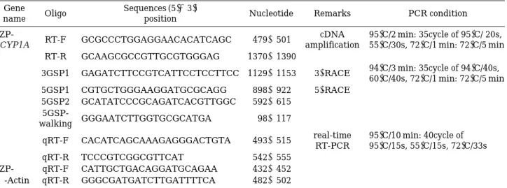
Table 1. Primer and PCR condition details.
Gene Oligo Sequences (5′→3′) Nucleotide Remarks PCR condition
name position
ZP- RT-F GCGCCCTGGAGGAACACATCAGC 479~501 cDNA 95�C/2 min: 35cycle of 95�C/ 20s,
CYP1A amplification 55�C/30s, 72�C/1 min: 72�C/5 min
RT-R GCAAGCGCCGTTGCGTGGGAG 1370~1390
3GSP1 GAGATCTTCCGTCATTCCTCCTTCC 1129~1153 3′-RACE 94�C/3 min: 35cycle of 94�C/40s, 60�C/40s, 72�C/1 min: 72�C/5 min 5GSP1 CGTGCTGGGAAGGATGCGCAGG 898~922 5′-RACE
5GSP2 GCATATCCCGCAGATCACGTTGGC 592~615 5GSP-
GGGAATCTTGGTGCGCATGA 98~117 walking
qRT-F CACATCAGCAAAGAGGGACTGTA 493~515 real-time 95�C/10 min: 40cycle of RT-PCR 95�C/15s, 55�C/15s, 72�C/33s
qRT-R TCCCGTCGGCGTTCAT 542~555
ZP- qRT-F CATTGCTGACAGGATGCAGAA 432~452
β-Actin qRT-R GGGCGATGATCTTGATTTTCA 482~502
Fig. 1. Representative DNA sequence of the Zacco platypus CYP1A gene (GenBank Accession No. JN648712). An asterisk (*) indicates a stop codon. Poly (A) signal sequence was underlined.
by a post-hoc multiple comparison (Duncan) test was used to compare each tissues. Student’s t-test was used to determine significances between ex- posure group at each time point and control. All data were analyzed using the SPSS statistical package (version 10.0; SPSS Inc., Chicago, IL, USA). Data are expressed as means±S.D. and differences are considered significant at p⁄0.05.
RESULTS AND DISCUSSION
We directly sequenced amplified clones of the
Z. platypus CYP1A gene and submitted the sequ-ence to GenBank (Accession no. JN648712) (Fig.
1). The Z. platypus CYP1A gene consisted of 1575 bp of the open reading frame (ORF) which encod- ing 524 amino acids of putative protein, 106 bp of the 5 ′- untranslated region (UTR) and 534 bp of the long 3 ′ -UTR. The Z. platypus CYP1A transcripts were ubiquitously distributed and showing high- est level of expression in the liver followed by gill, kidney and intestine (Fig. 2). Liver is the main site of xenobiotic metabolism and high level of CYP1A expression in observed in liver in most of the species (Arukwe, 2002; Lee et al., 2005; Miran- da et al., 2006, Kim et al., 2008a, b). Also in fish, gill is the important site of absorption and there- fore, activities of cytochrome P450-dependant en- zymes are associated with it to handle the toxic chemicals when they enter the system (Evans, 1987; Wilson and Laurent, 2002; Jönsson et al., 2003). These similar patterns of distribution of CYP1A are also reported in a few fish species such as hermaphrodite fish, Kryptolebias marmoratus (Lee et al., 2005), a yellow catfish, Pelteobagrus fulvidraco (Kim et al., 2008a) and a river puffer- fish, Takifugu obscurus (Kim et al., 2008b). On the other hand, Eye, heart, muscle and skin were weakly expressed. Concerning low level of expres- sion of CYP1A in these tissues, to our knowledge both these tissues have rarely been studied in fish as target organs for CYP1A induction. Otherwise, in mammals CYP1A in skin has been focused pri- marily because it has a function related to car- cinogen metabolism (Reiners et al., 1997; Marston et al., 2001; Smith et al., 2006). In general, the organs actively involved in the response to xeno- biotics or those involved in their absorption have high levels of CYP1A dependant enzyme activi- ties. While differential expression of the CYP1A gene in different tissues has also been reported in
case of Atlantic salmon (Salmo salar) by Arukwe (2002) and Rees et al. (2003). Thus, the tissue spe- cific induction of CYP1A gene expression requires further attention. Hahn (1998a) reviewed the tran- scriptional regulation of fish CYP1A through a transcription factor known as the aryl hydrocar- bon receptor (AhR). The transcriptional regulation of CYP1A in fish and mammals appears to be sim- ilar. CYP1A has been studied extensively due to its biological importance in the metabolism and toxicity of various xenobiotics (Hankinson, 1995).
CYP1A protein expression and activity, although constitutively present at significant levels, are up- regulated in response to the binding of AhR ago- nists. Binding of these agonists to the cytosolic AhR leads to translocation of the AhR to the nucle- us, dimerization of the AhR with ARNT (“aryl hydrocarbon receptor nuclear translocator,” a mis- nomer), and subsequent binding to an enhancer region termed the XRE (xenobiotic response ele- ment) or DRE (dioxin response element). CYP1A is one of several genes having at least one 59 DRE.
Also, there is evidence in mammals for regulation of CYP1A by other mechanisms by some chemicals (Hoffer et al., 1996; Ledirac et al., 1997). CYP1A, a monooxygenase, plays an important role in the Phase-I metabolism of many xenobiotic and endo- genous chemicals, including PAHs. We have so far not studied XREs in Z. platypus CYP1A but infor- mation on this aspect may elucidate the functional properties of the identified genes.
Fig. 2. Expression of Zacco platypus CYP1A gene in dif- ferent tissues (Br: Brain, Ey: Eye, Gi: Gill, Go:
Gonad, He: Heart, In: Intestine, Ki: Kidney, Li:
Liver, Mu: Muscle, Sk: Skin).
Relative CYP1A mRNA expression
0.6
0.4
0.2
0 c
d
d d
c
e
a a b
a
Br Ey Gi Go He In Ki Li Mu Sk
Tissue
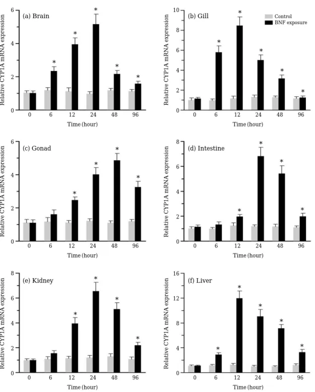
For study of BNF-induced expression of CYP1A we selected six vital organs, brain, gill, gonad, intestine, kidney and liver. Time-course studies revealed a different response in all the six tissues (Fig. 3). While brain, gills and liver showed sig- nificantly different levels of expression compare
with the control level at 6 h (p⁄0.05), the other organs showed a significant induction at 12 h (p⁄
0.05). Gill and liver showed the highest level at 12 h, others showed the highest level at 24 h or 48 h. In all the tissues, decreased expression levels were observed at 48 and 96 h when compared
Fig. 3. Relative mRNA expression of Zacco platypus CYP1A gene in different tissues after exposure to β-naphthoflavone (1μM) for 96 hours.
6
4
2
0
(a) Brain (b) Gill
(c) Gonad (d) Intestine
(e) Kidney (f) Liver
10
8
6
4
2
0
8
6
4
2
0
16
12
8
4
0 6
4
2
0
8
6
4
2
0
0 6 12 24 48 96
Time (hour)
0 6 12 24 48 96
Time (hour)
0 6 12 24 48 96
Time (hour)
0 6 12 24 48 96
Time (hour)
0 6 12 24 48 96
Time (hour)
0 6 12 24 48 96
Time (hour)
Relative CYP1A mRNA expression Relative CYP1A mRNA expressionRelative CYP1A mRNA expressionRelative CYP1A mRNA expression
Relative CYP1A mRNA expressionRelative CYP1A mRNA expression
*
*
* *
*
*
*
*
*
*
*
*
* *
*
*
*
*
*
*
*
*
*
*
*
* *
Control BNF exposure
with the levels at 24 h. BNF-induced expression of CYP1A has been reported in many fish species (Sarasquete and Segner, 2000; Chung-Davidson et al., 2004; Jönsson et al., 2007; Kim et al., 2008a, b). In contrast to our findings, Chung-Davidson et al. (2004) observed that brain CYP1A in juve- nile trout (Salvelinus namaycush) remained indu- ced for an extended period of time. However, sim- ilar to our results in brain, gill and liver, CYP1A mRNA induction in response to BNF exposure occurred rapidly and continued to rise in the BNF- treated lake trout after 4 h. Because of the large surface area, gills provide a suitable site for ab- sorption of toxicants and therefore role of CYP which catalyzes activation of xenobiotics has been reported from the gills of fish. In our study, gills were highly showed expression of BNF-induced CYP1A mRNA. A time-course study also showed high expression during the early phase of expo- sure (Fig. 3b). In gills of Atlantic salmon (Salmo salar), Rees et al. (2003) observed a peak response at 6 h and expression levels of gills were even high- er than in the liver. In our study kidney of Z. pla- typus also showed appreciable levels of CYP1A mRNA expression. However, muscle and skin showed negligible expression. Both these tissues are least important for metabolism of xenobiotics in fish (Monostory et al., 1996).
Overall, this is the first report of a full cDNA sequence of any cytochrome P450 gene from Z.
platypus. Concerning the fact that Z. platypus is ecologically important sentinel fish in vast parts of Asia, further intense studies on molecular gene expression of this fish are needed to be undertak- en. Therefore, present study on Z. platypus xeno- biotic metabolism could be extended to those areas involved in detoxification and antioxidant defense are underway in our laboratory.
ACKNOWLEDGEMENTS
This study was supported by the National Fish- eries Research & Development Institute (11-OE- 24).
LITERATURE CITED
Arukwe, A. 2002. Complementary DNA cloning, sequ- ence analysis and differential organ expression of β-naphthoflavone-inducible cytochrome P4501A
in Atlantic salmon (Salmo salar). Comparative Bio- chemistry and Physiology C 133: 613-624.
Billiard, S.M., N.C. Bols and P.V. Hodson. 2004. In vitro and in vivo comparisons of fishspecific CYP1A induction relative potency factors for selected poly- cyclic aromatic hydrocarbons. Ecotoxicology Envi- ronmental Safety 59: 292-299.
Campbell, P.M. and R.H. Devlin. 1996. Expression of CYP1A1 in livers and gonads of Pacific salmon:
quantitation of mRNA levels by RT-cPCR. Aquatic Toxicology 34: 47-69.
Cho, S.H., W.M. Sohn, S.S. Shin, H.J. Song, T.G. Choi, C.M. Oh, Y. Kong and T.S. Kim. 2006. Infection status of pond smelts, Hypomesus olidus, and other freshwater fishes with trematode metacercariae in 6 large lakes. The Korean Journal of Parasitology 44: 243-246.
Chung-Davidson, Y.W., C.B. Rees, H. Wu, S.S. Yun and W. Li. 2004. β-naphthoflavone induction of CYP1A in brain of juvenile lake trout (Salvelinus namaycush Walbaum). Journal of Experimental Biology 207: 1533-1542.
Cousinou, M., B. Nilsen, J. Lopez-Barea and G. Dora- do. 2000. New methods to use fish cytochrome P4501A to assess marine organic pollutants. Sci- ence of the Total Environment 247: 213-225.
Evans, D.H. 1987. The fish gill: Site of action and mo- del for toxic effects of environmental pollutants.
Environmental Health Perspective 71: 47-58.
Gao, K., I. Brandt, J.V. Goldstone and M.E. Jönsson.
2011. Cytochrome P450 1A, 1B, and 1C mRNA in- duction patterns in three-spined stickleback expo- sed to a transient and a persistent inducer. Com- parative Biochemistry and Physiology C 154: 42- 55.
Goldstone, H.M. and J.J. Stegeman. 2006. A revised evolutionary history of the CYP1A subfamily: Gene duplication, gene conversion, and positive selec- tion. Journal of Molecular Evolution 62: 708-717.
Hahn, M.E. 1998. The aryl hydrocarbon receptor: A comparative perspective. Comparative Biochemi- stry and Physiology C 121: 23-53.
Hahn, M.E. and J.J. Stegeman. 1994. Regulation of cytochrome P450 1A1 in teleosts: sustained induc- tion of CYP1A1 messenger-RNA, protein, and cat- alytic activity by 2,3,7,8 tetrachlorodibenzofuran in the marine fish Stenotomus chrysops. Toxicology and Applied Pharmacology 127: 187-198.
Hankinson, O. 1995. The aryl hydrocarbon receptor complex. Annual Review of Pharmacology and Toxicology 35: 307-340.
Hayashi, M., T. Ueda, K. Uyeno, K. Wada, N. Kinae, K. Saotome, N. Tanaka, A. Takai, Y.F. Sasaki, N.
Asano, T. Sofuni and Y. Ojima. 1998. Development of genotoxicity assay systems that use aquatic organisms. Mutation Research-Fundamental and Molecular Mechanisms of Mutagenesis 399: 125- 133.
Hoffer, A., C.Y. Chang and A. Puga. 1996. Dioxin in- duces transcription of fos and jun genes by Ah re- ceptor-dependent and -independent pathways. Tox- icology and Applied Pharmacology 141: 238-247.
Hu, M.C., H.J. Hsu, I.C. Guo and B.C. Chung. 2004.
Function of Cyp11a1 in animal models. Molecular and Cellular Endocrinology 215: 95-100.
Isin, E.M. and F.P. Guengerich. 2007. Complex reac- tions catalyzed by cytochrome P450 enzymes. Bio- chimica et Biophysica Acta 1770: 314-329.
Jönsson, M., A. Abrahamson, B. Brunstrom, I. Brandt, K. Ingebrigtsen and E.H. Jorgensen. 2003. EROD activity in gill filaments of anadromous and marine fish as a biomarker of dioxinlike pollutants. Com- parative Biochemistry and Physiology C 136: 235- 243.
Jönsson, M.E., B. Brunstrom, K. Ingebrigtsen and I.
Brandt. 2004. Cellspecific CYP1A expression and benzo[a]pyrene adduct formation in gills of rain- bow trout (Oncorhynchus mykiss) following CYP1A induction in the laboratory and in the field. Envi- ronmental Toxicology and Chemistry 23: 874-882.
Jönsson, M.E., R. Orrego, B.R. Woodin, J.V. Goldstone and J.J. Stegeman. 2007. Basal and 3,3′,4,4′,5-pen- tachlorobiphenyl-induced expression of cytochrome P450 1A, 1B and 1C genes in zebrafish. Toxicology and Applied Pharmacology 221: 29-41.
Kim, I.S. 1997. Illustrated encyclopedia of fauna and flora of Korea, Freshwater Fishes. 37, National Textbook Company.
Kim, J.H., D.S. Hwang, K.H. Son, S. Raisuddin, J.S.
Ki, J.S. Lee and K.N. Han. 2008a. cDNA cloning and expression of a xenobiotic metabolizing cyto- chrome P4501A (CYP1A) from the yellow catfish, Pelteobagrus fulvidraco (Siluriformes). Environ- mental Toxicology 23: 346-353.
Kim, J.H., S. Raisuddin, J.S. Ki, J.S. Lee and K.N.
Han. 2008b. Molecular cloning and betanaphtho- flavone-induced expression of a cytochrome P450 1A (CYP1A) gene from an anadromous river puffer- fish, Takifugu obscurus. Marine Pollution Bulletin 57: 433-440.
Ku, S.K., J.H. Lee and H.S. Lee. 2004. Immunohisto- chemical study on the endocrine cells in gut of the stomachless teleost, Zacco platypus (Cyprinidae).
Journal of Veterinary Medicine Series C 33: 212- 219.
Ledirac, N., C. Delescluse, G. de Sousa, M. Pravaloria, P. Lesca, M. Amichot, J.B. Berge and R. Rahmani.
1997. Carbaryl induces CYP1A1 gene expression in HepG2 and HaCaT cells but is not a ligand of the human hepatic Ah receptor. Toxicology and Appli- ed Pharmacology 144: 177-182.
Lee, Y.M., T.D. Williams, S.O. Jung and J.S. Lee. 2005.
cDNA cloning and expression of a cytochrome P450 1A (CYP1A) gene from the hermaphroditic fish Rivulus marmoratus. Marine Pollution Bulletin 51:
769-75.
Liu, Y., J. Wang, Y. Wei, H. Zhang, Y. Liu and J. Dai.
2008. Molecular characterization of cytochrome P450 1A and 3A and the effects of perfluorooctanoic acid on their mRNA levels in rare minnow (Gobio- cypris rarus) gills. Aquatic Toxicology 88: 183-190.
Livak, K.J. and T.D. Schmittgen. 2001. Analysis of relative gene expression data using real time quan- titative PCR and the 2-ΔΔCTmethod. Methods 25:
402-408.
Marston, C.P., C. Pereira, J. Ferguson, K. Fischer, O.
Hedstrom, W.M. Dashwood and W.M. Baird. 2001.
Effect of a complex environmental mixture from coal tar containing polycyclic aromatic hydrocar- bons (PAH) on the tumor initiation, PAH-DNA binding and metabolic activation of carcinogenic PAH in mouse epidermis. Carcinogenesis 22: 1077- 1086.
Miller, H., D.G. Bembo, J.A. Macdonald and C.W.
Evans. 1999. Induction of cytochrome P4501A (CYP1A) in Trematomus bernacchii as an indicator of environmental pollution in Antarctica: assess- ment by quantitative RT-PCR. Aquatic Toxicology 44: 183-193.
Miranda, C.L., W.G. Chung, J.L. Wang-Buhler, T. Mu- safia-Jeknic, W.M. Baird and D.R. Buhler. 2006.
Comparative in vitro metabolism of benzo[a]pyrene by recombinant zebrafish CYP1A and liver micro- somes from β-naphthoflavone-treated rainbow trout. Aquatic Toxicology 80: 101-108.
Monostory, K., K. Jemnitz and L. Vereczkey. 1996.
Xenobiotic metabolizing enzymes in fish: diversity, regulation and biomarkers for pollutant exposure.
Acta Physica Academiae Scientiarum Hungaricae 84: 369-381.
Nakamura, K. 1952. Environment, food habit, spawn- ing, development, growth and fishes of Zacco pla- typus in Chikuma River. Bulletin of Freshwater Fisheries Research Laboratory 1: 2-25.
Ortiz-Delgado, J.B. and C. Sarasquete. 2004. Toxicity, histopathological alterations and immunohistoche- mical CYP1A induction in the early life stages of the seabream, Sparus aurata, following waterborne exposure to B(a)P and TCDD. Journal of Molecular Histology 35: 29-45.
Park, J.H., S.M. Guk, T.Y. Kim, E.H. Shin, A. Lin, J.
Y. Park, J.L. Kim, S.T. Hong and J.Y. Chai. 2004.
Clonorchis sinensis metacercarial infection in the pond smelt Hypomesus olidus and the minnow Zacco platypus collected from the Soyang and Dae- chung Lakes. The Korean Journal of Parasitology 42: 41-44.
Rees, C.B., S.D. McCormick and W. Li. 2005. A non- lethal method to estimate CYP1A expression in laboratory and wild Atlantic salmon (Salmo salar).
Comparative Biochemistry and Physiology C 141:
217-224.
Rees, C.B., S.D. McCormick, J.P. Vanden Heuvel and W. Li. 2003. Quantitative PCR analysis of CYP1A
induction in Atlantic salmon (Salmo salar). Aquatic Toxicology 62: 67-78.
Reiners, J.J.J., C.L. Jones, N. Hong, R.E. Clift and C.
Elferink. 1997. Downregulation of aryl hydrocar- bon receptor function and cytochrome P450 1A1 induction by expression of Ha-ras oncogenes. Mole- cular Carcinogenesis 19: 91-100.
Sarasquete, C. and H. Segner. 2000. Cytochrome P4501A (CYP1A) in teleostean fishes. A review of immunohistochemical studies. Science of the Total Environment 247: 313-332.
Smith, G., S.H. Ibbotson, M.M. Comrie, R.S. Dawe, A.
Bryden, J. Ferguson and C.R. Wolf. 2006. Regula- tion of cutaneous drugmetabolizing enzymes and cytoprotective gene expression by topical drugs in human skin in vivo. The British Journal of Der- matology 155: 275-281.
Weber, L.P., S.L. Diamond, S.M. Bandiera and D.M.
Janz. 2002. Expression of HSP70 and CYP1A pro- tein in ovary and liver of juvenile rainbow trout exposed to β-naphthoflavone. Comparative Bioche- mistry and Physiology C 131: 387-394.
Williams, D.E., J.J. Lech and D.R. Buhler. 1998. Xeno- biotics and xenoestrogens in fish: modulation of cytochrome P450 and carcinogenesis. Mutation Research 399: 179-192.
Wilson, J.M. and P. Laurent. 2002. Fish gill morpho- logy: inside out. Journal of Experimental Zoology 293: 192-213.
Yamazaki, M., Y. Tanizaki and T. Shimokawa. 1996.
Silver and other trace elements in a freshwater fish, Carasius auratus langsdorfii, from the Asa- kawa River in Tokyo, Japan. Environmental Pol- lution 94: 83-90.
Yeom, D.H., S.A. Lee, G.S. Kang, J. Seo and S.K. Lee.
2007. Stressor identification and health assess- ment of fish exposed to wastewater effluents in Miho Stream, South Korea. Chemosphere 67: 2282 -2292.
(Manuscript received 21 October 2011, Revised 16 November 2011
Revision accepted 24 November 2011)