1
ABBREVIATIONS: ALT, alanine aminotransferase; AST, aspartate aminotransferase; DP complex, DNA/peptide complex; ECM, extracellular matrix; RiAS, ribbon-type antisense; TGF-β1, transforming growth factor-β1; NLS, nuclear localization signal;
HIV, human immunodeficiency virus.
Correspondence to: Kyung-Oh Doh, Department of Physiology, College of Medicine, Dongguk University, 707, Seokjang-dong, Gyeongju 780-714 Korea. (Tel) 82-54-770-2413, (Fax) 82-54-770- 2440, (E-mail) [email protected]
Effects of TGF-β1 Ribbon Antisense on CCl4-induced Liver Fibrosis
Kyung-Oh Doh
Department of Physiology, College of Medicine, Dongguk University, Gyeongju 780-714, Korea
Ribbon-type antisense oligonucleotide to TGF-β1 (TGF-β1 RiAS) was designed and tested to prevent or resolve the fibrotic changes induced by CCl4 injection. W hen Hepa1c1c7 cells were transfected with TGF-β1 RiAS, the level of TGF-β1 mRNA was effectively reduced. TGF-β1 RiAS, mismatched RiAS, and normal saline were each injected to mice via tail veins. When examined for the biochemical effects on the liver, TGF-β1 mRNA levels were significantly reduced only in the TGF-β1 RiAS-treated group.
The results of immunohistochemical studies showed that TGF-β1 RiAS prevented the accumulation of collagen and α-smooth muscle actin, but could not resolve established fibrosis. These results indicate that ribbon antisense to TGF-β1 with efficient uptake can effectively prevent fibrosis of the liver.
Key Words: Transforming growth factor-β1, Liver cirrhosis, Ribbon antisense, Cationic peptide
INTRODUCTION
Liver cirrhosis is the common pathological consequences of chronic liver injury caused by a variety of agents, including viruses, alcohol, hepatotoxins, and autoimmune disorders (Friedman, 2000). Fibrogenesis is characterized by an excessive accumulation of extracellular matrix (ECM), as the result of an imbalance of its synthesis versus degradation (Arthur, 2000). Although the mechanisms underlying the progression of liver cirrhosis have yet to be fully elucidated, cytokines have been implicated as mediators of fibrosis in the liver. Among these cytokines, transforming growth factor-β1 (TGF-β1) is particularly well-studied, and has been recognized as pro-fibrogenic in the case of liver injury (Kanzler et al, 1999; Bissell et al, 2001; Gressner et al, 2002). TGF-β1 is involved in the accumulation of ECMs for normal repair as a response to tissue injury, and is also responsible for fibrous changes due to aberrant overproduction of ECMs, including proteoglycans, collagens, fibronectin, and glycoproteins.
TGF-β1 also inhibits the degradation of newly synthesized matrix protein via an upregulation of the synthesis of protease inhibitors and a downregulation of the synthesis of matrix-degrading proteases (Knittel et al, 1999). Thus, an effective blockade of TGF-β1 synthesis or action appears to constitute a promising approach for the prevention of fibrous conditions, as suggested by previous reports (George et al, 1999; Qi et al, 1999; Ueno et al, 2000).
Antisense oligonucleotides (AS oligos) have proven to be valuable in the functional study of gene products, as they reduce the expression of genes in a sequence-specific manner. However, the use of AS oligos is still hindered by several critical problems, including instability to nuclease, sequence nonspecificity, and inadequate cellular uptake
(Wagner et al, 1993; Gryaznov et al, 1996). A variety of chemically modified oligos, including phosphorothioate and methylphosphonate oligos, have been developed as a measure to augment stability against nucleases. However, each of these modified oligonucleotides suffered from its own problems, which included a lack of sequence specificity, insensitivity to RNase H, and the prolongation of partial thrombosis time (Henry et al, 1997). Recently, it was reported that ribbon-type antisense (RiAS) oligos, which possess a covalently closed structure, are quite stable and effective in the specific ablation of target mRNA, and that they are associated with few of the problems of other modified AS oligos (Moon et al, 2000a; Moon et al, 2000b;
Bajpai et al, 2005).
The cellular uptake of AS oligos can be enhanced via the formation of complexes such as liposomes. Although liposomes exhibit several advantages, including low toxicity, lack of immunogenicity, and simple production, they tend to manifest relatively poor cellular uptake. Recent studies have demon- strated the utility of the cationic peptide as a delivery vehicle for biologically active drugs, including antisense oligonucleotides, both in cell cultures and in vivo (Schwarze et al, 1999; Fulda et al, 2002). These peptides are derived from the HIV Tat protein (Vives et al, 1997; Schwarze et al, 1999; Eguchi et al, 2001; Futaki et al, 2001), SV40 large T antigen (Zanta et al, 1999; Torchilin et al, 2001), Drosophila Antennapedia (Derossi et al, 1996), and histone H1 (Sorgi et al, 1997; Bharath et al, 2002). In this study, the modified nuclear localization signal (NLS) of human immunodeficiency virus (HIV)-1 Tat protein (Moon et al, 2007) was used as a vehicle both in vitro and in vivo. Thus, a RiAS to TGF-β1 was designed and tested to prevent or resolve CCl4-induced liver fibrosis and tissue damage. In
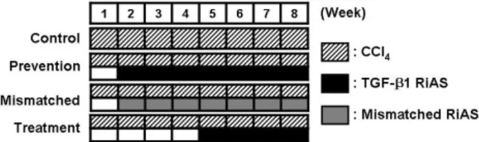
Fig. 1. Experimental schedule. For 8 weeks of experiment, 1 ml/kg (body weight) of CCl4 was intraperitoneally administered twice per week. RiAS to mouse TGF-β1 or mismatched RiAS (100μg/30 g body weight) was also intravenously administered twice per week from the second week of experiment in the prevention group and mismatched group, and from the 5th week of experiment in the treatment group.
an attempt to enhance cellular uptake, the TGF-β1 RiAS was added to the cationic peptide, thereby forming a DNA / peptide (DP) complex.
METHODS Cell line and animals
Hepa1c1c7 (mouse hepatoma cell line) was obtained from the American Type Culture Collection (ATCC) and main- tained in α-MEM containing 10% heat-inactivated fetal bovine serum (Welgene, Daegu, Korea) in a humidified 5%
CO2 incubator at 37oC. Sixty ICR male mice, weighing 30∼
35 g, were supplied by SLC (Hamamatsu, Shizuoka, Japan).
All animals received humane care. Animal experiments were performed according to international guidelines concerning the conduct of animal experimentation. In order to induce liver cirrhosis, 1 ml/kg (body weight) of CCl4 was intraperitoneally administered twice per week (Fig. 1). RiAS to mouse TGF-β1 (100μg/30 g body weight) was also intravenously administered twice per week from the second week of experiment in the prevention group (n=20), and 5th week of experiment in the treatment group (n=20).
Mismatched RiAS was intravenously administered twice per week at an identical dosage in the mismatched group (n=20). Normal saline was intravenously administered twice per week at an equal volume in the control group (n=20). At the administration of the drugs, RiAS was mixed with cationic peptide (DP complex) at a ratio of 1:3. Prior to sacrifice, the blood was collected in order to measure the serum parameters. Serum biochemical parameters were determined via standard spectrometric methods.
Construction of mouse TGF-β1 RiAS
Target sites for RiAS were selected via the sequential overlap simulation of secondary structures using the DNAsis program (Hitachi Software, San Bruno, CA, USA).
The antisense sequence was 5'-gatccaggccacatgttgctcc- acacttgattttaatctctgcaacctg-3' (from 49 bp on the TGF-β1 sequence), and the mismatched sequence was 5'-gatccaggc- cacatattactgcatacatgcttatattctctgcaacctg-3'. Oligonucleotides were synthesized as previously described (Moon et al, 2000a) via standard phosphoramidite chemistry using an automated DNA synthesizer, ExpediteTM8909 (Applied Biosystems, Foster City, CA, USA). Two molecules of the TGF-β1 antisense of the stem-loop structure were ligated
in order to form a ribbon-type antisense molecule by the complementary 4 base sequences at the 5' ends of the molecules at 16oC with T4 DNA ligase overnight.
Peptide synthesis and modifications
Peptide used in the present study was derived from the Tat protein of HIV-1 which has been reported as effective in DNA delivery (Moon et al, 2007). The Tat peptide corresponds to the nuclear localization signal (NLS) sequence of 9 amino acids (49-57: Arg-Lys-Lys-Arg-Arg-Gln-Arg-Arg- Arg). The peptide was modified at C-terminus by the addition of cysteine residue. Peptides were prepared in a solid phase synthesis by a peptide synthesizer ABI 433A (Applied Biosystems, Foster City, CA, USA), purified by preparative LC, and characterized using an analytical HPLC system (Shimadzu, Kyoto, Japan) comprised of a C18 column and a MALDI-TOF Mass spectrometer (Applied Biosystems, Foster City, CA, USA). Purified peptides were resuspended in ddH2O at a concentration of 10μg/μl, and kept at -70oC prior to further use.
Transfection efficiency of RiAS in vitro and in vivo FITC-labeled TGF-β1 RiAS was synthesized via the incorporation of fluorescein-11dUTP instead of TTP.
Hepa1c1c7 cells seeded in each well of a 24-well plate were treated with the DP complex, containing FITC-labeled TGF-β1 RiAS (0.3μg), in a 200μl volume. The DP complex, containing FITC-labeled TGF-β1 RiAS (0.3μg) in 600μl saline, was infused into the tail veins of mice for in vivo study. The liver was removed after 16 hours, and the tissue blocks of the fixed liver were cryosectioned to a thickness of 10μm for slide mounting. Gene transfer efficacy was evaluated via fluorescent microscopy.
RT-PCR for mouse TGF-β1 expression
After the transfection of the TGF-β1 RiAS, TGF-β1 expression was assessed via RT-PCR. RNA was prepared with WelprepTM RNA isolation reagent (Welgene, Daegu, Korea). The purified RNA was subjected to RT-PCR using the Access RT-PCR kit (Promega, Madison, WI, USA) and a thermal cycler (MJ Research, Watertown, MA, USA). The following primer pairs were used: forward 5-ggactctccacc- tgcaagac-3 and reverse 5'-gactggcgagccttagtttg-3' for mouse TGF-β1, and forward 5'-agtgtgacgttgacatccgta-3' and reverse 5'-gccagagcagtaatctccttct-3' for mouse β-actin. The PCR products were confirmed on 1% agarose gel, and quantitative analysis of the amplified DNA was conducted using the AlphaImager 1220, a gel documentation apparatus (Alpha Innotech, San Leandro, CA, USA).
Histological analysis
The fixation and embedding of liver tissues were conducted as previously described (Choi et al, 2005). The tissue sections were incubated overnight with anti-type I collagen antibody (Santa Cruz Biotechnology, Santa Cruz, CA, USA) and (-smooth muscle actin antibody (Dako, Glostrup, Denmark) in PBS containing 0.5% BSA and 2% FCS at 4oC.
The next day, the tissue sections were incubated with anti-rabbit HRP conjugates for 1 hour at room temperature.
The fibrous lesion areas were also determined via Masson's trichrome method, which is used to stain collagen fibers.
Fig. 2. (A) Schematic representation of ribbon-type antisense to TGF-β1 (TGF-β1 RiAS). The stem is formed by complementary sequences at both ends of each oligo. The 5' terminus of the stem has 4 bases of a single-stranded overhang of 5'-GATC-3'. Two TGF- β1 monomer molecules were ligated to generate a covalently closed molecule with diad symmetry. The RiAS oligos consist of two loops and an intervening stem. Each loop harbors an antisense sequence to TGF-β1. (B) Specific reduction of TGF-β1 mRNA by TGF-β1 RiAS. Hepa1c1c7 cells were transfected with DP complex and RT-PCR was conducted in order to determine the antisense activity of TGF-β1 RiAS. Transfection of TGF-β1 RiAS reduced TGF-β1 expression in Hepa1c1c7 cells. By way of contrast, however, when Hepa1c1c7 cells were treated with mismatched RiAS, TGF-β1 expression was not significantly affected. Mouse β-actin was as a control. Veh: vehicle, RiAS: TGF-β1 RiAS, MM: mismatched RiAS.
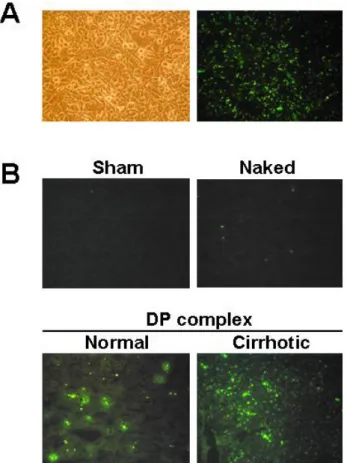
Fig. 3. (A) The DP complex mediated transfection in Hepa1c1c7 mouse hepatoma cells. Transfection of FITC-labeled TGF-β1 RiAS was conducted using cationic peptide. The DP complex was added to Hepa1c1c7 cells for 24 hours. Fluorescence signals are shown in the right panel. (B) Normal saline (sham) or 10 μg of FITC-labeled TGF-β1 RiAS as a form of naked DNA only and DP complex were injected through the tail veins of normal or cirrhotic mice. Tissue sections of mice liver were observed under a fluorescence microscope (×200).
Statistical analysis
Results are expressed as means±standard deviations (SD). Statistical significance was assessed via one-way ANOVA. p values of < 0.05 were considered to be significant.
RESULTS
Construction of ribbon antisense oligos to TGF-β1 and specific reduction of TGF-β1 mRNA
Two identical AS oligos possessing the stem-loop structure were covalently ligated to form a ribbon-antisense molecule, termed TGF-β1 RiAS (Fig. 2A). These TGF-β1 RiAS molecules possess a ribbon-type closed structure without an open end that would be attacked by exonu- cleases. We then determined whether TGF-β1 RiAS was effective in the elimination of target mRNA in a sequence- specific manner, when delivered as a DP complex as shown Fig. 2B. Hepa1c1c7 cells treated with 0.1μg TGF-β1 RiAS showed a reduction of TGF-β1 RNA by 71% and by 97%
at 0.3μg. On the other hand, when Hepa1c1c7 cells were treated with mismatched RiAS, TGF-β1 expression was not significantly affected.
Efficient cellular uptake of TGF-β1 RiAS when delivered as a DP complex
Nucleic acid including antisense oligomers shows poor cellular uptake, largely due to the charged polymeric backbone. In order to study the cellular uptake, the TGF-β1 RiAS was labeled with FITC-11dUTP incorporation during chemical synthesis and used for both in vitro and in vivo uptake study. When the FITC-labeled antisense oligos were used to form the DP complex and added to the Hepa1c1c7 cells, the cells showed strong fluorescence signals (Fig. 3A).
Thus, we attempted to determine whether the DP complex could also be employed for efficient delivery of RiAS into the liver after intravenous infusion. For in vivo tissue uptake, 10μg of FITC-labeled TGF-β1 RiAS were infused into the tail vein of mouse, and the liver was harvested 24 hours after treatment. Whereas the intravenously infused DP complex showed strong fluorescent signals in the liver cells, indicating efficient cellular uptake, the fluorescent signals were quite weak in tissues when only the FITC-labeled 'naked' RiAS was infused. Control livers treated with sham treatment showed no fluorescent signals.
Table 1. Effect of TGF-β1 RiAS on serum biochemical parameters
Control Mismatched Prevention Treatment
Aspartate aminotransferase (U/l) 605.0±199.9 578.1±217.4 147.4±50.2* 173.14±57.9*
Alanine aminotransferase (U/l) 316.3±100.7 367.7±125.8 104.3±17.5* 135.9±45.0*
Albumin (g/dl) 3.2±0.2 3.3±0.2 3.4±0.2 3.2±0.2
Total bilirubin (mg/dl) 0.3±0.2 0.2±0.2 0.2±0.1 0.2±0.1
After 8 weeks of experiments, blood was collected from each groups and analyzed. Data are expressed as mean±SD, n=5 to 8 per group (*p<0.05 vs. control).
Fig. 4. Histological observation of liver. Collagen deposition was detected as blue staining on Masson's trichrome staining (A).
Immunohistochemistry for type I collagen (B) and α-smooth muscle actin (C). Staining was conducted with fixed and dehydrated tissues from mice treated with normal saline (CON), mismatched RiAS (MM), and TGF-β1 RiAS (Prevention and Treatment group).
Stained tissues were mounted with a synthetic mounting solution for microscopic observation (×200).
We also observed that the DP complex could be employed for efficient RiAS uptake in cases of CCl4-induced liver cirrhosis (Fig. 3B).
Effects of TGF-β1 RiAS on liver cirrhosis
All mice in the prevention and treatment groups treated with TGF-β1 RiAS survived, whrereas the control group and the mismatched RiAS-treated group showed survival rates of 50% and 65%, respectively. At the end of 8 weeks of the experiment, TGF-β1 RiAS treated prevention and treatment groups showed lower levels of serum aspartate aminotransferase (AST) and alanine aminotransferase (ALT) than those in the control mice and the mice treated with mismatched RiAS. However, no differences were noted among the groups in terms of albumin and total bilirubin levels (Table 1).
Effect of TGF-β1 RiAS treatment on histology and collagen synthesis
CCl4 injected intraperitoneally induced massive accu- mulation of connective tissues, principally in the centri- lobular area and portal tract, when examined by Masson's trichrome-stained sections of the livers from both the control group and the mismatched RiAS-treated group.
However, TGF-β1 RiAS treatment significantly ameliorated the deposition of connective tissues (Fig. 4A). Positive collagen staining was detected at comparable levels in the
livers treated with either normal saline or mismatched RiAS. In contrast, type I collagen was found to be greatly reduced in the TGF-β1 RiAS-treated livers (Fig. 4B).
Immunohistochemistry of the (-smooth muscle actin, a marker for hepatic stellate cells, showed marked reductions in the livers of the TGF-β1 RiAS-treated group as compared with the control group and the mismatched RiAS-treated group (Fig. 4C). However, histological analysis showed no marked resolution of fibrosis in the treatment group. These data showed that TGF-β1 RiAS treatment can prevent the development of fibrosis.
DISCUSSION
In this study, we evaluated the ability of RiAS to TGF-β1 to eliminate target mRNA and to alleviate global tissue injuries in cases of liver cirrhosis. We introduced the FITC labeled RiAS / cationic peptide complex via the tail vein and found that it was delivered effectively to the cirrhotic livers, as well as the normal livers. RiAS to TGF-β1 was also efficiently delivered into the liver, and blocked CCl4- induced fibrosis, including collagen accumulation. TGF-β1 is the most potent profibrogenic factor in human fibrogenesis (Gressner et al, 2002). Thus, many studies on the blockage of TGF-β1 synthesis or action have been conducted in order to develop a method for the prevention of liver cirrhosis, and a variety of strategies, including the use of adenoviral vectors expressing truncated TGF-β type II receptor (Qi et al, 1999; Ueno et al, 2000; Nakamura et al, 2004), chimeric IgG harboring the extracellular portion of the TGF-β type II receptor (George et al, 1999), and adenoviral expression of a TGF-β1 antisense (Arias et al, 2002; Arias et al, 2003), have all been identified as effective strategies.
Antisense oligonucleotides in general show poor cellular uptake due to the charges on their polymeric backbone.
Cellular uptake of oligonucleotides can be improved when complexed with cationic vehicle. However, non-viral delivery vehicles, including liposomes, do not exhibit uptake efficiency satisfactory for many types of cells, particularly cells in primary cultures. Thus, an improved transfection reagent would clearly benefit both in vitro cell-line studies and in vivo applications. Some cationic peptides such as HIV Tat protein, SV40 large T antigen, Drosophila Antennapedia, and histone H1 have been found to show nucleic acid condensation, membrane penetration, and nuclear locali- zation activities (Derossi et al, 1996; Sorgi et al, 1997; Vives et al, 1997; Ludtke et al, 1999; Schwarze et al, 1999; Zanta et al, 1999; Dokka et al, 2000; Eguchi et al, 2001; Futaki et al, 2001; Torchilin et al, 2001; Bharath et al, 2002). In this study, we modified NLS of HIV-1 Tat protein and devised a simple DP complex composed of RiAS / cationic
peptide in an effort to augment cellular uptake. Although portal myofibroblasts (Ramadori and Saile, 2004) and cells from bone marrow origin (Forbes et al, 2004) have been demonstrated to have fibrogenic potential, the activation of the hepatic stellate cells is the most important event in liver fibrogenesis (Friedman et al, 1985). It has been shown that the quiescent hepatic stellate cells are transformed into active myofibroblasts expressing α-smooth muscle actin, and that these active hepatic stellate cells are the primary source of extracellular matrix proteins (Alcolado et al, 1997; Iredale et al, 1998; Friedman, 2000). Our present results indicated that TGF-β1 RiAS reduced the accu- mulation of collagen and α-smooth muscle actin, revealed by immunohistochemical staining. Contrary to what has traditionally been thought, it is now believed that even advanced liver fibrosis is reversible. Therefore, many scien- tists have attempted to discover new antifibrotic drugs (Hammel et al, 2001). In the present study, we attempted to treat liver fibrosis, which was formed after 4 weeks of intraperitoneal injection of CCl4 with TGF-β1 RiAS for 4 weeks, however, there was only a marginally resolution of established fibrosis. This insufficient resolution may be attributable to ECM cross-linking or insufficient treatment time, because it takes a long time to recover from fibrosis (Issa et al, 2004).
In conclusion, ribbon antisense to TGF-β1 mRNA coupled with enhanced transfection using the antisense oligos / cationic peptide complex was found to reduce the levels of target mRNA in a sequence-specific manner, and was able to alleviate global tissue fibrosis in the livers of CCl4-treated mice. As tissue fibrosis is a convergence point in the progression of many human diseases of the liver, kidney and lung, the potent antisense activity of RiAS TGF-β1 in the sequence-specific elimination of the target mRNA may prove to be useful in the development of antisense medicine against fibrous conditions in these human tissues.
REFERENCES
Alcolado R, Arthur MJ, Iredale JP. Pathogenesis of liver fibrosis.
Clin Sci (Lond) 92: 103-112, 1997
Arias M, Lahme B, Van de Leur E, Gressner AM, Weiskirchen R.
Adenoviral delivery of an antisense RNA complementary to the 3' coding sequence of transforming growth factor-beta1 inhibits fibrogenic activities of hepatic stellate cells. Cell Growth Differ 13: 265-273, 2002
Arias M, Sauer-Lehnen S, Treptau J, Janoschek N, Theuerkauf I, Buettner R, Gressner AM, Weiskirchen R. Adenoviral expression of a transforming growth factor-beta1 antisense mRNA is effective in preventing liver fibrosis in bile-duct ligated rats.
BMC Gastroenterol 3: 29, 2003
Arthur MJ, Fibrogenesis II. Metalloproteinases and their inhibitors in liver fibrosis. Am J Physiol Gastrointest Liver Physiol 279:
G245-249, 2000
Bajpai AK, Park JH, Moon IJ, Kang H, Lee YH, Doh KO, Suh SI, Chang BC, Park JG. Rapid blockade of telomerase activity and tumor cell growth by the DPL lipofection of ribbon antisense to hTR. Oncogene 24: 6492-6501, 2005
Bharath MM, Chandra NR, Rao MRS. Prediction of an HMG-box fold in the C-terminal domain of histone H1: insights into its role in DNA condensation. Proteins 49: 71-81, 2002
Bissell DM, Roulot D, George J. Transforming growth factor beta and the liver. Hepatology 34: 859-867, 2001
Choi YK, Moon IJ, Jung HK, Jang BC, Seo SI, Park JG. Prevention of tissue injury by ribbon antisense to TGF-beta1 in the kidney.
Int J Mol Med 15: 391-399, 2005
Derossi D, Calvet S, Trembleau A, Brunissen A, Chassaing G, Prochiantz A. Cell internalization of the third helix of the Antennapedia homeodomain is receptor-independent. J Biol Chem 271: 18188-18193, 1996
Dokka S, Toledo D, Shi X, Ye J, Rojanasakul Y. High-efficiency gene transfection of macrophages by lipoplexes. Int J Pharm 206: 97-104, 2000
Eguchi A, Akuta T, Okuyama H, Senda T, Yokoi H, Inokuchi H, Fujita S, Hayakawa T, Takeda K, Hasegawa M, Nakanishi M.
Protein transduction domain of HIV-1 Tat protein promotes efficient delivery of DNA into mammalian cells. J Biol Chem 276: 26204-26210, 2001
Forbes SJ, Russo FP, Rey V, Burra P, Rugge M, Wright NA, Alison MR. A significant proportion of myofibroblasts are of bone marrow origin in human liver fibrosis. Gastroenterology 126: 955
-963, 2004
Friedman SL. Molecular regulation of hepatic fibrosis, an integrated cellular response to tissue injury. J Biol Chem 275:
2247-2250, 2000
Friedman SL, Roll FJ, Boyles J, Bissell DM. Hepatic lipocytes: the principal collagen-producing cells of normal rat liver. Proc Natl Acad Sci U S A 82: 8681-8685, 1985
Fulda S, Wick W, Weller M, Debatin KM. Smac agonists sensitize for Apo2L/TRAIL- or anticancer drug-induced apoptosis and induce regression of malignant glioma in vivo. Nat Med 8: 808-
815, 2002
Futaki S, Suzuki T, Ohashi W, Yagami T, Tanaka S, Ueda K, Sugiura Y. Arginine-rich peptides. An abundant source of membrane-permeable peptides having potential as carriers for intracellular protein delivery. J Biol Chem 276: 5836-5840, 2001
George J, Roulot D, Koteliansky VE, Bissell DM. In vivo inhibition of rat stellate cell activation by soluble transforming growth factor beta type II receptor: a potential new therapy for hepatic fibrosis. Proc Natl Acad Sci U S A 96: 12719-12724, 1999 Gressner AM, Weiskirchen R, Breitkopf K, Dooley S. Roles of
TGF-beta in hepatic fibrosis. Front Biosci 7: d793-807, 2002 Gryaznov S, Skorski T, Cucco C, Nieborowska-Skorska M, Chiu CY,
Lloyd D, Chen JK, Koziolkiewicz M, Calabretta B. Oligonucleotide N3'→P5' phosphoramidates as antisense agents. Nucleic Acids Res 24: 1508-1514, 1996
Hammel P, Couvelard A, O'Toole D, Ratouis A, Sauvanet A, Flejou JF, Degott C, Belghiti J, Bernades P, Valla D, Ruszniewski P, Levy P. Regression of liver fibrosis after biliary drainage in patients with chronic pancreatitis and stenosis of the common bile duct. N Engl J Med 344: 418-423, 2001
Henry SP, Novotny W, Leeds J, Auletta C, Kornbrust DJ. Inhibition of coagulation by a phosphorothioate oligonucleotide. Antisense Nucleic Acid Drug Dev 7: 503-510, 1997
Iredale JP, Benyon RC, Pickering J, McCullen M, Northrop M, Pawley S, Hovell C, Arthur MJ. Mechanisms of spontaneous resolution of rat liver fibrosis. Hepatic stellate cell apoptosis and reduced hepatic expression of metalloproteinase inhibitors. J Clin Invest 102: 538-549, 1998
Issa R, Zhou X, Constandinou CM, Fallowfield J, Millward-Sadler H, Gaca MD, Sands E, Suliman I, Trim N, Knorr A, Arthur MJ, Benyon RC, Iredale JP. Spontaneous recovery from micronodular cirrhosis: evidence for incomplete resolution associated with matrix cross-linking. Gastroenterology 126: 1795-1808, 2004 Kanzler S, Lohse AW, Keil A, Henninger J, Dienes HP,
Schirmacher P, Rose-John S, zum Buschenfelde KH, Blessing M. TGF-beta1 in liver fibrosis: an inducible transgenic mouse model to study liver fibrogenesis. Am J Physiol 276: G1059-
1068, 1999
Knittel T, Mehde M, Kobold D, Saile B, Dinter C, Ramadori G.
Expression patterns of matrix metalloproteinases and their inhibitors in parenchymal and non-parenchymal cells of rat liver: regulation by TNF-alpha and TGF-beta1. J Hepatol 30:
48-60, 1999
Ludtke JJ, Zhang G, Sebestyen MG, Wolff JA. A nuclear localization signal can enhance both the nuclear transport and expression of 1 kb DNA. J Cell Sci 112: 2033-2041, 1999
Moon IJ, Choi K, Choi YK, Kim JE, Lee Y, Schreiber AD, Park JG. Potent growth inhibition of leukemic cells by novel ribbon-type antisense oligonucleotides to c-myb1. J Biol Chem 275: 4647-4653, 2000a
Moon IJ, Kang H, Seu YB, Chang BC, Song DK, Park JG. Marked transfection enhancement by the DPL (DNA/peptide/lipid) complex. Int J Mol Med 20: 429-437, 2007
Moon IJ, Lee Y, Kwak CS, Lee JH, Choi K, Schreiber AD, Park JG. Target site search and effective inhibition of leukaemic cell growth by a covalently closed multiple anti-sense oligonucleotide to c-myb. Biochem J 346 Pt 2: 295-303, 2000b
Nakamura T, Ueno T, Sakamoto M, Sakata R, Torimura T, Hashimoto O, Ueno H, Sata M. Suppression of transforming growth factor-beta results in upregulation of transcription of regeneration factors after chronic liver injury. J Hepatol 41: 974
-982, 2004
Qi Z, Atsuchi N, Ooshima A, Takeshita A, Ueno H. Blockade of type beta transforming growth factor signaling prevents liver fibrosis and dysfunction in the rat. Proc Natl Acad Sci U S A 96: 2345-2349, 1999
Ramadori G, Saile B. Portal tract fibrogenesis in the liver. Lab Invest 84: 153-159, 2004
Schwarze SR, Ho A, Vocero-Akbani A, Dowdy SF. In vivo protein transduction: delivery of a biologically active protein into the
mouse. Science 285: 1569-1572, 1999
Sorgi FL, Bhattacharya S, Huang L. Protamine sulfate enhances lipid-mediated gene transfer. Gene Ther 4: 961-968, 1997 Torchilin VP, Rammohan R, Weissig V, Levchenko TS. TAT peptide
on the surface of liposomes affords their efficient intracellular delivery even at low temperature and in the presence of metabolic inhibitors. Proc Natl Acad Sci U S A 98: 8786-8791, Ueno H, Sakamoto T, Nakamura T, Qi Z, Astuchi N, Takeshita 2001 A, Shimizu K, Ohashi H. A soluble transforming growth factor beta receptor expressed in muscle prevents liver fibrogenesis and dysfunction in rats. Hum Gene Ther 11: 33-42, 2000 Vives E, Brodin P, Lebleu B. A truncated HIV-1 Tat protein basic
domain rapidly translocates through the plasma membrane and accumulates in the cell nucleus. J Biol Chem 272: 16010-16017, Wagner RW, Matteucci MD, Lewis JG, Gutierrez AJ, Moulds C, 1997 Froehler BC. Antisense gene inhibition by oligonucleotides containing C-5 propyne pyrimidines. Science 260: 1510-1513, Zanta MA, Belguise-Valladier P, Behr JP. Gene delivery: a single 1993 nuclear localization signal peptide is sufficient to carry DNA to the cell nucleus. Proc Natl Acad Sci U S A 96: 91-96, 1999