Comparison of Drainage Volume of Chronic Subdural Hematoma According to Drainage Catheter Type
Gun-Young Lee,
1Chang Hyun Oh,
2Yu Shik Shim,
1Seung Hwan Yoon,
1Hyeong-Chun Park,
1Chong Oon Park,
1and Dongkeun Hyun
11Department of Neurosurgery, School of Medicine, Inha University, Incheon;
2Department of Neurosurgery, Guro Teun Teun Hospital, Seoul, Korea.
Received: January 18, 2013 Revised: April 11, 2013 Accepted: April 11, 2013
Corresponding author: Dr. Dongkeun Hyun, Department of Neurosurgery,
School of Medicine, Inha University, 27 Inhang-ro, Jung-gu, Incheon 400-711, Korea.
Tel: 82-32-890-2370, Fax: 82-32-890-3099 E-mail: [email protected]
∙ The authors have no financial conflicts of interest.
© Copyright:
Yonsei University College of Medicine 2013 This is an Open Access article distributed under the terms of the Creative Commons Attribution Non- Commercial License (http://creativecommons.org/
licenses/by-nc/3.0) which permits unrestricted non- commercial use, distribution, and reproduction in any medium, provided the original work is properly cited.
Purpose: To assess the therapeutic value of two different drainage catheters in treating chronic subdural hematoma (CSDH). Materials and Methods: Two types of drainage catheters can be used to treat CSDH according to the position of holes in the catheter: open-type or closed-type catheter. In this retrospective study, 199 total patients with CSDH were reviewed according to catheter type. Among them, 84 patients were and 113 in the closed-type group (holes positioned within the dis- tal-most 1 cm of the catheter). The surgeon selected the catheter type. Total drain- age volume, initial drainage volume within 2 days, percentage of initial drainage volume per total drainage volume, duration of catheter insertion, and reoperation rate were compared. Results: Total drainage volume was not different between the two groups (p=0.333). The initial drainage volume within 2 days was larger in the open-type group than closed-type group (p=0.024), but the percentage of initial drainage volume per total drainage volume was not different (p=0.354). The dura- tion of catheter insertion was shorter in the open-type group than closed-type group (p=0.015). The reoperation rate of CSDH was also higher in the open-type group than closed-type group (p=0.004). Conclusion: CSDH drainage with an open-type catheter is faster compared with a closed-type catheter, but total drain- age volume is similar and reoperation rate is higher. Therefore, the open-type cath- eter for CSDH drainage has limited clinical value.
Key Words: Chronic subdural hematoma, catheter, drainage
INTRODUCTION
Chronic subdural hematomas (CSDHs) usually occur in the neurosurgical disease in the elderly, particularly after minor head injury.1,2 In patients older than 50 years, the brain parenchyme is reduced by almost 200 g, which results in an increased subdural space and arachnoid space up to 11%.3 Hematomas can occupy this extra volume without increasing intracranial pressure. Furthermore, a slowly progress- ing hematoma allows the brain to adjust by compressing the venous channels, thus providing further space for the expanding hematoma.3 In addition, the outer subdu- ral membrane, which functions to absorb the subdural fluid, produces a layer of
text of the current literature.
MATERIALS AND METHODS
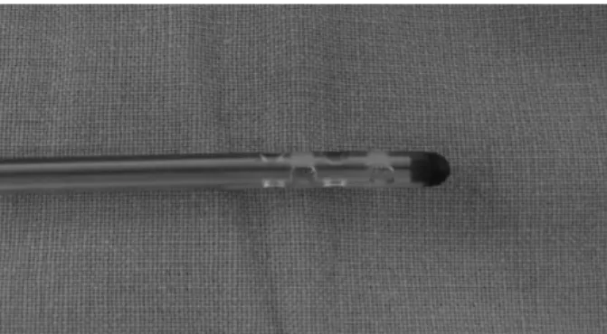
From March 2010 to May 2012, 199 consecutive patients (33 females and 166 males) with CSDH underwent burr- hole craniostomy with closed-system drainage at the De- partment of Neurosurgery, Inha University School of Medi- cine. The drainage catheter was categorized as either the open type or closed type according to the hole position on the catheter, with the open type having holes positioned within the distal-most 5 cm of the catheter and a large hole at the end (Fig. 1), and the closed type having holes posi- tioned within the distal-most 1 cm of the catheter (Fig. 2).
Total 199 patients with CSDH were retrospectively reviewed according to the catheter type; 84 patients were included in the open-type group (Group A) and 113 in the closed-type group (Group B), while 2 patients with both types of cathe- ters were excluded from this study. CSDH was defined as the presence of a typical neomembrane, liquefied blood with- in the hematoma cavity, and at least a 3-week interval since the head trauma. Diagnosis was confirmed by CT and/or magnetic resonance imaging in all patients. They were also subdivided according to their CT and clinical findings. We analyzed the type of hematoma in CSDH which is a impor- tant factor that affects drainage. Nomura, et al.8 classified the CSDH into two groups: non-mixed density (hematoma observed as purely uniformed density such as low-density, isodensity or high-density) and mixed density. According to Markwalder, et al.,9 clinical condition was classified as fol- lowed (Table 1). As a result, the neurological deficit or any absence of symptom was graded “0”; minimal deficit ac- companied by nausea and headache was graded as “1”;
neurological deficit like hemiparesis related to drowsiness or disorientation was graded as 2; further neurological defi- thin-walled sinusoidal vascular channels that are liable to
bleed because of the presence of loose cell junctions. As long as there is a balance between the expanding and ab- sorptive forces, however, the size of the hematoma remains constant and the patient remains asymptomatic.
The pressure gradient between the sinusoidal channels in the capsule and hematoma’s cavity was disrupted by multi- ple head injury or position changes of the head in the verti- cal. And, disruption of the pressure gradiet induce addition- al bleeding. After Valsalva maneuver temporarily reduce intracranial pressure. And, that can lead to an expansion of the hematoma.4 In addition, the activation of the kallikrein- kinin cascade increases vascular permeability and may cause blood extravasation and plasma exudation from the capillaries into both the outer membrane and the hematoma cavity, thereby enlarging the hematoma.5,6
It is a clinical entity with a decreasing mortality and mor- bidity, especially since the advent of computed tomography (CT) and advances in surgical technique. There is still debate, however, on the pathophysiology, methodology of manage- ment, and surgical treatment of CSDH.7 Although many surgical techniques have been explored for the treatment of CSDH, most neurosurgeons favor a burr-hole craniostomy and closed-system drainage without irrigation because it has low morbidity and mortality rates and is simple. In neu- rosurgical field, there are different types of drainage cathe- ter such as open or closed type. These catheters are catego- rized by the position of holes in the catheter, therefore, the function for drainage could be different by the types of cath- eters. However, there has been no study comparing the cath- eter type used in surgery.
In this study, 197 patients with CSDH underwent surgery and were analyzed prospectively for radiological, clinical, and surgical features. Results in the open-type catheter group were compared with those in the closed-type catheter group, and the surgical treatment options are discussed in the con-
Fig. 1. The open-type catheter has 5 holes within the distal-most 5 cm, with
a large hole at the end position of the catheter. Fig. 2. The closed-type catheter’s hole configuration has 8 holes within the distal-most 1 cm.
pared with each other in total drainage volume, initial drain- age volume within 2 days, percentage of initial drainage vol- ume per total drainage volume, duration of catheter insertion, and reoperation rate. For statistical analysis, we used the chi- square test (Fisher’s extract test) to determine significant differences between the groups. Differences were consid- ered statistically significant at p<0.05.
RESULTS
In this study, the selection of the catheter was made, based only on personal preference without considering age, gen- der, type of hematoma, and consciousness of patients.
Patient population
The series consists of 165 men and 32 women with a mean age of 70.9 years (range, 2 months to 98 years). In Group A, mean age was 72.5±10.2 years and 69.6±13.9 years in Group B. Group A (84 patients) was made up of 74 men and 10 women (88.1% men), whereas Group B (113 patients) was made up of 91 men and 22 women (80.5% men). There was no statistically significant difference in age between the 2 groups (age, p=0.111; sex, p=0.156) (Table 2).
Drainage volume
The total drainage volumes were 192.1±140.6 mL for open- type and 173.2±131.5 mL for closed-type catheters (p=0.333).
The catheters were maintained for 2.75±1.40 days with open-type and 3.35±1.90 days with closed-type catheters, which was statistically significant (p=0.015).
For the first 2 days, the drainage volumes were 163.8
±101.6 mL for open-type and 133.1±87.4 mL for closed-type catheters, and the drainage volume was larger in open-type catheters (p=0.020). The recurrence rate was 22.35%±37.49 for open-type and 8.06%±17.45 for closed-type catheters, and this difference was especially statistically significant (p=0.24).
Pre- and postoperative grading
According to the clinical Markwalder grading scale, 22 pa- cit with stupor and hemiplegia was graded as “3”; decere-
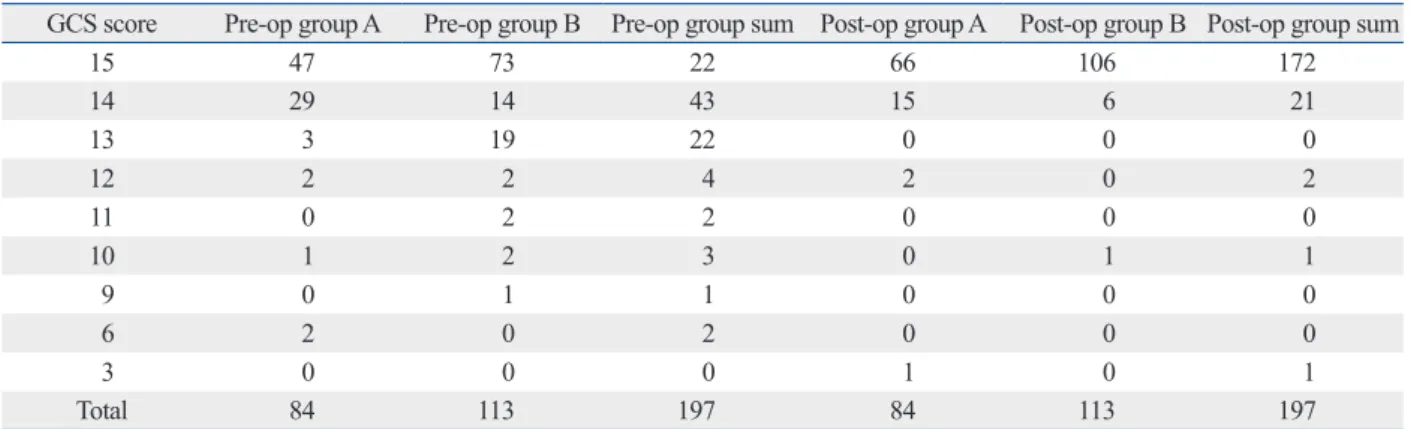
brate or decorticate posture associated with coma was grad- ed as “4”. Additionally, Glasgow Coma Scale (GCS) score was used. Full recovery, minimal neurological deficit, se- vere neurological deficit, or death.10 That were classified for the postoperative clinical status.
Most operations were performed by either general or lo- cal anesthesia. All patients were undergone by surgical pro- cedures of 1 burr hole over the area of maximal hematoma width and closed-system drainage with a commercial sili- cone catheter and bag. The type of catheter was selected to the attending neurosurgeon. Right after subdural hematoma was spontaneously evacuated after burr-hole trephination, the drainage catheter was inserted. In all cases, warm saline irrigation through the silicone catheter was never applied to the procedure. The drainage bags maintained below the head level were removed in accordance with the change of the drainage volume and radiological findings; however, they were generally removed within 6 days to avoid the complications associated with a drainage catheter. Brain CT for pre-operation and post-operation was performed respec- tively and even follow-up CT was done for all cases. Clini- cal and/or radiological criteria were used for evaluation of CSDH recurrence and to determine the necessity for reop- eration. The clinical criteria finally included a change in mental status, worsening of preexisting neurological deficit, and new onset or aggravation of headache for recurrence.
For radiological criteria for recurrence, there was an increase in subdural collection and/or compressed brain parenchy- ma. They were all determined by comparing CT findings for the follow-up period with the postoperative CT findings.
As defined above, all patients underwent reoperation if there are neurological symptoms recurred or if there are new neu- rological symptoms developed with the increase in hemato- ma thickness and volume on follow-up CT. Someone who showed no or minimal neurological deficit postoperatively, or who had a small amount of residual hematoma, were discharged and followed up as outpatients.
Follow-up CT was performed on the 2nd, 5th, and 30th postoperative days. The differently treated groups were com- Table 1. Markwalder Chronic Subdural Hematoma Scale
Grade 0 Patient neurologically normal
Grade 1 Patient alert and orientated; absence of mild symptoms such as headache, or mild neurological deficit such as reflex asymmetry
Grade 2 Patient drowsy or disorientated, or variable neurological deficit such as hemiparesis
Grade 3 Patient stuporous but responding appropriately to noxious stimuli; severe focal signs such as hemiplegia Grade 4 Patient comatose with absent motor responses to painful stimuli; decerebrate or decorticate posturing
ference was statistically significant (p=0.034).
Type of hematoma
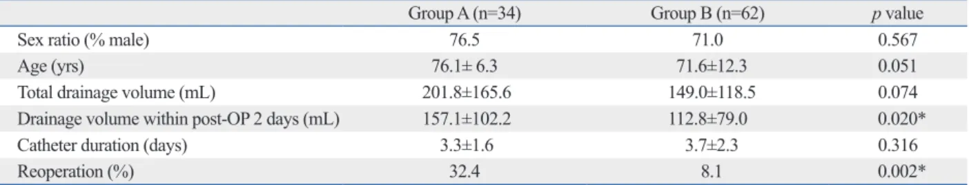
The type of hematoma was classified into mixed- and non- mixed type. The total drainage volumes were 201.79 (±165.5) mL for open-type and 149.0 (±118.47) mL for closed-type catheters. The catheters were maintained for 3.29 (±1.60) days with open-type, and 3.74 (±2.29) days with closed- type catheters, which was not statistically significant.
For the first 2 days, the drainage volumes were 157.06 (±102.24) mL for open-type and 112.80 (±78.98) mL for closed-type catheters, and the drainage volume was larger in open-type catheters (p=0.020). The recurrence rate was 22.35% (±37.49) for open-type and 8.06% (±17.45) for closed-type catheters, and this difference, was statistically significant (p=0.012) (Table 5).
For the non-mixed type of hematomas (n=101), the total tients were in grade 0, 98 patients in grade 1, 65 patients in
grade 2, 10 patients in grade 3, and 2 patients in grade 4 (Ta- ble 3). The preoperative GCS grade for each catheter group is shown in Table 4, but it did not reach statistical signifi- cance (p=0.521). The change of postoperative grade was rel- atively milder on the Markwalder scale compared to the GCS scale; however, it was not statistically significant (p=0.373).
Reoperation
Hematoma progress was tracked through follow-up brain CT in order to assess when patients could be discharged (hematoma fully resolved) and whether reoperation would be necessary (postoperative hematoma recurrence). The re- operation was performed during the hospitalization if the hematoma was not resolving. About 20.9% of patients in Group A required reoperation (n=84), but only 14.2% of pa- tients in Group B (n=113) underwent reoperation; this dif-
Table 4. The Preoperative and Postoperative Glasgow Coma Scale (GCS) Grade in Catheter Groups
GCS score Pre-op group A Pre-op group B Pre-op group sum Post-op group A Post-op group B Post-op group sum
15 47 73 22 66 106 172
14 29 14 43 15 6 21
13 3 19 22 0 0 0
12 2 2 4 2 0 2
11 0 2 2 0 0 0
10 1 2 3 0 1 1
9 0 1 1 0 0 0
6 2 0 2 0 0 0
3 0 0 0 1 0 1
Total 84 113 197 84 113 197
Table 2. Summary of Results between 2 Groups
Group A (n=84) Group B (n=113) p value
Sex ratio (% male) 88.1 80.5 0.156
Age (yrs) 72.5±10.2 69.6±13.9 0.111
Total drainage volume (mL) 192.1±140.6 173.2±131.5 0.333
Drainage volume within post-OP 2 days (mL) 163.8±101.6 133.1±87.4 0.024*
Catheter duration (days) 2.75±1.40 3.35±1.90 0.015*
Reoperation (%) 20.9 14.2 0.034*
*Indicates significance at the p<0.05 level.
Table 3. The Preoperative and Postoperative Markwalder Chronic Subdural Hematoma Scale in Catheter Groups
Markwalder score Pre-op group A Pre-op group B Pre-op group sum Post-op group A Post-op group B Post-op group sum
0 12 10 22 59 90 149
1 34 64 98 21 22 43
2 31 34 65 1 1 2
3 5 5 10 2 0 2
4 2 0 2 1 0 1
Total 84 113 197 84 113 197
sion after surgery has also been suggested to contribute to the recurrence of CSDH.11,19 Interestingly, the literature in- dicates that the rate of recurrence is not significantly differ- ent for different surgical methods. It has been observed, however, that a prolonged period of maintenance in drain- age reduces the recurrence of CSDH, but no study has in- vestigated the drainage method in recovery from surgery.
After the burr-hole drainage, there are adverse effects on the cerebral swelling that include the thick and fibrous mem- brane of the hematoma, a decline of cerebral blood flow due to functional abnormality, degenerative cerebral shrinking, and lowered intracranial pressure due to the flowing out of cerebrospinal fluid.10,20,21 The faster drainage enables the in- tracranial pressure to lower before cerebral swelling fully recovers, and this has detrimental effects on cerebral swell- ing in the subdural space.
The drainage theory of CSDH was not confirmed, never- theless, the expansion of CSDH due to the oncotic pressure leads to an increase in cranial pressure and results in neuro- logical symptoms.22 Moreover, the drainage occurs via po- tential energy after drainage catheter insertion and the dif- ference in drainage can be explained by fluid mechanics.
The diameter of the catheter and the size of catheter en- trance can affect the result of this study; however, the vol- ume of drainage was controlled in this study by using cath- eters of the same diameter. Furthermore, the size of catheter entrance, depth of the same catheter, and the number of catheters could all influence CSDH drainage.23
The major difference between the 2 drainage methods drainage volume was 185.52 (±122.12) for open-type and
202.55 (±141.44) for closed-type catheters. In the first 2 days, the drainage volume for each catheter type was not different; 168.32 (±101.93) mL for open-type and 157.82 (±91.39) mL for closed-type catheters. The recurrence rates were 30.00% (±36.29) for open-type and 21.57% (±31.54) for closed-type catheters, which was also not significant;
however, the maintenance of catheter was 2.38 (±1.12) days for open-type and 2.88 (±1.11) days for closed-type cathe- ters, which was statistically significant (p=0.026) (Table 6).
DISCUSSION
Treatment of CSDH has improved dramatically in recent years because of advances in diagnostic tools and surgical techniques. However, there is still some debate regarding the best strategy for treatment. Use of a burr-hole craniosto- my and closed-system drainage without irrigation to man- age symptomatic CSDH is the initial treatment because it is associated with lower recurrence rates than other treatment methods.11,12 After performing a burr-hole drainage, the rate of reoperation varies between 2.7% and 30%.5,9,13-16 In this study, reoperation rate was 17.3% (Group A=20.9%; Group B=14.2%), which is in keeping with that reported in many other studies. Debate has ensued over the mechanisms un- derlying the recurrence of CSDH. Many postulate that poor brain re-expansion and recurrent bleeding from the outer membrane are the major causes.6,17,18 Intracapsular air intru-
Table 5. Summary of Results between 2 Groups for Subjects with Mixed-Type Hematoma
Group A (n=34) Group B (n=62) p value
Sex ratio (% male) 76.5 71.0 0.567
Age (yrs) 76.1± 6.3 71.6±12.3 0.051
Total drainage volume (mL) 201.8±165.6 149.0±118.5 0.074
Drainage volume within post-OP 2 days (mL) 157.1±102.2 112.8±79.0 0.020*
Catheter duration (days) 3.3±1.6 3.7±2.3 0.316
Reoperation (%) 32.4 8.1 0.002*
*Indicates significance at the p<0.05 level.
Table 6. Summary of Results between 2 Groups for Subjects with Non-Mixed-Type Hematoma
Group A (n=50) Group B (n=51) p value
Sex ratio (% male) 96 92.1 0.419
Age (yrs) 70.1±11.7 67.2±15.4 0.295
Total drainage volume (mL) 185.5±122.1 202.5±141.4 0.519
Drainage volume within post-OP 2 days (mL) 168.3±101.9 157.8±91.4 0.587
Catheter duration (days) 2.4±1.1 2.9±1.1 0.026*
Reoperation (%) 30.0 21.5 0.337
*Indicates significance at the p<0.05 level.
chronic subdural hematoma. Neurosurg Clin N Am 2000;11:399- 406.
2. Hamilton MG, Frizzell JB, Tranmer BI. Chronic subdural hema- toma: the role for craniotomy reevaluated. Neurosurgery 1993;
33:67-72.
3. Tabaddor K, Shulmon K. Definitive treatment of chronic subdural hematoma by twist-drill craniostomy and closed-system drainage.
J Neurosurg 1977;46:220-6.
4. Markwalder TM, Reulen HJ. Influence of neomembranous organ- isation, cortical expansion and subdural pressure on the post-oper- ative course of chronic subdural haematoma--an analysis of 201 cases. Acta Neurochir (Wien) 1986;79:100-6.
5. Fujisawa H, Ito H, Kashiwagi S, Nomura S, Toyosawa M. Kalli- krein-kinin system in chronic subdural haematomas: its roles in vascular permeability and regulation of fibrinolysis and coagula- tion. J Neurol Neurosurg Psychiatry 1995;59:388-94.
6. Aung TH, Wong WK, Mo HP, Tsang CS. Management of chronic subdural haematoma: burr hole drainage, replacement with Hart- mann’s solution, and closed-system drainage. Hong Kong Med J 1999;5:383-6.
7. Kwon TH, Park YK, Lim DJ, Cho TH, Chung YG, Chung HS, et al. Chronic subdural hematoma: evaluation of the clinical signifi- cance of postoperative drainage volume. J Neurosurg 2000;93:
796-9.
8. Nomura S, Kashiwagi S, Fujisawa H, Ito H, Nakamura K. Char- acterization of local hyperfibrinolysis in chronic subdural hemato- mas by SDS-PAGE and immunoblot. J Neurosurg 1994;81:910-3.
9. Markwalder TM, Steinsiepe KF, Rohner M, Reichenbach W, Markwalder H. The course of chronic subdural hematomas after burr-hole craniostomy and closed-system drainage. J Neurosurg 1981;55:390-6.
10. Benzel EC, Bridges RM Jr, Hadden TA, Orrison WW. The single burr hole technique for the evacuation of non-acute subdural he- matomas. J Trauma 1994;36:190-4.
11. Oku Y, Takimoto N, Yamamoto K, Onishi T. Trial of a new opera- tive method for recurrent chronic subdural hematoma. J Neuro- surg 1984;61:269-72.
12. Kuroki T, Katsume M, Harada N, Yamazaki T, Aoki K, Takasu N.
Strict closed-system drainage for treating chronic subdural haema- toma. Acta Neurochir (Wien) 2001;143:1041-4.
13. Okada Y, Akai T, Okamoto K, Iida T, Takata H, Iizuka H. A com- parative study of the treatment of chronic subdural hematoma-- burr hole drainage versus burr hole irrigation. Surg Neurol 2002;
57:405-9.
14. Cameron MM. Chronic subdural haematoma: a review of 114 cases. J Neurol Neurosurg Psychiatry 1978;41:834-9.
15. Eggert HR, Harders A, Weigel K, Gilsbach J. [Recurrence follow- ing burr hole trephination of chronic subdural hematomas]. Neu- rochirurgia (Stuttg) 1984;27:141-3.
16. Robinson RG. Chronic subdural hematoma: surgical management in 133 patients. J Neurosurg 1984;61:263-8.
17. Camel M, Grubb RL Jr. Treatment of chronic subdural hematoma by twist-drill craniotomy with continuous catheter drainage. J Neurosurg 1986;65:183-7.
18. Rosenørn J, Gjerris F. Long-term follow-up review of patients with acute and subacute subdural hematomas. J Neurosurg 1978;48:
345-9.
19. Nakaguchi H, Tanishima T, Yoshimasu N. Relationship between drainage catheter location and postoperative recurrence of chronic subdural hematoma after burr-hole irrigation and closed-system
used in the present study is the position and size of the cath- eter holes. Specifically, the open-type catheter has a large hole located in the end portion. The open-type catheter is expected to drain much faster initially than the closed-type catheter, because it has more theoretical advantage to drain.
As expected, the observed drainage for the first 2 days in this study was much faster for the open-type catheter.
This study was designed to classify hematoma into mixed type and non-mixed type. According to previous study, he- matoma of non-mixed type is relatively larger,8 and hemato- ma of mixed type showed higher recurrent rate.24 In this study, the difference of drainage is relevant to the result which was not classified hematoma with mixed and non- mixed type. In mixed type, there was significantly higher drainage and re-op rate for the first two days when the open catheter was used. On the other hand, closed type of catheter showed relatively long maintenance period which was statis- tically significant, indicating that the difference of catheter can affect the drainage of hematoma as well as the type of hematoma.
This faster drainage is related to the observed higher inci- dence of reoperation due to hematoma recurrence after CSDH in Group A and to the shorter maintenance period of the open-type catheter, which was 2.75 (±1.40) days for Group A and 3.35 (±1.90) days for Group B.
There have been several studies investigating the effect of anticoagulant therapies on the recurrence and treatment of CSDH. However, there was no study assessing the im- pact of drainage according to the type of catheter and the re- sult of treatment for CSDH. The present study strongly indi- cates that the prognosis depends on the type of catheter. The use of the open-type catheter allowed for faster drainage of hematoma following surgery; however, the use of the closed- type catheter resulted in a lower rate of reoperation.
In conclusion, the use of open-type catheter allowed for faster drainage of hematoma following surgery; however, the use of closed-type catheter resulted in a lower rate of reoper- ation. When using closed-system drainage to treat CSDH, careful catheter selection is imperative. These data show that closed-type catheter is as reliable and effective as open- type catheter, and offers the advantage of a lower reopera- tion rate.
REFERENCES
1. Chen JC, Levy ML. Causes, epidemiology, and risk factors of
53:512-5.
23. Fox RW, Pritchard PJ, McDonald AT. Introduction to Fluid Me- chanics. 7th ed. New York: John Wiley & Sons Inc; 2009.
24. Ito H, Yamamoto S, Saito K, Ikeda K, Hisada K. Quantitative esti- mation of hemorrhage in chronic subdural hematoma using the 51Cr erythrocyte labeling method. J Neurosurg 1987;66:862-4.
drainage. J Neurosurg 2000;93:791-5.
20. Arbit E, Patterson RH Jr, Fraser RA. An implantable subdural drain for treatment of chronic subdural hematoma. Surg Neurol 1981;15:175-7.
21. Obana WG, Pitts LH. Management of head injury. Extracerebral lesions. Neurosurg Clin N Am 1991;2:351-72.
22. Weir B. Oncotic pressure of subdural fluids. J Neurosurg 1980;