INTRODUCTION
Breast cancer is one of the leading cancers among females, resulting in approximate 1.7 million cases and 522 thousands deaths in 2012.1 Based on hormone receptor, as well as human
epidermal growth factor receptor type 2 (HER2) status, breast cancer could be classified into several subtypes as follows: lu- minal A, luminal B, HER2/neu+, and triple-negative.2 Triple- negative breast cancer (TNBC) is defined as tumors that lack expression of estrogen receptor (ER), progesterone receptor (PR), and HER2.3 Due to its specific receptor status, TNBC usually accepts chemotherapy rather than endocrine therapy or trastuzumab.4 However, a sensitive and specific non-inva- sive biomarker to detect clinical prognosis of TNBC is needed.
MicroRNAs (miRNAs), endogenous RNAs with about 23 nu- cleotides, regulate protein expression by paring with mRNAs of protein-coding genes and guiding their post-transcriptional repression.4 Deviated expression of the microRNA-34 (miR- 34) family has been reported to play an important role in the pathogenesis of many cancers.5 Expression analysis of miR-34 family has suggested that its members play critical roles in
Low Expression of Circulating MicroRNA-34c is Associated with Poor Prognosis
in Triple-Negative Breast Cancer
Zhihao Zeng
1, Xiaowu Chen
2, Dajian Zhu
2, Zhongran Luo
1, and Min Yang
1Departments of 1Cardiothoracic Surgery and 2General Surgery, ShunDe Hospital of Southern Medical University, Foshan, China.
Purpose: The microRNA-34 (miR-34) family is important in tumor regulation. This study aimed to investigate the association of circulating miR-34 family proteins with clinicopathological features and their prognostic value in triple-negative breast cancer (TNBC) patients.
Materials and Methods: In this cohort study, 173 TNBC patients admitted to First People’s Hospital of Shunde from May 1, 2009 to April 30, 2013 were enrolled. Meanwhile, 75 age-matched healthy women volunteers were identified as healthy controls (HCs).
We examined the expression of miR-34 family (miR-34a/b/c) proteins in plasma collected from TNBC patients before any treat- ment was performed and from age-matched HCs using qPCR methods.
Results: The expressions of miR-34a/34b/34c were significantly lower in TNBC patients than in HC (p<0.001, p=0.027, p<0.001, respectively). miR-34a was correlated with tumor grade (p=0.038), lymph node positive (p=0.027), distant metastasis (p=0.004), and surgery (p=0.023); miR-34b was correlated with lymph node positivity (p=0.027); and miR-34c was correlated with tumor grade (p=0.017) and distant metastasis (p<0.001). Kaplan-Meier curve analysis displayed low expression of miR-34a as associated with worse overall survival (OS) (p=0.011), as well as miR-34c low expression (p=0.002). In addition, univariate and multivariate Cox proportional hazards regression was performed, and low expression of miR-34c (p=0.011) was found to be an independent risk factor for OS, as well as tumor grade (p=0.013), lymph node positive (p=0.050), and distant metastasis (p=0.021).
Conclusion: In conclusion, this study demonstrated reduced miR-34a/c expression is highly associated with tumor progression and indicated worse prognosis. Also, miR-34c was an independent risk factor for OS in TNBC patients.
Key Words: Circulating microRNA, triple-negative breast cancer, miR-34 family
pISSN: 0513-5796 · eISSN: 1976-2437
Received: February 6, 2017 Revised: April 13, 2017 Accepted: April 15, 2017
Corresponding author: Dr. Xiaowu Chen, Department of General Surgery, ShunDe Hospital of Southern Medical University, Penglai Road, Daliang District, Foshan 528300, China.
Tel: 86-757-22318000, Fax: 86-757-22318000, E-mail: [email protected]
•The authors have no financial conflicts of interest.
© Copyright: Yonsei University College of Medicine 2017
This is an Open Access article distributed under the terms of the Creative Com- mons Attribution Non-Commercial License (http://creativecommons.org/licenses/
by-nc/4.0) which permits unrestricted non-commercial use, distribution, and repro- duction in any medium, provided the original work is properly cited.
Yonsei Med J 2017 Jul;58(4):697-702 https://doi.org/10.3349/ymj.2017.58.4.697
many aspects of cancer biology, including invasion, metasta- sis, proliferation, cell survival, apoptosis, cell growth, cell cycle, migration, senescence, angiogenesis, and silencing, by regu- lating the expression of their target genes.5-7 However, most of these studies are tissue- and cell-based miR-34 family expres- sions. To exploit a noninvasive prognostic biomarker, the asso- ciation between circulating miR-34 family expressions and clini- cal prognosis needs to be discovered.
This study aimed to investigate the association of circulating miR-34 family expression with clinicopathological features and their prognostic value in patients with TNBC.
MATERIALS AND METHODS
Patients
In this prospective cohort study, 173 TNBC patients admitted to First People’s Hospital of Shunde from May 1, 2009 to April 30, 2013 were enrolled. All patients were diagnosed and assessed by clinical assessment, radiographic examination, and patho- logical confirmation through needle biopsy or during surgery.
TNBC status was defined as ER negative (ER-) [below 10% stain- ing by immunohistochemistry (IHC)], PR negative (PR-) (be- low 10% staining by IHC) and HER2 negative (HER2-) (0 or 1 by IHC defined as below 10% staining or slight and incomplete stain- ing above 10% cells). Meanwhile, data of 75 age-matched healthy women volunteers were enrolled as healthy controls (HCs).
Peripheral blood samples were obtained from all TNBC pa- tients before any treatments were performed, such as surgery, chemotherapy, and radiotherapy, and during the physical ex- amination in HCs. After sampling, TNBC patients received ap- propriate treatments according to disease condition and clini- cal practice policy, and were followed up regularly until August 31, 2016. All clinical and pathological characteristics at base- line, as well as treatments post-sampling, were collected. This study was approved by the Ethics Committee of First People’s Hospital of Shunde, and all participants provided written in- formed consent.
Sample acquisition and handling
An 8 mL aliquot of blood was obtained from all participants directly into sodium citrate tubes. The whole blood was al- lowed to stand for ~3 h at -4C° before centrifuging at 1500 g for 10 min at room temperature. The resultant plasma was aliquot- ed into Eppendorf tubes and stored at -70C°.
RNA extraction
Total RNA was isolated with TRIzol (Invitrogen, Carlsbad, CA, USA) and purified by RNeasy mini kit (QIAGEN, Hilden, Ger- many) accordingly. Quality, quantity, and integrity of RNA was measured by a nanodrop spectrophotometer (ND-1000, Nano- drop Technologues, Wilmington, Delware, USA) and Ry gel elec- trophoresis, respectively. The total RNA was subsequently stored
at -70C° for further detection.
Quantitative reverse-transcription polymerase chain reaction (qRT-PCR)
To confirm the findings obtained by analyzing the miRNA pro- filing, we measured the expression of upregulated miRNAs us- ing TaqManqRT-PCR. All reactions were carried out using Gene Amp PCR System 9700 (Applied Biosystems, Foster City, CA, USA). Briefly, the reverse transcription reaction was performed in a 20-μL mixture consisting of 0.3 μL of 10 μM RT primer, 100 ng RNA sample, 2 μL of 2.5 mM dNTPs, 0.2 μL MMLV reverse transcriptase, 2 μL of 10× reverse transcription buffer, 0.3 μL RNase inhibitor, and nuclease-free water. The reaction mix- ture was incubated for 30 min in 16°C, 40 min in 42°C, and 5 min in 85°C, and then kept in -20°C. Next, qPCR was performed in a final volume of 10 μL containing 1 μL PCR probe, 2 μL prod- uct from the RT reaction, 5 μL PCR Master Mix (2×), and 2 μL nuclease-free water. The thermal cycle started with 10 min at 95°C, followed by 40 cycles of 95°C for 10 s and 60°C for 1 min.
The threshold cycle (Ct) values obtained for each miRNA were then normalized to obtain ΔCt values and eventually used to plot relative expression values. Data are presented as mean±
standard deviation, and miRNA values are presented as fold changes relative to controls. U6 was used as internal reference.
The miRNA expression fold changes were calculated using the arithmetic formula 2 -∆∆Ct.
Statistical analysis
Data were analyzed using SPSS software (version 20.0, SPSS, Inc., Chicago, IL, USA). Categorical variables were presented as counts and proportions, and were compared using chi-square test or Fisher’s exact test. The Kaplan-Meier method and log-rank test was used to evaluate differences in survival according to miR-34 expression. Univariate and multivariate Cox proportion- al hazards regression analysis were used to identify risk factors for overall survival (OS). All statistical tests were two-sided, and p-value <0.05 was considered statistically significant.
RESULTS
miR-34 expression in TNBC patients and HCs
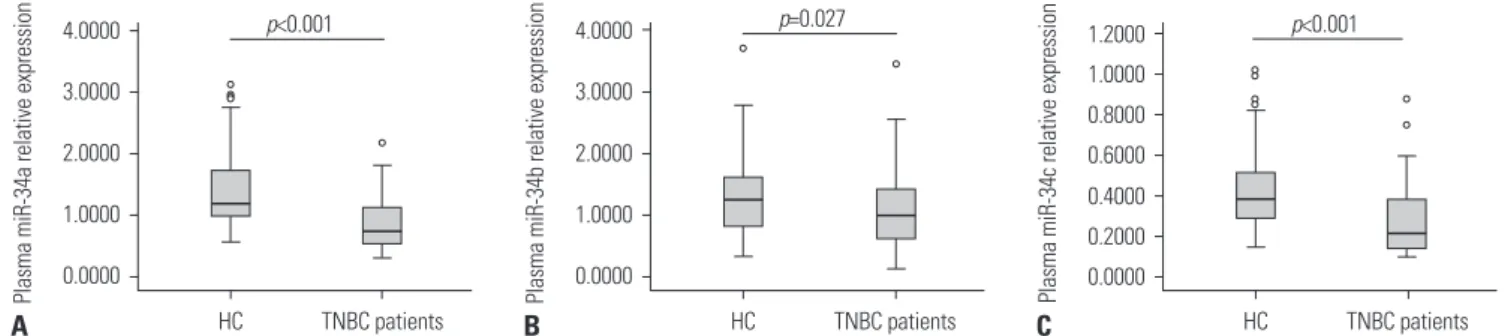
As demonstrated in Fig. 1, the expressions of miR-34a/b/c in plasma were significantly lower in TNBC patients, compared with HCs: miR-34a: 0.738 (0.533–1.141) vs. 1.197 (0.984–1.797), p<0.001; miR-34b: 0.980 (0.621–1.424) vs. 1.246 (0.780–1.611), p=0.027; miR-34c: 0.215 (0.143–0.384) vs. 0.390 (0.284–0.516), p<0.001.
Correlation of miR-34a/b/c expressions with clinical and pathological features
To reveal to correlation between miR-34a/b/c expression and clinicopathological characteristics, TNBC patients were divid-
ed into high (n=87) and low (n=86) expression groups by the median value of miR-34a/b/c. As shown in Table 1, miR-34a was correlated with tumor grade (p=0.038), lymph node positive (p=0.027), distant metastasis (p=0.004), and surgery (p=0.023);
miR-34b was correlated with lymph node positive (p=0.027);
and miR-34c was correlated with tumor grade (p=0.017) and
distant metastasis (p<0.001). There were no other differences be- tween clinicopathological features with miR-34a/b/c expression.
miR-34a/c low expressions were associated with worse prognosis
Next, Kaplan-Meier curve analysis was performed to demon-
Fig. 1. Expressions of miR-34a/b/c in TNBC patients and HCs. (A) miR-34a. (B) miR-34b. (C) miR-34c. TNBC, triple-negative breast cancer; HC, health control; miR-34, microRNA-34.
1.2000 1.0000 0.8000 0.6000 0.4000 0.2000 0.0000
TNBC patients p<0.001
Plasma miR-34c relative expression HC C 4.0000
3.0000 2.0000 1.0000
0.0000
TNBC patients p=0.027
Plasma miR-34b relative expression HC B 4.0000
3.0000 2.0000 1.0000
0.0000
TNBC patients p<0.001
Plasma miR-34a relative expression HC A
Table 1. Demographic, Clinical, and Pathological Characteristic of TNBC Patients at Baseline
Parameters Total miR-34a miR-34b miR-34c
p value* p value† p value‡
Low High Low High Low High
Age (yr), n (%) 0.253 0.593 0.080
≤50 89 (51) 41 (46) 48 (54) 43 (48) 46 (52) 39 (44) 50 (56)
>50 84 (49) 46 (55) 38 (45) 44 (52) 40 (48) 48 (57) 36 (43)
Menstrual status, n (%) 0.404 0.937 0.291
Premenopause 83 (48) 39 (47) 44 (53) 42 (51) 41 (49) 38 (46) 45 (54)
Postmenopause 90 (52) 48 (53) 42 (47) 45 (50) 45 (50) 49 (54) 41 (46)
Tumor grade, n (%) 0.038 0.445 0.017
I/II 118 (68) 53 (45) 65 (55) 57 (48) 61 (52) 52 (44) 66 (56)
III 55 (32) 34 (62) 21 (38) 30 (55) 25 (45) 35 (64) 20 (36)
Tumor size, n (%) 0.110 0.324 0.057
≤2 cm 76 (44) 33 (43) 43 (57) 35 (46) 41 (54) 32 (42) 44 (58)
>2 cm 97 (56) 54 (56) 43 (44) 52 (54) 45 (46) 55 (57) 42 (43)
Lymph node status, n (%) 0.027 0.027 0.110
Negative 88 (51) 37 (42) 51 (58) 37 (42) 51 (58) 39 (44) 49 (56)
Positive 85 (49) 50 (59) 35 (41) 50 (59) 35 (41) 48 (56) 37 (44)
Distant metastasis, n (%) 0.004 0.130 <0.001
Negative 128 (74) 56 (44) 72 (56) 60 (47) 68 (53) 53 (41) 75 (59)
Positive 45 (26) 31 (69) 14 (31) 27 (60) 18 (40) 34 (76) 11 (24)
TNM stage, n (%) 0.136 0.393 0.618
I 23 (13) 11 (33) 12 (67) 11 (33) 12 (67) 11 (33) 12 (67)
II 57 (33) 22 (39) 35 (61) 24 (42) 33 (58) 25 (44) 32 (56)
III 48 (28) 27 (56) 21 (44) 28 (58) 20 (42) 26 (54) 22 (46)
IV 45 (26) 27 (60) 18 (40) 24 (53) 21 (47) 25 (56) 20 (44)
Treatments, n (%)
Surgery 111 (64) 48 (43) 63 (57) 52 (47) 59 (53) 52 (47) 59 (53) 0.023 0.313 0.313
Chemotherapy 171 (99) 86 (50) 85 (50) 86 (50) 85 (50) 86 (50) 85 (50) 0.157 0.157 0.157
Radiotherapy 122 (71) 63 (52) 59 (48) 59 (48) 63 (52) 62 (51) 60 (49) 0.433 0.583 0.652
TNBC, triple-negative breast cancer; miR-34, microRNA-34.
Data are presented as counts (%). Significance of the comparison was determined by chi-squared test. A p value <0.05 was considered statistically significant.
*Difference of miR-34a, †Difference of miR-34b, ‡Difference of miR-34c.
strate the correlation between miR-34 family expressions and OS. As shown in Fig. 2, patients with low expression of miR-34a (p=0.011) (Fig. 2A) and miR-34c (p=0.002) (Fig. 2C) had shorter OS than high expression group, while no differences were ob- served in miR-34b (p=0.138) (Fig. 2B).
miR-34c low expression was an independent factor for worse OS
To better identify the impact of factors at baseline on prognosis, univariate Cox proportional hazards regression were performed, and all factors with a p value <0.1 were further analyzed by multi- variate Cox’s proportional hazards regression.
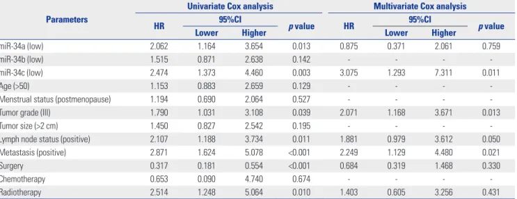
Univariate Cox’s proportional hazards regression analysis in- dicated low expression of miR-34a (p=0.013) and miR-34c (p=
0.003), tumor grade III (p=0.039), lymph positive (p=0.011), dis- tant metastasis positive (p<0.001), and radiotherapy (p=0.010) and predicted worse OS, while surgery treatment could predict prolonged OS (p<0.001). Further multivariate Cox proportional
hazards regression demonstrated only low expression of miR- 34c (p=0.011), tumor grade (p=0.013), lymph positive (p=0.050), and distant metastasis positive (p=0.020) were independent risk factors for OS (Table 2).
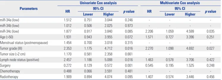
Subgroup (TNM stage I–III patients and stage IV patients) analysis of prognosis
In TNM stage I–III subgroups (Table 3), tumor grade (p=0.016), lymph positive (p=0.016), and surgery treatment (p=0.001) were found to be correlated with OS by univariate Cox proportional hazards regression, while miR-34c low expression (p=0.035) and tumor grade (p=0.027) were independent risk factors for worse OS in multivariate analysis. In TNM stage IV subgroup (Table 4), miR-34a (p=0.029), miR-34b (p=0.020), and miR-34c (p=0.010) expressions were shown to be associated with short- er OS by univariate Cox proportional hazards regression, while no independent predictive factors for OS were found by multi- variate analysis.
Table 2. Univariate and Multivariate Cox Proportional Hazards Regression Analysis of Risk Factors for Overall Survival in TNBC Patients
Parameters
Univariate Cox analysis Multivariate Cox analysis
HR 95%CI
p value HR 95%CI
p value
Lower Higher Lower Higher
miR-34a (low) 2.062 1.164 3.654 0.013 0.875 0.371 2.061 0.759
miR-34b (low) 1.515 0.871 2.638 0.142 - - - -
miR-34c (low) 2.474 1.373 4.460 0.003 3.075 1.293 7.311 0.011
Age (>50) 1.153 0.883 2.659 0.129 - - - -
Menstrual status (postmenopause) 1.194 0.690 2.064 0.527 - - - -
Tumor grade (III) 1.790 1.031 3.108 0.039 2.071 1.168 3.671 0.013
Tumor size (>2 cm) 1.450 0.827 2.542 0.195 - - - -
Lymph node status (positive) 2.107 1.188 3.734 0.011 1.881 0.979 3.612 0.050
Metastasis (positive) 2.871 1.624 5.078 <0.001 2.249 1.129 4.480 0.021
Surgery 0.317 0.181 0.554 <0.001 0.684 0.319 1.468 0.330
Chemotherapy 0.653 0.090 4.740 0.674 - - - -
Radiotherapy 2.514 1.248 5.064 0.010 1.403 0.605 3.256 0.431
TNBC, triple-negative breast cancer; HR, hazard ratio; CI, confidence interval; miR-34, microRNA-34.
Data are presented as HR, 95% CI and p value. A p value <0.05 was considered statistically significant. Significance was determined by univariate Cox propor- tional hazards regression analysis, and all factors with a p value <0.1 were further analyzed by multivariate Cox proportional hazards regression.
1.0 0.8 0.6 0.4 0.2 0.0
Months Log-rank test: p=0.011
miR-34a low miR-34a high miR-34a low-censored miR-34a high-censored
0 20 40 60 80 100
Accumulative overall survival
A
1.0 0.8 0.6 0.4 0.2 0.0
Months Log-rank test: p=0.002
miR-34c low miR-34c high miR-34c low-censored miR-34c high-censored
0 20 40 60 80 100
Accumulative overall survival
C 1.0
0.8 0.6 0.4 0.2 0.0
Months Log-rank test: p=0.138
miR-34b low miR-34b high miR-34b low-censored miR-34b high-censored
0 20 40 60 80 100
Accumulative overall survival
B
Fig. 2. Kaplan-Meier curve analysis of miR-34a/b/c for overall survival. (A) miR-34a. (B) miR-34b. (C) miR-34c. miR-34, microRNA-34.
DISCUSSION
This publication was designed to study the relationship of cir- culating miR-34 family expressions with prognosis in TNBC patients and to evaluate its possibility of becoming a novel non- invasive prognostic biomarker. Our results demonstrated the expressions of miR-34a/34b/34c to be significantly decreased in TNBC patients. Moreover, low expressions of miR-34a and miR-34c indicated shorter OS, while miR-34c low level was an independent factor for worse prognosis.
Due to its specific receptor status, TNBC usually accepts che- motherapy rather than endocrine therapy or trastuzumab. There is no standard effective chemotherapy regimen for TNBC pa- tients, which makes an biomarker to evaluate effectiveness of the therapy critical. Studies have demonstrated that miRNAs regulate many oncogenes, tumor suppressor genes, and dis- semination and chemoresistance of tumors, which suggest that these miRNAs play critical roles in the pathogenesis of numer-
ous cancers, including TNBC.8,9 Previous studies demonstrat- ed the miR-34 family to be activated to transcript by high mu- tations rate of p53 gene production in TNBC patients,10 and involved in cancer cell proliferation, invasion, metastases, and apoptosis, as well as cell survival, cell cycle progression, cell senescence, cell migration, and angiogenesis.5 All these char- acteristics of the miR-34 family make it a promising biomarker of TNBC. Our study first focused on the relationship between circulating miR-34 family and prognosis in TNBC, which pro- vided data for further development of novel noninvasive prognostic biomarkers.
Previous studies demonstrated the miR-34 family as tumor suppressive miRNA participating in the p53-driven apoptotic pathways.10,11 However, the role of miR-34a in tumor suppres- sion is controversial in previous reports. MiR-34a has also been pointed out as a potential tumor suppressor by inducing apop- tosis in neuroblastoma cells.12 Controversially, overexpression of miR-34a has also been reported as an indicator of an aggres- Table 3. Risk Factors Analysis for Overall Survival in TNM Stage I–III Patients
Parameters
Univariate Cox analysis Multivariate Cox analysis
HR 95% CI
p value HR 95% CI
p value
Lower Higher Lower Higher
miR-34a (low) 1.512 0.751 3.044 0.246 - - - -
miR-34b (low) 1.012 0.506 2.025 0.973 - - - -
miR-34c (low) 1.877 0.917 3.840 0.085 2.206 1.059 4.599 0.035
Age (>50) 1.931 0.943 3.955 0.072 1.571 0.727 3.396 0.251
Menstrual status (postmenopause) 1.454 0.700 3.018 0.315 - - - -
Tumor grade (III) 2.353 1.175 4.712 0.016 2.270 1.098 4.692 0.027
Tumor size (>2 cm) 1.170 0.581 2.358 0.661 - - - -
Lymph node status (positive) 2.457 1.186 5.088 0.016 1.463 0.578 3.706 0.422
Surgery 0.272 0.129 0.572 0.001 0.545 0.195 1.525 0.248
Chemotherapy 0.488 0.066 3.591 0.481 - - - -
Radiotherapy 1.909 0.894 4.074 0.095 1.407 0.574 3.446 0.456
HR, hazard ratio; CI, confidence interval; miR-34, microRNA-34.
Data are presented as HR, 95% CI and p value. A p value <0.05 was considered statistically significant. Significance was determined by univariate Cox propor- tional hazards regression analysis, and all factors with a p value <0.1 were further analyzed by multivariate Cox proportional hazards regression.
Table 4. Risk factors Analysis for Overall Survival in TNM Stage IV Patients
Parameters
Univariate Cox analysis Multivariate Cox analysis
HR 95% CI
p value HR 95% CI
p value
Lower Higher Lower Higher
miR-34a (low) 3.414 1.133 10.283 0.029 0.919 0.151 5.591 0.927
miR-34b (low) 3.328 1.204 9.202 0.020 1.734 0.434 6.924 0.436
miR-34c (low) 4.264 1.416 12.838 0.010 3.175 0.588 17.133 0.179
Age (>50) 1.083 0.448 2.614 0.860 - - - -
Menstrual status (postmenopause) 2.004 0.801 5.011 0.137 - - - -
Tumor grade (III) 1.311 0.490 3.505 0.589 - - - -
Tumor size (>2 cm) 2.067 0.728 5.868 0.172 - - - -
Lymph node status (positive) 1.255 0.491 3.207 0.636 - - - -
HR, hazard ratio; CI, confidence interval; miR-34, microRNA-34.
Data are presented as HR, 95% CI and p value. A p value <0.05 was considered statistically significant. Significance was determined by univariate Cox propor- tional hazards regression analysis, and all factors with a p value <0.1 were further analyzed by multivariate Cox proportional hazards regression.
sive breast tumor phenotype,13 and low expression of miR-34a suppressed breast cancer cells to proliferate.14 As for circulating miR-34a, a previous study showed it was significantly reduced in breast cancer without association with stage or grade of tu- mors.15 However, our results showed downregulation of miR- 34a to be associated with worse tumor grade. Whether the dif- ferences in patient population (general breast cancer vs. TNBC) and sources of miR-34a (tissue vs. blood sample) led to these dis- parities deserves further investigation. As for the metastatic- condition, we found upregulation of miR-34a to be significant- ly correlated with non-metastatic condition in TNBC, which was similar with a previous report in breast cancer.16 Additionally, we also found miR-34a was related to lymph node status, and most importantly, was a risk factor of the OS in TNBC patients.
Our study revealed that miR-34c downregulation was corre- lated with tumor grade, metastasis, and was an independent risk factor of OS in TNBC patients. The mechanism of miR-34c involving in breast cancer by suppressing breast cancer migra- tion and invasion by targeting GIT1 and Fra-1,17,18 inducing G2/
M cell cycle arrest in breast cancer cells.19 The down-regulation of miR-34c was induced by epithelial-mesenchymal transition and DNA methylation promoting self-renewal.20
In our study, there was no significant correlation between circulating expression of miR-34b and tumor status or OS in TBNC. However, in a study detected by formalin-fixed paraffin- embedded tissues from breast cancer, miR-34b, but not miR- 34a/c, expression was reported to be negatively correlated with disease free survival and OS in TNBC patients.21 These diverse results might result from the different distribution of the deviat- ed miR-34b in breast cancer tissues and blood samples, and fur- ther investigation is needed.
In conclusion, this study demonstrated that reduced miR-34a/
c expression is highly associated with tumor progression and indicated worse prognosis and that miR-34c was an indepen- dent risk factor for OS in TNBC patients.
REFERENCES
1. Torre LA, Bray F, Siegel RL, Ferlay J, Lortet-Tieulent J, Jemal A.
Global cancer statistics, 2012. CA Cancer J Clin 2015;65:87-108.
2. Perou CM, Sørlie T, Eisen MB, van de Rijn M, Jeffrey SS, Rees CA, et al. Molecular portraits of human breast tumours. Nature 2000;406:
747-52.
3. Foulkes WD, Smith IE, Reis-Filho JS. Triple-negative breast cancer.
N Engl J Med 2010;363:1938-48.
4. Bartel DP. MicroRNAs: target recognition and regulatory functions.
Cell 2009;136:215-33.
5. Maroof H, Salajegheh A, Smith RA, Lam AK. MicroRNA-34 family, mechanisms of action in cancer: a review. Curr Cancer Drug Targets 2014;14:737-51.
6. Misso G, Di Martino MT, De Rosa G, Farooqi AA, Lombardi A, Cam- pani V, et al. Mir-34: a new weapon against cancer? Mol Ther Nu- cleic Acids 2014;3:e194.
7. Rokavec M, Li H, Jiang L, Hermeking H. The p53/miR-34 axis in de- velopment and disease. J Mol Cell Biol 2014;6:214-30.
8. Garcia AI, Buisson M, Bertrand P, Rimokh R, Rouleau E, Lopez BS, et al. Down-regulation of BRCA1 expression by miR-146a and miR- 146b-5p in triple negative sporadic breast cancers. EMBO Mol Med 2011;3:279-90.
9. Radojicic J, Zaravinos A, Vrekoussis T, Kafousi M, Spandidos DA, Stathopoulos EN. MicroRNA expression analysis in triple-negative (ER, PR and Her2/neu) breast cancer. Cell Cycle 2011;10:507-17.
10. He L, He X, Lim LP, de Stanchina E, Xuan Z, Liang Y, et al. A microR- NA component of the p53 tumour suppressor network. Nature 2007;447:1130-4.
11. Corney DC, Hwang CI, Matoso A, Vogt M, Flesken-Nikitin A, God- win AK, et al. Frequent downregulation of miR-34 family in human ovarian cancers. Clin Cancer Res 2010;16:1119-28.
12. Welch C, Chen Y, Stallings RL. MicroRNA-34a functions as a poten- tial tumor suppressor by inducing apoptosis in neuroblastoma cells.
Oncogene 2007;26:5017-22.
13. Peurala H, Greco D, Heikkinen T, Kaur S, Bartkova J, Jamshidi M, et al. MiR-34a expression has an effect for lower risk of metastasis and associates with expression patterns predicting clinical out- come in breast cancer. PLoS One 2011;6:e26122.
14. Dutta KK, Zhong Y, Liu YT, Yamada T, Akatsuka S, Hu Q, et al. As- sociation of microRNA-34a overexpression with proliferation is cell type-dependent. Cancer Sci 2007;98:1845-52.
15. Nugent M, Miller N, Kerin MJ. Circulating miR-34a levels are re- duced in colorectal cancer. J Surg Oncol 2012;106:947-52.
16. Javeri A, Ghaffarpour M, Taha MF, Houshmand M. Downregula- tion of miR-34a in breast tumors is not associated with either p53 mutations or promoter hypermethylation while it correlates with metastasis. Med Oncol 2013;30:413.
17. Tao WY, Wang CY, Sun YH, Su YH, Pang D, Zhang GQ. MicroRNA- 34c suppresses breast cancer migration and invasion by targeting GIT1. J Cancer. 2016;7:1653-62.
18. Yang S, Li Y, Gao J, Zhang T, Li S, Luo A, et al. MicroRNA-34 sup- presses breast cancer invasion and metastasis by directly targeting Fra-1. Oncogene 2013;32:4294-303.
19. Achari C, Winslow S, Ceder Y, Larsson C. Expression of miR-34c induces G2/M cell cycle arrest in breast cancer cells. BMC Cancer 2014;14:538.
20. Yu F, Jiao Y, Zhu Y, Wang Y, Zhu J, Cui X, et al. MicroRNA 34c gene down-regulation via DNA methylation promotes self-renewal and epithelial-mesenchymal transition in breast tumor-initiating cells.
J Biol Chem 2012;287:465-73.
21. Svoboda M, Sana J, Redova M, Navratil J, Palacova M, Fabian P, et al.
MiR-34b is associated with clinical outcome in triple-negative breast cancer patients. Diagn Pathol 2012;7:31.