Assessment of Blood-Brain Barrier Permeability by Dynamic Contrast-Enhanced MRI in Transient Middle Cerebral Artery Occlusion Model after Localized Brain Cooling in Rats
Eun Soo Kim, MD, PhD
1, Seung-Koo Lee, MD, PhD
2, Mi Jung Kwon, MD, PhD
3, Phil Hye Lee, MD, PhD
4, Young-Su Ju, MD, PhD
5, Dae Young Yoon, MD, PhD
6, Hye Jeong Kim, MD
7, Kwan Seop Lee, MD
1Departments of 1Radiology, 3Pathology, and 5Industrial Medicine, Hallym University Sacred Heart Hospital, Hallym University College of Medicine, Anyang 14068, Korea; Departments of 2Radiology and 4Neurology, Yonsei University College of Medicine, Seoul 03722, Korea; 6Department of Radiology, Hallym University Kangdong Sacred Heart Hospital, Hallym University College of Medicine, Seoul 05355, Korea; 7Department of Radiology, Kangnam Sacred Heart Hospital, Hallym University College of Medicine, Seoul 07441, Korea
Objective: The purpose of this study was to evaluate the effects of localized brain cooling on blood-brain barrier (BBB) permeability following transient middle cerebral artery occlusion (tMCAO) in rats, by using dynamic contrast-enhanced (DCE)- MRI.Materials and Methods: Thirty rats were divided into 3 groups of 10 rats each: control group, localized cold-saline (20°C) infusion group, and localized warm-saline (37°C) infusion group. The left middle cerebral artery (MCA) was occluded for 1 hour in anesthetized rats, followed by 3 hours of reperfusion. In the localized saline infusion group, 6 mL of cold or warm saline was infused through the hollow filament for 10 minutes after MCA occlusion. DCE-MRI investigations were performed after 3 hours and 24 hours of reperfusion. Pharmacokinetic parameters of the extended Tofts-Kety model were calculated for each DCE-MRI. In addition, rotarod testing was performed before tMCAO, and on days 1–9 after tMCAO. Myeloperoxidase (MPO) immunohisto-chemistry was performed to identify infiltrating neutrophils associated with the inflammatory response in the rat brain.
Results: Permeability parameters showed no statistical significance between cold and warm saline infusion groups after 3-hour reperfusion 0.09 ± 0.01 min-1 vs. 0.07 ± 0.02 min-1, p = 0.661 for Ktrans; 0.30 ± 0.05 min-1 vs. 0.37 ± 0.11 min-1, p = 0.394 for kep, respectively. Behavioral testing revealed no significant difference among the three groups. However, the percentage of MPO-positive cells in the cold-saline group was significantly lower than those in the control and warm-saline groups (p < 0.05).
Conclusion: Localized brain cooling (20°C) does not confer a benefit to inhibit the increase in BBB permeability that follows transient cerebral ischemia and reperfusion in an animal model, as compared with localized warm-saline (37°C) infusion group.
Keywords: Brain; Ischemia; Middle cerebral artery; Blood-brain barrier; Permeability; Dynamic contrast-enhanced-MRI; DCE-MRI
Received February 22, 2016; accepted after revision May 17, 2016.
This study was supported by a faculty research grant of Yonsei University College of Medicine #6-2012-0091.
Corresponding author: Seung-Koo Lee, MD, PhD, Department of Radiology, Yonsei University College of Medicine, 50-1 Yonsei-ro, Seodaemun-gu, Seoul 03722, Korea.
• Tel: (822) 2228-2373 • Fax: (822) 393-3035
• E-mail: [email protected]
This is an Open Access article distributed under the terms of the Creative Commons Attribution Non-Commercial License (http://creativecommons.org/licenses/by-nc/3.0) which permits unrestricted non-commercial use, distribution, and reproduction in any medium, provided the original work is properly cited.
Korean J Radiol 2016;17(5):715-724
INTRODUCTION
Hypothermia is very effective at preventing ischemia- induced neuronal damage (1-7). However, the use of whole body surface cooling for hypothermia therapy is associated with management problems and complications such as pneumonia in 40% of patients (8). Recently, localized brain cooling has been reported as a more effective technique than whole-body cooling, to inhibit or at least delay neuronal damage (3, 9-14). In addition, localized brain cooling before reperfusion significantly reduces the infarct
pISSN 1229-6929 · eISSN 2005-8330
area in multiple animal models of stroke (15). Localized brain cooling following cerebral ischemia in animal models also markedly reduces inflammatory reactions and endothelial expression of intracellular adhesion molecule-1 (ICAM-1), which is strongly associated with blood-brain barrier (BBB) breakdown of micro-vessels in ischemic brain tissue (16-18). Therefore, increased BBB permeability is one of the cerebral neuronal damage mechanisms for reperfusion injury (19, 20). Permeability imaging using dynamic contrast-enhanced (DCE)-MRI can measure the integrity of BBB (21-23). Permeability parameters using DCE-MRI have been assessed in recent stroke studies related to BBB dysfunction (24-26). To our best knowledge, the effects of localized brain cooling on BBB permeability following cerebral ischemia have not been evaluated in a rat model.
The aim of the current study was to investigate the effect of localized brain cooling on BBB permeability, after transient focal cerebral ischemia reperfusion in rats, using DCE-MRI.
MATERIALS AND METHODS
Ethics Statement
This animal study was approved by and performed in accordance with the Institutional Animal Care and Use Committee guidelines. Yonsei Medical Center animal experimentation Ethics Committee monitored the scientific and ethical proceedings of animal experiments under the management and use program of experimental animals based on Guide for the care and use of laboratory animal (National Research Council, USA).
Subjects
A total of 30 adult Sprague-Dawley rats (303 ± 16.2 g [mean ± SD]) were used in the study. A modified filament technique was used to produce transient middle cerebral artery occlusion (tMCAO) in a rat model, as previously described (27). Rats were anesthetized by intraperitoneal injection of 2:3 mixture of xylazine (Rompun, Bayer, Berlin, Germany) and zoletil (0.3 mg; tiletamine/zolazepam, Virbac, Carros, France), and the left external carotid artery (ECA) was exposed. A length of 18.5–19.0 mm modified PE- 50 catheter (with 0.2-mm outer diameter and 0.1-mm inner diameter) was inserted into the intracranial circulation via the left ECA. The filament was lodged in the narrow proximal anterior cerebral artery (ACA), blocking the middle cerebral artery (MCA) at its origin. After 1 hour of MCA occlusion, rats in the control group were re-anesthetized
and reperfused for 3 hours by withdrawal of the hollow filament from the left MCA.
The animals were divided into control, cold-saline infusion, and warm-saline infusion groups, with 10 rats in each group. The rats in the control group received no treatment. The rats in the cold-saline infusion group received an intra-arterial infusion of 6 mL cold (20°C) saline to the brain after 1 hour of MCA occlusion. The rats in the warm-saline infusion group were infused with 6 mL warm (37°C) saline using the same method as the cold- saline group. The warm-saline infusion group served as another control group, to observe the effects of a localized saline infusion on brain injury resulting from transient ischemia and reperfusion (16-18). In the saline infusion groups, the catheter was withdrawn 1 mm from the origin of the MCA after 1 hour of MCA occlusion. During and after withdrawal of the catheter, 6 mL, cold or warm saline was slowly injected continuously into the junction of the MCA and ACA, using an infusion pump to maintain a rate of 0.6 mL, over 10 minutes after the catheter was completely withdrawn and reperfusion established (approximately 0.25 mL/g brain tissue per minute).
Brain Temperature
Brain temperature in the rats was monitored in the ipsilateral area supplied by the MCA. Needle thermistor probes (Harvard Apparatus) were placed into the ipsilateral cortex through a hole 3 mm lateral to the bregma and into the striatum through a hole 3 mm posterior and 4 mm lateral to the bregma. The local brain temperature in the cortex and striatum supplied by the MCA remained unchanged from 37°C in control and localized warm-saline infusion groups. In the cold-saline infusion group, localized brain temperature was reduced to 33–34°C after 10 minutes of cold (20°C) saline infusion. After stopping the cold- saline infusion, the brain temperature gradually increased to 37°C. The rectal temperature was maintained at 37°C using a circulating heating pad.
MRI Protocol
Animal MRI was performed using a 3.0-tesla system (Achieva, Philips, Best, the Netherlands) with an 8-channel SENSE wrist coil. The first DCE-MRI was performed
immediately after 3 hours of MCA reperfusion. The second DCE-MRI was performed after 24 hours of reperfusion. All images were obtained in the coronal plane with a 60-mm field of view. Pre- and post-contrast T1-weighted (repetition
time [TR]/echo time [TE], 625/18 ms) and T2-weighted (TR/
TE, 2006/80 ms) images were acquired with 2-mm section thickness, 0.2-mm section gap, and 192 x 192 matrix.
Diffusion-tensor imaging was performed by applying 6 diffusion-encoding directions with a b value of 600 s/mm2 and with x b0 images (TR/TE, 3327/52 ms; 2-mm section thickness; 0.2-mm section gap; 128 x 128 matrix) (28). To achieve quantitative hemodynamic measurements of cerebral permeability and perfusion, we injected 2 boluses via tail veins. The first bolus of contrast was administered to measure permeability and served as a preload bolus for the perfusion scans. For DCE-MRI, precontrast 3D T1-weighted images were obtained with the following parameters: TR/
TE, 13.2/6.5 ms; 112 x 112 mm matrix; 2-mm section thickness; 0.2-mm section gap; and flip angle, 5°. After the precontrast scan, 60 DCE T1-weighted images were obtained with the same MRI parameters except for an increased flip angle of 15°. After acquisition of the fifth image volume, gadolinium-based contrast, gadobutrol (Gadavist, 0.2 mmol/kg; Bayer Healthcare, Berlin, Germany) was injected.
The total scan time for DCE-MRI was 4 minutes 30 seconds with a temporal resolution of 4.5 seconds. Perfusion- weighted imaging (the rapid principles of echo shifting with a train of observations; TR/TE, 26.6/38.2 ms; 64 x 64 matrix) with 60 dynamic scans was performed following injection of the second bolus of Gadavist (0.2 mmol/kg) 4 hours after reperfusion.
Post Processing and Image Analysis
Permeability parameters such as volume transfer constant
(Ktrans), rate transfer coefficient (kep), volume fraction of
extravascular extracellular space (EES) (ve), and volume fraction of blood plasma (vp) were calculated using off-line Philips research imaging development environment (PRIDE) tools provided by Philips Medical System. This software is based on the pharmacokinetic model of extended Tofts- Kety (29, 30). The two-compartment model of extended Tofts and Kety assumes that the intravascular space and EES are divided by the BBB. The degree of contrast leakage from the intravascular space to the EES is referred to as the volume transfer constant; and the reflux leakage of contrast from the EES to the intravascular space (plasma) is referred to as the rate transfer coefficient (kep). All computation procedures were automatically performed by PRIDE tools except drawing the region of interest (ROI) for the arterial input function. Arterial input function was measured several times at the area of the left internal carotid artery, and
the proper arterial input function showing high amplitude, early sharp rise, and fast decay was selected for processing.
MRI sequences were reviewed for the presence of ischemic lesions at each time point. To evaluate the patterns of change in permeability parameters after reperfusion of the MCA, we used the average values for the basal ganglia and cortex. Acute infarction showed in the left basal ganglia and cortex, corresponding to contrast-enhancing area on MRIs after 3 hours of reperfusion. To measure the permeability changes at ROIs of acute infarction in DCE- MRI, two different ROIs were first placed in an enhancing portion of the infarct area of the cortex and basal ganglia in hemispheres ipsilateral to the MCA occlusion on DCE- MRI. Secondly, two different ROIs were placed in the normal cortex and basal ganglia to determine the baseline permeability parameters. Thirdly, the permeability of infarct brain tissue was measured in the follow-up DCE-MRI in areas corresponding to the first MRI. Serial changes in permeability were evaluated for the three ROIs in normal and infarct areas on DCE-MRI.
Rotarod Performance Test
All rats were subjected to rotarod behavioral testing before tMCAO, and on days 1–9 after tMCAO, by an
investigator who was blinded to the group assignments. For the rotarod test, the rat was placed on a rotarod cylinder, and the time the animal remained on the cylinder was measured in seconds. The speed was slowly increased from 4 to 40 rpm within 5 minutes (31). A trial ended if the animal fell off the rungs or gripped the device and spun around for two consecutive revolutions without attempting to walk on the rungs. The animals were trained for 3 days before tMCAO. The mean duration on the device was calculated from 10 trials conducted 1 day before surgery (the baseline value). Motor testing data were recorded for 9 days, and compared with the internal baseline control (before surgery).
Immunohistochemistry and Histological Assay After completion of the second MRI, the rats were sacrificed to obtain brain tissue. Rats were deeply anaesthetized and perfused with 0.9% sodium chloride followed by 4% paraformaldehyde. Following decapitation, brains were removed, fixed in 10% formalin, and embedded in paraffin. Coronal sections (5-µm thick) were stained with hematoxylin and eosin (H&E). Neutrophils are reportedly the first leukocyte subpopulation to be recruited to the
ischemic brain; and an extensive infiltration of neutrophils is observed 24 hours after transient ischemic changes or infarct in rats, which is associated with BBB breakdown (32, 33). Immunohistochemistry for myeloperoxidase (MPO) was performed to identify infiltrating neutrophils on sections of rat brains subjected to no treatment vs. treatment (cold- or warm-saline infusion) after tMCAO, in order to detect BBB breakdown. Tissue sections were deparaffinized in xylene, rehydrated, and heated at 100°C in citrate buffer (pH 6.0) for 5 minute for antigen retrieval. The sections were incubated with a rabbit polyclonal antibody against MPO (1:500 dilution; A0398, Dako, Glostrup, Denmark) for 1 hour at room temperature, followed by incubation with secondary antibody, donkey anti-rabbit IgG (1:500 dilution; Molecular Probes, Eugene, OR, USA) for 1 hour at room temperature.
Staining was developed by reaction with diaminobenzidine chromogen, and sections were counterstained with hematoxylin. For quantitative analysis of cell numbers in the infarcted regions, the slides were digitally photographed using a confocal microscope at a 400 x magnification (BX50,
Olympus, Tokyo, Japan). Ten fields of view were randomly selected and photographed to count the number of MPO- positive cells in each section (version 4.6, Spot Software, Diagnostic Instruments Inc., Sterling Heights, MI, USA). All analyses were performed by a pathologist blinded to the treatment conditions.
Statistical Analysis
The comparison of permeability parameters at different time points among the three groups were assessed using mixed modeling. Correlations between permeability parameters for each group in ROIs in the cortex and basal ganglia were analyzed. Difference analysis between baseline control and mean duration (of 10 trials) at each time point was plotted for rotarod tests. The differences in MPO- positive neutrophil infiltration between the three groups were assessed using the Mann-Whitney U test. All statistical analyses were performed using the statistical software package, SPSS (version 21, SPSS Inc., IBM Company, Chicago, IL, USA). A p value of < 0.05 was considered
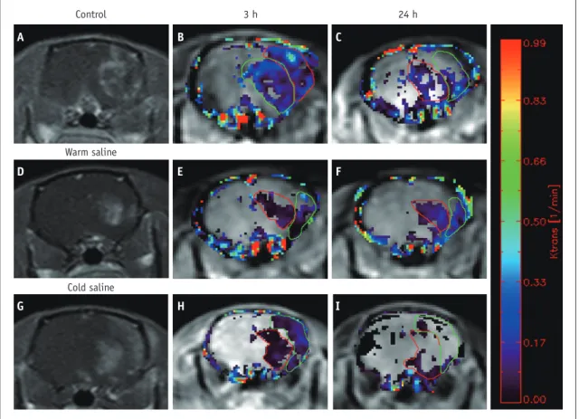
Fig. 1. Brain MRI of mouse.
Contrast enhancement can be seen at infarcted area of left brain (A, D, G). Color coded permeability maps of Ktrans obtained 3 hours (B, E, H) and 24 hours (C, F, I) after reperfusion show increased permeability in infracted area. Mean Ktrans values were 0.15 ± 0.03 min-1, 0.12 ± 0.02 min-1 in control groups, 0.07 ± 0.02 min-1, 0.09 ± 0.02 min-1 in warm saline group and 0.09 ± 0.01 min-1, 0.05 ± 0.02 min-1 in cold saline group.
Control
Warm saline
Cold saline
3 h 24 h
A
D
G
B
E
H
C
F
I
statistically significant.
RESULTS
Permeability Parameters
None of the cases showed an increase in permeability parameters in the contralateral (normal-appearing) hemisphere on DCE-MRI. Although the number of samples was small for each group, data for each of the permeability parameters were normally distributed. Areas of contrast enhancement showed variable patterns for each group. Most rats showed dominant enhancement in the basal ganglia
and others showed dominant enhancement in both the basal ganglia and cortex (Fig. 1). In addition, an increase
of Ktrans in permeability was noted on DCE-MRI (Fig. 1). DCE-
MRI showed an increase in permeability parameters in the ipsilateral hemisphere of the MCA occlusion. Among the permeability parameters, Ktrans and kep were significantly lower in the cold saline infusion group, as compared with the control group after 3-hour reperfusion: 0.15 ± 0.03 min-1 vs.
0.09 ± 0.01 min-1, p = 0.010 for Ktrans and 0.60 ± 0.20 min-1 vs. 0.03 ± 0.05 min-1, p = 0.046 for kep, respectively (Fig. 2).
Ktrans showed a significant decrease in the warm-saline group,
as compared with the control group after 3-hour reperfusion
Fig. 2. Permeability changes at 2 different time points after reperfusion (mixed model).
A. Ktrans (min-1) shows significant decrease in control group, as compared with warn and cold saline group (control vs. cold saline infusion [p =
0.0095], control vs. warm saline infusion [p = 0.017]). Permeability parameters show no statistical significance between cold and warm saline infusion groups (p = 0.661). B. Kep (min-1) is significantly lower in cold saline infusion group, as compared with control group (p = 0.046).
Permeability parameters show no statistical significance between cold and warm saline infusion groups (p = 0.394), and between control and warm saline groups (p = 0.163).
0.20 0.18 0.16 0.14 0.12 0.10 0.08 0.06 0.04 0.02 0.00 Ktrans (min-1)
3 h 24 h
p = 0.0095
p = 0.0166 Control Warm saline Cold saline
p = 0.6609
0.90 0.80 0.70 0.60 0.50 0.40 0.30 0.20 0.10 0.00 Kep (min-1)
3 h 24 h
p = 0.1633 p = 0.3945 p = 0.0462
Control Warm saline Cold saline
A B
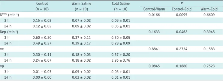
Table 1. Permeability Imaging Parameters Measured 3 and 24 Hours after Reperfusion*
Control (n = 10)
Warm Saline (n = 10)
Cold Saline (n = 10)
P
Control-Warm Control-Cold Warm-Cold
Ktrans (min-1) 0.0166 0.0095 0.6609
3 h 0.15 ± 0.03 0.07 ± 0.02 0.09 ± 0.01
24 h 0.12 ± 0.02 0.09 ± 0.02 0.05 ± 0.01
Kep (min-1) 0.1633 0.0462 0.3945
3 h 0.60 ± 0.20 0.37 ± 0.11 0.30 ± 0.05
24 h 0.49 ± 0.27 0.39 ± 0.17 0.28 ± 0.09
ve 0.8841 0.2734 0.1583
3 h 0.30 ± 0.11 0.18 ± 0.03 0.57 ± 0.20
24 h 0.24 ± 0.07 0.18 ± 0.02 3.96 ± 3.76
vp 0.0845 0.1680 0.7523
3 h 0.01 ± 0.03 0.05 ± 0.02 0.05 ± 0.01
24 h 0.00 ± 0.00 0.03 ± 0.02 0.01 ± 0.01
*Data are means ± SD.
h = hours, Kep = rate transfer coefficient, ve = volume fraction of extravascular extracellular space, vp = volume fraction of blood plasma
(control vs. warm-saline infusion group: 0.15 ± 0.03 min-1 vs.
0.07 ± 0.02 min-1, p = 0.017 for Ktrans).
In the control group, Ktrans showed a marked initial increase followed by a slight decrease. In the warm- saline infusion group, Ktrans showed an initial increase that persisted. In the cold-saline infusion group, serial follow-up of Ktrans showed an initial increase followed by a later decrease. Other permeability parameters showed no consistent pattern of change over follow-up (Table 1).
However, permeability parameters showed no statistical significance between cold and warm saline infusion groups after 3-hour reperfusion: 0.09 ± 0.01 min-1 vs. 0.07 ± 0.02 min-1, p = 0.661 for Ktrans; 0.30 ± 0.05 min-1 vs. 0.37 ± 0.11 min-1, p = 0.394 for kep, respectively (Table 1).
Rotarod Test
Twenty-four hours after MCA occlusion, the rotarod performance decreased markedly in the three groups; but
the changes in the cold-saline and warm-saline groups were not significant, as compared with baseline values.
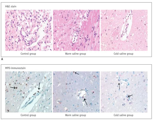
Histopathological and Immunohistochemical Findings On high-power views (400 x) of the H&E-stained sections, multiple foamy macrophages and infiltration of inflammatory cells were seen in the perimicrovascular area in the control group. In the warm-saline group, foamy macrophages and inflammatory cells were decreased, as compared with the control group. In the cold-saline group, foamy macrophages and inflammatory cells were markedly decreased. On high-power views (400 x) of the rat brain sections, MPO-positive neutrophil (arrow) infiltrate was observed in the perimicrovascular area in the control group.
The number of MPO-positive cells (arrows) were significantly decreased in warm-saline and cold-saline groups, as compared with the control group (Fig. 3).
The overall numbers of inflammatory cells were 52.6
Fig. 3. Immunohistochemical staining of mouse brain.
A. High-power views (400 x) of hematoxylin and eosin (H&E)-stained sections. In cool-saline group, foamy macrophages and inflammatory cells in perimicrovascular area are decreased, as compared with control and warm saline group. B. High-power views (400 x) of myeloperoxidase (MPO)-immunostained sections of rat brain. In control group, MPO-positive neutrophils (arrow) infiltrate perimicrovascular area. There are decreases in number of MPO-positive cells (arrows) in warm-saline and cold-saline groups, as compared with control group.
H&E stain
MPO-immunostain
Control group Control group
Warm saline group Warm saline group
Cold saline group Cold saline group A
B
± 27.3 (mean ± SD) in the control group, 7.2 ± 2.6 in the cold-saline group, and 29.4 ± 30.9 in the warm- saline group, respectively. The numbers of MPO-positive neutrophils were 37.8 ± 27.3 in the control group, 37.6
± 11.5 in the cold-saline group, and 65.2 ± 44.5 in the warm-saline group, respectively. The percentage of the total number of inflammatory cells that were MPO-positive was significantly decreased in the cold- and warm-saline infusion groups, as compared with the control group (p
= 0.008 and 0.032, respectively, p < 0.05). Notably, the percentage of MPO-positive cells in the cold-saline group was significantly lower than that of the warm-saline group (p
= 0.009) (Fig. 4).
DISCUSSION
In this study, we evaluated BBB permeability of infarction site among control, cold-saline infusion, and warm-
saline infusion groups. Several reports indicate that brain hypothermia can reduce the size of the infarct area and damage due to cerebral ischemic after tMCAO in an animal model (12, 34, 35). Although the underlying mechanisms are not completely understood, it is generally accepted that brain hypothermia has protective effects against BBB disruption (36). Some studies suggest that the post- ischemic protective effects of brain hypothermia may be
related to other mechanisms, such as the reduction of leukotrienes, improvements in glucose utilization and blood flow, and slowing of reactions involving free radicals and the propagation of lipid-peroxidation cascades (36-38).
A significant decrease in numbers of inflammatory cells was observed in the area affected by ischemia on 3-hour and 24-hour follow-up in the cold-saline group, as compared with the warm-saline group. However, parameters related to BBB permeability showed no significant differences between the cold- and warm-saline groups; likewise, the behavioral testing did not reveal significant differences among the three groups.
There are several possible explanations for these effects.
Firstly, these findings suggest that the contrast leakage in vessels was reduced by a more stable BBB after localized infusion of either cold or warm saline to the ischemic area.
Some studies have reported that the infusion of saline into the ischemic territory plays a very important role in the prevention of reperfusion injuries in addition to the effects of hypothermia (39). Infusion with saline at 37°C can reduce the expression of ICAM-1 and leukocyte infiltration into the ischemic area. ICAM-1, a mediator of the acute inflammatory reaction, causes circulating leukocytes to infiltrate the ischemic brain parenchyma. Secondly, the duration of localized brain cooling and the follow-up period might have been too short for the specific effects of cooling to be observed. Most animal experiments are based on the assumption that timing is critical for the therapeutic window of mild hypothermia after tMCAO. Several studies investigating systemic hypothermia and localized brain cooling in animals and humans have indicated a cooling duration of only 10 minutes, because oxygen extraction remains unchanged and the feasibility and safety of the experiment are still unclear (6, 20, 34, 40). Our study design was based on a similar assumption and it was difficult to determine the exact time window for localized brain cooling. One report has indicated that localized cold- saline infusion (20°C) for 2.5 hours after brain ischemia significantly reduces infarction volume, as compared to reperfusion without localized cold-saline infusion (41).
The therapeutic time for localized saline infusion could be extended by at least 30 minutes. Third, although an isolated and selective brain-cooling technique by infusion of cooled saline infusion into the carotid artery provides precise control of the systemic circulatory blood temperature, precise control of brain tissue temperature using this technique is very difficult at the rewarming stage. This is Fig. 4. Comparison of percentage of MPO-positive cells in total
mixed population of inflammatory cells. Each box plot represents mean and standard deviation and line through box plot indicates range. Percentage of MPO-positive cells is significantly higher in control group than in cold-saline and warm-saline groups (p < 0.05).
In addition, percentage of MPO-positive cells in cold-saline group is significantly lower than that of warm-saline group (p < 0.05). MPO = myeloperoxidase
100
80
60
40
20
0
MPO-positive cells/total cells (%)
Control group
p = 0.008 p = 0.009 p = 0.032
20°C saline
infusion group 37°C saline infusion group
because the cessation of cooled saline infusion into the brain produces an immediate and rapid elevation of brain tissue temperature caused by recirculation of warm blood.
Also, infusion of saline for brain cooling is a negative factor for the prevention of brain edema (42).
Cytotoxic edema is the predominant type of edema in acute infarction (43). Subsequently, brain ischemia and hypoxia would deteriorate due to brain swelling and intracranial hypertension, which would cause neuron dysfunction and further aggravate the cytotoxic brain edema. At this time, cytotoxic brain edema would predominate, and EES is expected to decrease. However, cytotoxic edema alone is not enough to aggravate the lesion because water content is not increased; when BBB is compromised, water is driven from the vessels into the EES and then enters the cells (44). Thus, the BBB may be a target for treatments to relieve brain edema and swelling.
Dynamic contrast enhanced-MRI is primarily recommended for pharmacodynamic assessment of antiangiogenic and antivascular therapies associated with BBB breakdown and is ideal for the quantification of permeability parameters (45-50). Gadolinium-DTPA does not cross an intact BBB and can therefore be used to detect BBB disruption. After focal cerebral ischemia or infarction, the disturbance of BBB integrity has been confirmed by gadolinium-DTPA-enhanced T1-weighted image.
Our study had several limitations. A limited number of animal subjects were used in the experiments; therefore, further investigations with a large number of animals will be necessary in the future. Secondly, the ROI was drawn manually on DCE-MRI images in contrast-enhancing areas of the basal ganglia and cortex. Therefore, ROIs were drawn several times in each subject, and the average value was calculated. Thirdly, we were limited by a general lack of knowledge regarding delayed or secondary results at a temperature of 20°C for the cold saline infusion to make a clear distinction from the warm-saline group. Further clinical investigations will be required in future to decide the optimal temperature for localized brain cooling in order to ensure the most viable and efficient results.
In conclusion, our trial does not provide evidence that localized brain cooling confers a protective effect on BBB integrity following transient focal cerebral ischemia and reperfusion in rats. Further study of this approach remains to be evaluated.
Acknowledgments
Thanks to researcher Ah-Ream Yang (Department of Anatomy, Yonsei University College of Medicine, Seoul, Korea) for making the animal experimental models from the beginning to the end of this research.
REFERENCES
1. Kataoka K, Yanase H. Mild hypothermia--a revived
countermeasure against ischemic neuronal damages. Neurosci Res 1998;32:103-117
2. Chopp M, Knight R, Tidwell CD, Helpern JA, Brown E, Welch KM. The metabolic effects of mild hypothermia on global cerebral ischemia and recirculation in the cat: comparison to normothermia and hyperthermia. J Cereb Blood Flow Metab 1989;9:141-148
3. Colbourne F, Sutherland G, Corbett D. Postischemic
hypothermia. A critical appraisal with implications for clinical treatment. Mol Neurobiol 1997;14:171-201
4. Barone FC, Feuerstein GZ, White RF. Brain cooling during transient focal ischemia provides complete neuroprotection.
Neurosci Biobehav Rev 1997;21:31-44
5. Ginsberg MD, Sternau LL, Globus MY, Dietrich WD, Busto R. Therapeutic modulation of brain temperature: relevance to ischemic brain injury. Cerebrovasc Brain Metab Rev 1992;4:189-225
6. Schwab S. Therapy of severe ischemic stroke: breaking the conventional thinking. Cerebrovasc Dis 2005;20 Suppl 2:169- 178
7. Kallmünzer B, Kollmar R. Temperature management in stroke - an unsolved, but important topic. Cerebrovasc Dis 2011;31:532-543
8. Schwab S, Schwarz S, Spranger M, Keller E, Bertram M, Hacke W. Moderate hypothermia in the treatment of patients with severe middle cerebral artery infarction. Stroke 1998;29:2461- 2466
9. Huang FP, Zhou LF, Yang GY. The effect of extending mild hypothermia on focal cerebral ischemia and reperfusion in the rat. Neurol Res 1998;20:57-62
10. Colbourne F, Sutherland GR, Auer RN. An automated system for regulating brain temperature in awake and freely moving rodents. J Neurosci Methods 1996;67:185-190
11. Kawai N, Okauchi M, Morisaki K, Nagao S. Effects of delayed intraischemic and postischemic hypothermia on a focal model of transient cerebral ischemia in rats. Stroke 2000;31:1982- 1989; discussion 1989
12. Maier CM, Sun GH, Kunis D, Yenari MA, Steinberg GK. Delayed induction and long-term effects of mild hypothermia in a focal model of transient cerebral ischemia: neurological outcome and infarct size. J Neurosurg 2001;94:90-96 13. Xue D, Huang ZG, Smith KE, Buchan AM. Immediate or
delayed mild hypothermia prevents focal cerebral infarction.
Brain Res 1992;587:66-72
14. Yanamoto H, Hong SC, Soleau S, Kassell NF, Lee KS. Mild postischemic hypothermia limits cerebral injury following transient focal ischemia in rat neocortex. Brain Res 1996;718:207-211
15. Hewawasam P, Ding M, Chen N, King D, Knipe J, Pajor L, et al.
Synthesis of water-soluble prodrugs of BMS-191011: a maxi-K channel opener targeted for post-stroke neuroprotection.
Bioorg Med Chem Lett 2003;13:1695-1698
16. Ding Y, Li J, Luan X, Lai Q, McAllister JP 2nd, Phillis JW, et al.
Local saline infusion into ischemic territory induces regional brain cooling and neuroprotection in rats with transient middle cerebral artery occlusion. Neurosurgery 2004;54:956- 964; discussion 964-965
17. Luan X, Li J, McAllister JP 2nd, Diaz FG, Clark JC, Fessler RD, et al. Regional brain cooling induced by vascular saline infusion into ischemic territory reduces brain inflammation in stroke. Acta Neuropathol 2004;107:227-234
18. Ding Y, Li J, Rafols JA, Phillis JW, Diaz FG. Prereperfusion saline infusion into ischemic territory reduces inflammatory injury after transient middle cerebral artery occlusion in rats.
Stroke 2002;33:2492-2498
19. Zhang RL, Chopp M, Zhang ZG, Jiang Q, Ewing JR. A rat model of focal embolic cerebral ischemia. Brain Res 1997;766:83-92 20. Busto R, Dietrich WD, Globus MY, Ginsberg MD. Postischemic
moderate hypothermia inhibits CA1 hippocampal ischemic neuronal injury. Neurosci Lett 1989;101:299-304
21. Kassner A, Roberts TP, Moran B, Silver FL, Mikulis DJ.
Recombinant tissue plasminogen activator increases blood-brain barrier disruption in acute ischemic stroke:
an MR imaging permeability study. AJNR Am J Neuroradiol 2009;30:1864-1869
22. Larsson HB, Courivaud F, Rostrup E, Hansen AE. Measurement of brain perfusion, blood volume, and blood-brain barrier permeability, using dynamic contrast-enhanced T(1)-weighted MRI at 3 tesla. Magn Reson Med 2009;62:1270-1281
23. Choi HS, Ahn SS, Shin NY, Kim J, Kim JH, Lee JE, et al.
Permeability parameters measured with dynamic contrast- enhanced MRI: correlation with the extravasation of evans blue in a rat model of transient cerebral ischemia. Korean J Radiol 2015;16:791-797
24. Durukan A, Marinkovic I, Strbian D, Pitkonen M, Pedrono E, Soinne L, et al. Post-ischemic blood-brain barrier leakage in rats: one-week follow-up by MRI. Brain Res 2009;1280:158- 165
25. Ding G, Jiang Q, Li L, Zhang L, Gang Zhang Z, Ledbetter KA, et al. Detection of BBB disruption and hemorrhage by Gd- DTPA enhanced MRI after embolic stroke in rat. Brain Res 2006;1114:195-203
26. Jahng GH, Li KL, Ostergaard L, Calamante F. Perfusion magnetic resonance imaging: a comprehensive update on principles and techniques. Korean J Radiol 2014;15:554-577 27. Longa EZ, Weinstein PR, Carlson S, Cummins R. Reversible
middle cerebral artery occlusion without craniectomy in rats.
Stroke 1989;20:84-91
28. Kim SJ, Choi CG, Kim JK, Yun SC, Jahng GH, Jeong HK, et
al. Effects of MR parameter changes on the quantification of diffusion anisotropy and apparent diffusion coefficient in diffusion tensor imaging: evaluation using a diffusional anisotropic phantom. Korean J Radiol 2015;16:297-303 29. Tofts PS, Kermode AG. Measurement of the blood-brain barrier
permeability and leakage space using dynamic MR imaging. 1.
Fundamental concepts. Magn Reson Med 1991;17:357-367 30. Tofts PS, Brix G, Buckley DL, Evelhoch JL, Henderson E,
Knopp MV, et al. Estimating kinetic parameters from dynamic contrast-enhanced T(1)-weighted MRI of a diffusable tracer:
standardized quantities and symbols. J Magn Reson Imaging 1999;10:223-232
31. Gupta YK, Sinha K, Chaudhary G. Transient focal ischemia induces motor deficit but does not impair the cognitive function in middle cerebral artery occlusion model of stroke in rats. J Neurol Sci 2002;203-204:267-271
32. Chu HX, Kim HA, Lee S, Moore JP, Chan CT, Vinh A, et al.
Immune cell infiltration in malignant middle cerebral artery infarction: comparison with transient cerebral ischemia. J Cereb Blood Flow Metab 2014;34:450-459
33. Zhou W, Liesz A, Bauer H, Sommer C, Lahrmann B, Valous N, et al. Postischemic brain infiltration of leukocyte
subpopulations differs among murine permanent and transient focal cerebral ischemia models. Brain Pathol 2013;23:34-44 34. Walter B, Bauer R, Kuhnen G, Fritz H, Zwiener U. Coupling
of cerebral blood flow and oxygen metabolism in infant pigs during selective brain hypothermia. J Cereb Blood Flow Metab 2000;20:1215-1224
35. Choi JH, Marshall RS, Neimark MA, Konstas AA, Lin E, Chiang YT, et al. Selective brain cooling with endovascular intracarotid infusion of cold saline: a pilot feasibility study.
AJNR Am J Neuroradiol 2010;31:928-934
36. Huang ZG, Xue D, Preston E, Karbalai H, Buchan AM. Biphasic opening of the blood-brain barrier following transient focal ischemia: effects of hypothermia. Can J Neurol Sci 1999;26:298-304
37. Nguyen GT, Coulthard A, Wong A, Sheikh N, Henderson R, O’Sullivan JD, et al. Measurement of blood-brain barrier permeability in acute ischemic stroke using standard first- pass perfusion CT data. Neuroimage Clin 2013;2:658-662 38. Na DG, Sohn CH, Kim EY. Imaging-based management of
acute ischemic stroke patients: current neuroradiological perspectives. Korean J Radiol 2015;16:372-390
39. Ding Y, Young CN, Li J, Luan X, McAllister JP 2nd, Clark JD, et al. Reduced inflammatory mediator expression by pre- reperfusion infusion into ischemic territory in rats: a real- time polymerase chain reaction analysis. Neurosci Lett 2003;353:173-176
40. Schwartz AE, Stone JG, Pile-Spellman J, Finck AD, Sandhu AA, Mongero LB, et al. Selective cerebral hypothermia by means of transfemoral internal carotid artery catheterization. Radiology 1996;201:571-572
41. Zhao WH, Ji XM, Ling F, Ding YC, Xing CH, Wu H, et al.
Local mild hypothermia induced by intra-arterial cold saline infusion prolongs the time window of onset of reperfusion
injury after transient focal ischemia in rats. Neurol Res 2009;31:43-51
42. Okamoto K, Nagao K, Miki T, Nitobe E, Arima K, Hayashi N.
New hypothermia method using blood cooling system: MONAN and KANEM method. In: Hayashi N, ed. Brain Hypothermia.
Tokyo: Springer-Verlag Tokyo, 2000:203-209
43. Wei XE, Zhang YZ, Li YH, Li MH, Li WB. Dynamics of rabbit brain edema in focal lesion and perilesion area after traumatic brain injury: a MRI study. J Neurotrauma 2012;29:2413-2420 44. Beaumont A, Marmarou A, Hayasaki K, Barzo P, Fatouros P,
Corwin F, et al. The permissive nature of blood brain barrier (BBB) opening in edema formation following traumatic brain injury. Acta Neurochir Suppl 2000;76:125-129
45. Bisdas S, Naegele T, Ritz R, Dimostheni A, Pfannenberg C, Reimold M, et al. Distinguishing recurrent high-grade gliomas from radiation injury: a pilot study using dynamic contrast- enhanced MR imaging. Acad Radiol 2011;18:575-583 46. Calamante F, Gadian DG, Connelly A. Delay and dispersion
effects in dynamic susceptibility contrast MRI: simulations
using singular value decomposition. Magn Reson Med 2000;44:466-473
47. Thacker NA, Scott ML, Jackson A. Can dynamic susceptibility contrast magnetic resonance imaging perfusion data be analyzed using a model based on directional flow? J Magn Reson Imaging 2003;17:241-255
48. Kassner A, Annesley DJ, Zhu XP, Li KL, Kamaly-Asl ID, Watson Y, et al. Abnormalities of the contrast re-circulation phase in cerebral tumors demonstrated using dynamic susceptibility contrast-enhanced imaging: a possible marker of vascular tortuosity. J Magn Reson Imaging 2000;11:103-113 49. Weisskoff RM, Zuo CS, Boxerman JL, Rosen BR. Microscopic
susceptibility variation and transverse relaxation: theory and experiment. Magn Reson Med 1994;31:601-610
50. Leach MO, Brindle KM, Evelhoch JL, Griffiths JR, Horsman MR, Jackson A, et al. The assessment of antiangiogenic and antivascular therapies in early-stage clinical trials using magnetic resonance imaging: issues and recommendations. Br J Cancer 2005;92:1599-1610