Yonsei Med J http://www.eymj.org Volume 51 Number 1 January 2010 141
Diseases attributable to nontuberculous mycobacteria (NTM) are on the rise, and NTM is responsible for an increasing proportion of mycobacterial diseases in many developed countries.1,2 In South Korea, the number of NTM isolates increased from 448 in 1992 to 1,737 in 2002, while the total number of myco- bacterial colonies referred to the Korean Institute of Tuberculosis has decreased from 18,970 to 5,181 in the same period.3NTM can cause lung disease in an immunocompetent host as well as in an immunocompromised host such as an acquired immune deficiency syndrome (AIDS) patient.2Decisions regarding the diagnosis and treatment of NTM pulmonary disease can be problematic because clinical findings, radiographic characteristics, and microbiological culture results must be carefully considered to exclude other possible etiologies.1-4
Most species of NTM are resistant to first-line anti-tuberculosis drugs, but they do respond to newer macrolides such as azithromycin and clarithromycin. In the
Case Report
DOI 10.3349/ymj.2010.51.1.141pISSN: 0513-5796, eISSN: 1976-2437 Yonsei Med J 51(1): 141-144, 2010
Sequential Bilateral Lung Resection in a Patient with Mycobacterium Abscessus Lung Disease Refractory
to Medical Treatment
Seung Heon Lee,* Joo-Won Min, Sang-Won Um,
�Seon-Sook Han,
�Sung Koo Han, Young-Soo Shim, and Jae-Joon Yim
Division of Pulmonary and Critical Care Medicine, Department of Internal Medicine and Lung Institute, Seoul National University College of Medicine, Seoul, Korea.
Mycobacterium abscessus (M. abscessus) is the second most common nontuberculous mycobacteria (NTM) in South Korea. Nevertheless, the diagnosis and treatment of M. abscessus lung disease can be problematic.
Surgical resection has been tried for patients with localized M. abscessus lung disease refractory to medical treatment. Here, we report on a 25-year-old woman with M. abscessus lung disease who had been diagnosed and treated three times for pulmonary tuberculosis. She was initially diagnosed as having M. intracellulare lung disease; however, M. abscessus was isolated after several months of medication. She had multiple bronchiectatic and cavitary lesions bilaterally, and M. abscessus was repeatedly isolated from her sputa despite prolonged treatment with clarithromycin, ethambutol, moxifloxacin, and amikacin. She improved only after sequential bilateral lung resection.
Based on the experience with this patient, we suggest that, if medical treatment fails, surgical resection of a diseased lung should be considered even in patients with bilateral lesions.
Key Words: Atypical mycobacteria, surgery, therapeutics
Received: January 22, 2008 Revised: June 11, 2008 Accepted: June 11, 2008
Corresponding author: Dr. Jae-Joon Yim, Division of Pulmonary and Critical Care Medicine, Department of Internal Medicine and Lung Institute, Seoul National University College of Medicine, 101 Daehak-ro, Jongno-gu, Seoul 110-744, Korea.
Tel: 82-2-2072-2059, Fax: 82-2-762-9662 E-mail: [email protected]
*Current address: Seung Heon Lee, Division of Pulmonary and Critical Care Medicine, Department of Internal Medicine, Busan Paik Hospital, Inje University College of Medicine, Busan, Korea.
�Current address: Sang-Won Um, Division of Pulmonary and Critical Care Medicine, Department of Medicine, Samsung Medical Center, Sungkyunkwan University School of Medicine, Seoul, Korea.
�Current address: Seon-Sook Han, Department of Internal Medicine, School of Medicine, Kangwon National University Hospital, Chuncheon, Korea.
∙The authors have no financial conflicts of interest.
© Copyright:
Yonsei University College of Medicine 2010
INTRODUCTION
case of a rapidly growing mycobacterium such as Myco- bacterium abscessus (M. abscessus), antimicrobial treatment is further complicated by high levels of in vitro resistance.
Thus, injectable antimicrobial drugs and longer durations of treatment are needed.5Despite the use of these poten- tially toxic drugs, studies have reported treatment success rates of only 56.3% and 55.6%.6,7In this context, surgical resection has been tried for localized M. abscessus lung disease.1,8,9In the present work, we report a case involving a patient with M. abscessus lung disease, initially diagnosed as having M. intracellulare, who improved with sequential bilateral lung resection and anti-mycobacterial medications.
A 25-year-old woman arrived at our hospital citing fever and chills for 2 days. She also complained of a productive cough, night sweats, and general weakness for several weeks. She had been treated as if she had pulmonary tuberculosis (TB) three times previously, 17, 12, and 5 years earlier. Although the results from the mycobacterial studies of the first two episodes of TB were not available, the anti-TB treatments for the first and second episodes of TB had each continued for 6 months, and she reported that she took every medication as scheduled. Five years ago, she was diagnosed as having recurrent pulmonary TB based on an acid-fast staining of sputum. A mycobacterial culture was not performed. Treatment with isoniazid, rifampicin, ethambutol, and pyrazinamide was again started. However, the regimen was transiently changed to non-hepatotoxic drugs, including ethambutol, streptomy- cin, and cycloserine. She experienced several adverse drug effects: pancytopenia of rifampicin, ototoxicity of strepto-
mycin, and neurotoxicity of cycloserine.
On admission to our hospital, her vitals were as follows:
blood pressure, 110/50 mmHg; body temperature, 38.4˚C;
pulse rate, 126 bpm; and respiration rate, 20 breaths per minute. A physical examination revealed crackles in both lower lobes and inspiratory wheezing in the right middle lobe and the left upper lobe, and there was no scarring indicating a BCG vaccination on either arm. A chest roent- genogram showed air space consolidation in both lower lung zones with nodules and a right middle lobe collapse.
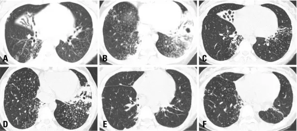
A computerized tomography (CT) scan of the chest revealed bronchiectasis with a collapse of the right middle lobe and air space consolidation with cavity formation in the left lower lobe. In addition, in the right upper lobe and both lower lobes, there was branching linear opacity, revealing bronchogenic spread (Fig. 1). Laboratory findings included a white blood cell count of 20,500 cells/mm3(segment form, 83.7%), hemoglobin of 11.6 g/dL, and platelet cell count of 241,000 cells/mm3. Arterial blood gas analysis performed while breathing without oxygen supplements revealed a pH of 7.39, partial pressure of oxygen of 82.2 mmHg, partial pressure of carbon dioxide of 37.8 mmHg, and oxygen saturation of 95.9%. Hepatic function was normal, except for a mildly elevated aspartate aminotrans- ferase (AST) level of 62 U/L. Renal function was normal, and an human immunodeficiency virus (HIV) test was negative. Acid-fast staining of her sputa revealed numerous bacilli (1-9/10 HPF).
Assuming that she had drug-resistant pulmonary TB, we started a regimen that included isoniazid, ethambutol, levofloxacin, prothionamide, pyrazinamide, and p-amino- salicylic acid. We did not use rifampicin or aminoglyco- side because of previous adverse effects. P-aminosalicylic acid was also discontinued within 2 weeks due to abdominal Seung Heon Lee, et al.
Yonsei Med J http://www.eymj.org Volume 51 Number 1 January 2010 142
CASE REPORT
Fig. 1. High-resolution computed tomography (HRCT) on admission showed the collapse with bronchiectasis of right middle lobe, cavitary lesion of left lower lobe, and multiple branching linear opacity on both lower lobes (A and B). Follow-up HRCT performed one year following the medical treatment revealed minimal improvement (C and D). No newly developed lesion was observed on HRCT taken eight months after bilateral lung resection (E and F).
A B C
D E F
pain. Colonies of mycobacteria were identified in her sputa after 40 days of culture in the Lowenstein-Jensen medium.
These bacilli were proved to be NTM, instead of M. tuber- culosis complex, by a Gen-Probe test (Gen-Probe Inc., San Diego, CA, USA). These mycobacteria were identified as M. intracellulare by a previously reported polymerase chain reaction-restriction fragment length polymorphism (PCR-RFLP) method using cultured mycobacterial colonies at the Korean Institute of Tuberculosis.10Under the diag- nosis of M. intracellulare lung disease, the regimen was changed to clarithromycin, ethambutol, and moxifloxacin.
Despite the regular intake of medication, her symptoms did not improve, and the results of smears for acid-fast bacilli were repeatedly positive (1-9/100 HPF). After 8 months of medication, M. abscessus was isolated instead of M. intracellulare (Table 1). A chest CT performed 1 year after the treatment began revealed minimal improve- ment (Fig. 1). Amikacin was added to the regimen after careful evaluation of her hearing function. However, she refused to try cefoxitin and imipenem because of possible leukopenia and high cost. We performed a bronchoscopic lavage to identify causative mycobacteria, and M. abscessus was isolated from the cultures of lavaged fluid from the right middle and left lower lobes.
Considering the poor symptomatic response to anti- mycobacterial medications and the persistence of a cavi- tary lesion, we decided to conduct a surgical resection. She
underwent a video-assisted thoracoscopic right middle lobe lobectomy, right lower lobe wedge resection, and right upper lobe wedge resection. She was discharged 6 days after the operation without postoperative complication.
On the second operation, which was conducted 1 month later, a video-assisted thoracoscopic left lower lobe basal segmentectomy and left upper lobe wedge resection were performed. She was discharged 11 days after the operation without postoperative complication. After the second operation, negative sputum conversion was achieved. At 11 months after the operation, the results of the smears for acid-fast bacilli were negative, and the amount of sputum had decreased. She was being kept on chemotherapy regimens with clarithromycin, ethambutol, and moxifloxa- cin without complaining of any respiratory symptoms (Table 1). Amikacin was discontinued after 12 months because of increasing tinnitus. A CT of the chest performed 9 months after the second operation revealed no new lesions (Fig. 1).
Approximately 40% of patients with M. abscessus lung disease have an underlying medical condition, such as previous mycobacterial infections, cystic fibrosis, lipoid pneumonia, lung transplantation, achalasia, or other Bilateral Resection for M. Abscessus Lung Disease
Yonsei Med J http://www.eymj.org Volume 51 Number 1 January 2010 143
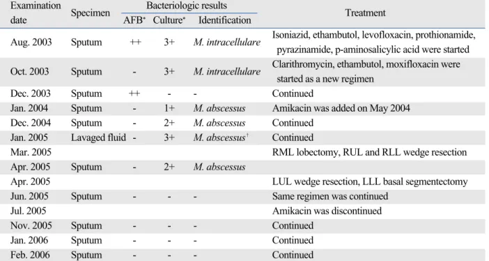
Table 1. Sequential Results of Acid Fast Smear, Mycobacterial Culture, and Identification of Sputa or Lavaged Fluid Examination
Specimen Bacteriologic results
Treatment
date AFB* Culture* Identification
Aug. 2003 Sputum ++ 3+ M. intracellulare Isoniazid, ethambutol, levofloxacin, prothionamide, pyrazinamide, p-aminosalicylic acid were started Oct. 2003 Sputum - 3+ M. intracellulare Clarithromycin, ethambutol, moxifloxacin were
started as a new regimen
Dec. 2003 Sputum ++ - - Continued
Jan. 2004 Sputum - 1+ M. abscessus Amikacin was added on May 2004
Dec. 2004 Sputum - 2+ M. abscessus Continued
Jan. 2005 Lavaged fluid - 3+ M. abscessus� Continued
Mar. 2005 RML lobectomy, RUL and RLL wedge resection
Apr. 2005 Sputum - 2+ M. abscessus
Apr. 2005 LUL wedge resection, LLL basal segmentectomy
Jun. 2005 Sputum - - - Same regimen was continued
Jul. 2005 Amikacin was discontinued
Nov. 2005 Sputum - - - Continued
Jan. 2006 Sputum - - - Continued
Feb. 2006 Sputum - - - Continued
RML, right middle lobe; RUL, right upper lobe; RLL, right lower lobe; LUL, left upper lobe; LLL, left lower lobe.
*Quantitation scale for Acid-Fast Bacillus smears and mycobacterial growth on agar plates followed diagnostic standards of American Thoracic Society.13
�M. abscessus was isolated from lavaged fluids right middle lobe and left lower lobe.
DISCUSSION
Seung Heon Lee, et al.
Yonsei Med J http://www.eymj.org Volume 51 Number 1 January 2010 144
conditions associated with recurrent vomiting.4,5,8These patients often develop the disease at a younger age com- pared with patients without an underlying illness. In patients without associated medical conditions, the lung disease progresses very slowly, and the chest radiograph usually shows multilobar, patchy, reticulonodular or mixed inter- stitial-alveolar infiltrates with an upper lobe predominance.1
This patient was initially diagnosed as having M. intra- cellulare pulmonary disease; however, M. abscessus was isolated instead after the 8-month anti-mycobacterial treat- ment. Mycobacterium abscessus likely caused the patient’s lung disease because it was isolated from the lavaged fluid from both lungs. In fact, alternative isolations of M. abs- cessus and M. intracellulare have been reported to occur in as many as 15% of patients with M. avium complex pul- monary diseases and 31% of patients with M. abscessus pulmonary disease.8,11Clinicians should carefully decide which organism needs to be the target for treatment based on serial isolations of NTM.
The reported success rate of medical treatment in patients with M. abscessus pulmonary disease has been low.
Griffith, et al.8reported that only 10 of the 119 patients with M. abscessus were cured. Among these 10 patients, seven received antibiotic treatment followed by surgical resection of localized lesions, whereas only three were successfully treated with antibiotics alone.8After the introduction of macrolides, the prognosis of medical treatment in patients with M. abscessus improved. In two Korean case series, nine out of 16 patients and five out of nine patients with M.
abscessus were cured with medical treatment alone.6,7 Although surgical resection for localized mycobacterial disease can be associated with significant morbidity and mortality12and the history of a underlying previous lung disease can limit the surgical approach,1surgical resection can be used in select patients with localized M. abscessus lung disease. As far as we know, the patient in this report was the first case to undergo bilateral sequential lung resection for the treatment of M. abscessus lung disease.
The fact that sputum conversion was obtained only after the second resection underscores the necessity for the bilateral resection in this patient. Based on our experience
with this patient, we suggest that, if medical treatment fails, resection of a diseased lobe should be considered even in patients with bilateral lesions.
1. American Thoracic Society. Diagnosis and treatment of disease caused by nontuberculous mycobacteria. Am J Respir Crit Care Med 1997;156(2 Pt 2):S1-25.
2. British Thoracic Society. Management of opportunist mycobac- terial infections: Joint Tuberculosis Committee Guidelines 1999.
Thorax 2000;55:210-8.
3. Yim JJ, Park YK, Lew WJ, Bai GH, Han SK, Shim YS. Myco- bacterium kansasii pulmonary diseases in Korea. J Korean Med Sci 2005;20:957-60.
4. Koh WJ, Kwon OJ, Lee KS. Diagnosis and treatment of nontu- berculous mycobacterial pulmonary diseases: a Korean perspec- tive. J Korean Med Sci 2005;20:913-25.
5. Daley CL, Griffith DE. Pulmonary disease caused by rapidly growing mycobacteria. Clin Chest Med 2002;23:623-32.
6. Kim EK, Shim TS, Lim CM, Lee SD, Koh YS, Kim WS. Clinical manifestation of pulmonary infection due to rapidly growing nontuberculous mycobacteria. Tuberc Respir Dis 2003;54:283-94.
7. Koh WJ, Kwon OJ, Suh GY, Chung MP, Kim H, Lee NY, et al.
Treatment outcome of Mycobacterium abscessus pulmonary disease. Tuberc Respir Dis 2003;55 Suppl 2:107.
8. Griffith DE, Girard WM, Wallace RJ Jr. Clinical features of pulmonary disease caused by rapidly growing mycobacteria. An analysis of 154 patients. Am Rev Respir Dis 1993;147:1271-8.
9. Kwon YS, Kwon OJ, Lee NY, Han J, Lee KS, Shim YM. Suc- cessful treatment of Mycobacterium abscessus lung disease with pneumonectomy combined with antibiotic therapy: a case report.
Korean J Med 2005;69:424-7.
10. Lee H, Park HJ, Cho SN, Bai GH, Kim SJ. Species identification of mycobacteria by PCR-restriction fragment length polymor- phism of the rpoB gene. J Clin Microbiol 2000;38:2966-71.
11. Wallace RJ Jr, Zhang Y, Brown BA, Dawson D, Murphy DT, Wilson R, et al. Polyclonal Mycobacterium avium complex infections in patients with nodular bronchiectasis. Am J Respir Crit Care Med 1998;158:1235-44.
12. Pomerantz M, Madsen L, Goble M, Iseman M. Surgical manage- ment of resistant mycobacterial tuberculosis and other mycobac- terial pulmonary infections. Ann Thorac Surg 1991;52:1108-11.
13. American Thoracic Society. Diagnostic Standards and Classi- fication of Tuberculosis in Adults and Children. Am J Respir Crit Care Med 2000;156(4 Pt 1):1376-95.