대한장애인치과학회지 7(2) 2011
- 103 -
Ⅰ. Introduction
Down syndrome(DS) is a consequence of trisomy 21 and the most common congenital chromosomal abnormality with a prevalence of approximately 8.5- 10 cases/10000 live births1,2). Acute lymphoblastic leukemia(ALL) is the most common childhood malig- nancy and children with DS have a markedly in-
creased risk of developing leukemia when compared with the general population, such that DS is recog- nized as the most common risk factor for the devel- opment of both ALL and acute myeloid leukemia(AML)3). The association of DS and leukemia was recognized quite early, with the first published series being reported over 75 years ago4). This initial observation has been confirmed and re- fined, and it is now generally held that children with DS have a 10- to 20-fold increased risk of developing leukemia when compared with their non-DS counter- parts5,6).
◆ 증 례
Dental treatment of a Down syndrome patient with acute lymphoblastic leukemia : a case report
Jung-Woo Kim, Chong-Chul Kim, Sang-Hoon Lee, Ki-Taeg Jang, Jung-Wook Kim, Young-Jae Kim, Teo-Jeon Shin, Hong-Keun Hyun
Department of Pediatric Dentistry, School of Dentistry, Seoul National University
급성 림프구성 백혈병을 앓고 있는 Down syndrome 환자의 치과치료: 증례보고
김정우∙김종철∙이상훈∙장기택∙김정욱∙김영재∙신터전∙현홍근 서울대학교 치의학대학원 소아치과학교실
다운 증후군(Down syndrome, Trisomy 21)은 가장 흔하게 나타나는 증후군으로 대략 1/1000의 확률로 태어난다.
세 개의 유전적 유형이 있지만, 95% 정도가 3개의 21번 염색체를 갖는 비분리 염색체 유형이다. 주된 구강 증상으로는 거대설, 균열설, 과잉치, 결손치, 왜소치, 유치와 영구치의 맹출지연 그리고 이에 따른 부정교합 등이 있다. 75%의 환 자에서는 어린 나이에 치주질환에 이환되기 쉬우며, 치아우식증에는 낮은 이환율을 보인다는 보고가 있다.
급성 림프구성 백혈병(Acute Lymphoblastic Leukemia)은 주로 어린이에서 나타나며, 특히 다운 증후군 환자에서 의 발병율은 정상인에 비해 20배 높다. 치은비대와 출혈의 양상이 주로 나타나며, 간혹 상악골과 구개골에 종괴가 관찰 되기도 한다.
본 증례에서는 서울대학교치과병원 소아치과에 내원한 환자 중 급성림프구성 백혈병을 앓고 있는 다운 증후군 환아 가 있어 이를 보고하고자 한다.
Key words :다운 증후군, 급성 림프구성 백혈병, 선천적 결손치, 치은 염증 Abstract
교신저자: 현 홍 근
110-768 서울특별시 종로구 창경궁로 62-1 서울대학교 치과병원 소아치과
Tel: 02-2072-0112 Fax: 02-744-3599 E-mail: hege1@snu.ac.kr
원고접수일: 2011.09.09 / 원고최종수정일: 2011.10.17 / 원고채택일: 2011.12.06
Korean Association for Disability and Oral Health 7(2) 2011
- 104 - Common oral symptoms of ALL-DS patients in- clude macroglossia, fissured tongue, supernumerary teeth, congenital missing teeth, microdontia, delayed eruption, gingival enlargement and bleeding.
Ⅱ. Case report
A boy aged 7 years and 1 month was referred from a medical center to the Seoul National University dental hospital department of pediatric dentistry.
This Down syndrome patient was diagnosed with ALL and wanted general dental examination and treatment. He was on multiple ALL medication (Prednisone, Vincristine and Methotrexate).
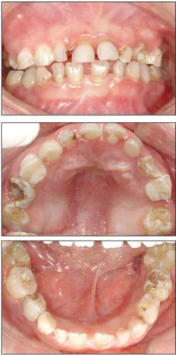
Intraoral and radiographic examination revealed poor oral hygiene, multiple severe dental caries, one con- genital missing tooth(upper left second primary mo- lar) and generalized gingival enlargement(Fig. 1).
Dental treatment was carried out under general anesthesia mainly due to the lack of patient coopera- tion. Extraction of multiple teeth(upper right second primary molar, upper left primary canine, lower left second primary molar and lower right first primary molar) with severe caries was carried out.
Pulpectomy of the upper right first primary molar, upper left first primary molar, lower left primary ca- nine, lower left first primary molar and lower right second primary molar was performed and restored with stainless steel crown. Four band and loop space maintainers were delivered in order to preserve the open spaces(Fig. 2). 3-month follow-up visit was car- ried out for 7 years and the patient is now 14 years old. Peg lateralis, two congenital missing second pre- molars and slight gingival enlargement can be seen(Fig. 3). Oral hygiene and patient’s cooperation has improved dramatically.
Fig. 1.Initial intraoral photo before treatment. (Age: 7 years and 1 month)
Fig. 2. Intraoral photo after caries treatment under general anesthesia. (Age: 7 years and 1 month)
대한장애인치과학회지 7(2) 2011
- 105 -
Ⅲ. Discussion
The basis for the increased occurrence of ALL in children with DS is not known. Extrinsic factors thought to play a role in the development of child- hood leukemia include exposures to radiation, med- ications infection, diet and carcinogens7). However, epidemiologic studies to date have provided no in- sights unique to the DS population8). The presence of an additional chromosome 21 is the underlying basis for the increased risk of ALL in DS. A role of trisomy 21 as a causative factor in the increased risk of ALL in DS patients is supported by the observation that abnormalities of chromosome 21 are the most com- mon acquired numerical abnormality in ALL9).
Leukemic patients are prone to develop gingival enlargement, ulceration, and oral infection. Localized or generalized gingival enlargement is caused by in-
flammation and infiltration of atypical and immature white blood cells. The gingival is boggy and bleeds easily, and multiple tooth sites are typically affected.
Generalized gingival enlargement is more common and is particularly prevalent when oral hygiene is poor. The combination of poor oral hygiene and gingi- val enlargement contributes to gingival bleeding and fetor oris. Plaque control measures, chlorhexidine, and chemotherapy promote resolution of the condition.
Supportive care must be maintained from an oral perspective because many of these patients experi- ence infections of the oral mucosa during the course of their disease. Optimal oral hygiene should be en- couraged, and aggressive investigation of any oral complaint should be performed as soon as possible to prevent potentially serious oral infectious complica- tions. It is more important to prevent than to treat mainly because treatment is usually carried out un- Fig. 3.Facial, intraoral photo and panoramic radiograph after 7 years of follow-up. (Age: 14 years and 1 month)
Korean Association for Disability and Oral Health 7(2) 2011
- 106 - der general anesthesia due to lack of patient coopera- tion and this is not easy for both the patient and parent. The role of both parent and caregiver is very important because most DS patients with ALL can- not solely perform the basic actions in managing oral health. The importance of toothbrushing, routine dental appointments and interest on the patient’s oral hygiene should be instructed to the caregiver.
Professional fluoride application, oral prophylaxis and preventive care will result in an optimal oral hygiene.
References
1. Edmonds LD, James LM: Temporal trends in the birth prevalence of selected congenital malforma- tions in the birth defects monitoring program/commission on professional and hospital activities. Teratology 48:647-649, 1993.
2. Cocchi G, Gualdi S, Bower C et al.:International trends of Down syndrome 1993-2004: births in relation to maternal age and terminations of pregnancies. Clin and Molec Teratology 88:474- 479, 2010.
3. Lange B: The management of neoplastic disor-
ders of haematopoiesis in children with Down’s syndrome. Brit J of Haematol, 110:512-524, 2000.
4. Krivit W, Good RA: Simultaneous occurrence of mongolism and leukemia; report of a nationwide survey. AMA J Dis Child, 94:289-293, 1957.
5. Stewart A, Webb J, Hewitt D: A survey of child- hood malignancies. British Med J, 30:1495- 1508, 1958.
6. Rajante J, Siimes MA: Long-term prognosis of children with Down’s syndrome and leukemia: a 34-year nation-wide experience. J Intell Disability Res, 47:617-621, 2003.
7. Ross JA, Davies SM, Potter JD et al.:
Epidemiology of childhood leukemia, with a focus on infants. Epidemiologic Rev, 16:243-272, 1994.
8. Ross JA, Spector LG, Robison LL et al.:
Epidemiology of leukemia in children with Down syndrome. Pediatr Blood & Cancer, 44:8-12, 2005.
9. Berger R: Acute lymphoblastic leukemia and chromosome 21. Cancer Gene and Cytogene, 94:8-12, 1997.