Ⅰ. Introduction
Moebius syndrome is a congenital neurological dis- ease characterized by partial, unilateral or bilateral palsy of the facial and abducens cranial nerves. This syndrome was defined by Paul Moebius in 1888. It is an extremely rare disorder and estimated to be pre- sent in 0.002% of births
1).
The etiology remains unclear, but genetic and envi- ronmental factors such as drugs use during pregnan-
cy are possible etiological hypotheses
2).
The typical characteristics of Moebius syndrome in- clude impairment of eye movement and loss of facial expression. Also, this syndrome often involves facial deformities and anomalies of the extremities and brachial musculature
3).
From a dental standpoint, the most frequent find- ings include microstomia, micrognathia, cleft palate, high arched palate, tongue deformity, bifid uvula, multiple congenitally missing teeth, hypoplasia of the teeth, nursing bottle caries, hypoplastic upper lip, and lack of lip seal
2,4-8). These manifestations can cause many dental problems.
The purpose of this case report is to present the general characteristics, oral manifestations, and den-
◆ 증 례
Oral findings and dental management of a patient with Moebius syndrome: a case report
Eunkyoung Lee, Youngjin Kim, Hyunjung Kim, Soonhyeun Nam*
Department of Pediatric Dentistry, School of Dentistry, Kyungpook National University, Daegu, Korea
Moebius syndrome is a rare, congenital neurological disease involving facial paralysis and limitation of eye movements. It results from maldevelopment of the sixth and seventh cranial nerves. Dental features of this syndrome include micrognathia, microstomia, tongue deformity, cleft palate, hypoplasia of the teeth, and congenital missing teeth.
A 7-year-old female with Moebius syndrome was referred from a local dental clinic for caries treatment.
She presented with facial paralysis and microstomia. Oral findings included multiple caries with enamel hypoplasia, congenital missing teeth, and tongue deformity. Dental treatments including restorative and preventive procedures were performed.
Oral findings and management aspects of Moebius syndrome for this case are discussed. Early evaluation and multidisciplinary care are needed for children with Moebius syndrome. [ J Korean Dis Oral Health Vol.10, No.2: 101-105, December 2014]
Key words : Moebius syndrome, Palsy of facial nerve, Palsy of abducens nerve, Oral findings Abstract
Corresponding author : Soonhyeun Nam
2177 Dalgubeol-daero, Jung-gu, Daegu, 700-412, Korea
Department of Pediatric Dentistry, School of Dentistry, Kyungpook National University
Tel: +82-53-600-7211, Fax: +82-53-426-6608
E-mail: [email protected]
tal management of a child diagnosed with Moebius syndrome.
Ⅱ. Case Report
A 7-year-old female patient visited our clinic for treatment of multiple caries. The patient was born after 36 weeks’gestation with 2.7 kg weight by ce- sarean delivery. At birth, general hypotonia, small mandible, and cleft palate were observed. She was fed by gavage due to a sucking problem in the neona- tal period. She was diagnosed Moebius syndrome with no limb malformations and normal intelligence.
She had plastic surgery to treat cleft palate at ages 3 years. Because of chronic otitis media, she is followed periodically by pediatricians.
In extraoral examination, the patient exhibited loss of facial expression (mask-like face) and incapacity to move the eyes from side to side. Many features of Moebius syndrome - including micrognathia, lack of lip seal, everted and thin upper lip, broad and flat nasal bridge, thin eyebrows, protruding ears, and in- distinct philtrum - were present (Fig. 1).
Oral findings included microstomia, narrow arch, limited mouth opening, restricted tongue movement with short lingual frenum, bifid tongue, and dry lips.
She also had enamel hypoplasia in upper incisors and
all permanent first molars, and poor oral hygiene with multiple caries and several residual roots. And anterior crossbite (functional classⅢ malocclusion) related with early loss of maxillary primary incisors was observed (Fig. 2).
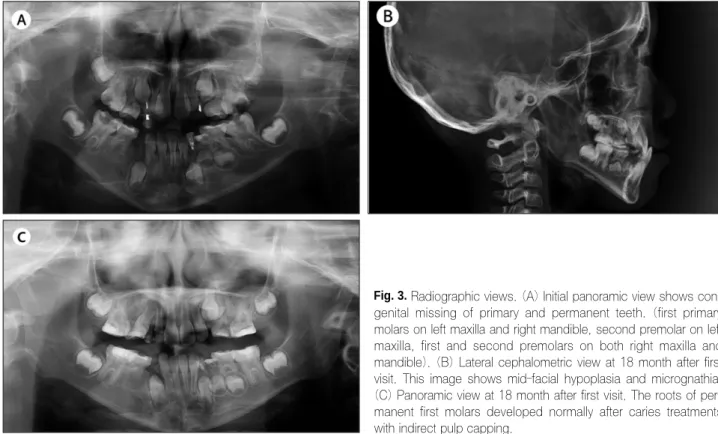
In radiographic views, congenitally missing teeth including the first primary molars on the left maxilla and right mandible, the first and second premolars on the right maxilla and mandible, and the second premolar on the left maxilla were observed (Fig. 3A).
Lateral cephalogram showed micrognathia and slight midfacial hypoplasia (Fig. 3B).
Treatment of caries, tongue-tie operation, preven- tive procedure such as fluoride application and fis- sure sealing were planned. Especially, preservation of primary teeth without permanent successors would be important for the patient. And removable appli- ance would be helpful for anterior crossbite.
Indirect pulp capping of all permanent first molars
with calcium hydroxide and glass ionomer restoration
was performed. Eighteen months later, normal root
development of these teeth was observed. But sec-
ondary caries of both maxillary first molar were pre-
sented, so additional treatment will be needed (Fig
3C). Now, the patient is checked regularly with oral
prophylaxis, and other remaining treatments will be
performed .
Fig. 2. (A, B, C) Initial intraoral photographs of a patient aged 7 years. These images show microstomia, narrow arch, limitation of
mouth opening, dry lips, bifid tongue, restriction of tongue movement (failure of upward movement), dental caries of all first permanent molar with hypoplastic enamel and early exfoliation of upper primary incisors. (D, E, F) Intraoral photographs at 18 months after first vis- it. Anterior crossbite, enamel hypoplasia of upper incisors, dental caries of left mandibular lateral incisor and ectopic eruption of right upper canine are observed.Fig. 3. Radiographic views. (A) Initial panoramic view shows con-
genital missing of primary and permanent teeth. (first primary molars on left maxilla and right mandible, second premolar on left maxilla, first and second premolars on both right maxilla and mandible). (B) Lateral cephalometric view at 18 month after first visit. This image shows mid-facial hypoplasia and micrognathia.(C) Panoramic view at 18 month after first visit. The roots of per- manent first molars developed normally after caries treatments with indirect pulp capping.