https://doi.org/10.5468/ogs.2020.63.3.278 pISSN 2287-8572 · eISSN 2287-8580
Introduction
Congenital heart defects (CHDs) are the most common fe- tal congenital abnormality, independently or in association with other fetal anatomical defects. Prenatal identification of CHDs is important because these account for 30% to 50%
of cases of infant death [1]. An optimal gestational age for screening for fetal cardiac anomalies is between 18 and 22 weeks of gestation [2]. However, early detection of CHDs in the first trimester is possible owing to the recent develop- ment of high-resolution ultrasonography [3]. In a 2006 sys- tematic review of the first-trimester ultrasound examination for detecting major CHDs using transabdominal or trans-
Efficacy of fetal cardiac axis evaluation in the first
trimester as a screening tool for congenital heart defect or aneuploidy
Youn-Joon Jung, MD
*, Bo-Ra Lee, MD
*, Gwang Jun Kim, MD, PhD
Department of Obstetrics and Gynecology, School of Medicine, Chung-Ang University, Seoul, Korea
Objective
To prove the efficacy of determining the abnormal fetal cardiac axis for screening congenital heart defects (CHDs) and predicting fetal aneuploidy at 11.0 to 13.6 weeks of pregnancy.
Methods
This retrospective study was performed at a single high-risk pregnancy center. The fetal cardiac axis was evaluated between 11.0 and 13.6 weeks of gestation in 142 fetuses. The cardiac axis in a 4-chamber view was measured as the angle between the line tracing the long axis of the heart and the line bisecting the thorax in the anteroposterior direction. A CHD was confirmed based on the second- to third-trimester fetal status or postnatal imaging. Aneuploidy was diagnosed using chorionic villus sampling, amniocentesis, or genetic testing after birth. Fisher’s exact test was performed to assess the association between the fetal cardiac axis and the abnormal fetal status. A 2-way contingence table analysis was performed to confirm the efficacy of the fetal cardiac axis as a screening tool.
Results
Among the 142 fetuses, 10 had a CHD while 17 had aneuploidy. The abnormal fetal cardiac axis was significantly associated with CHDs (P=0.013) and aneuploidy (P=0.010). None of the fetuses with CHDs or aneuploidy had an isolated abnormal cardiac axis alone without other sonographic findings. The sensitivity of the fetal cardiac axis was 50.0% for CHDs and 41.2% for aneuploidy.
Conclusion
The fetal cardiac axis can be an additional helpful tool for prenatal screening of CHDs and aneuploidy in the first trimester.
Keywords: Fetal heart; Axis; Congenital heart defect; Aneuploidy; First trimester
Received: 2019.06.21. Revised: 2019.11.07. Accepted: 2019.11.12.
Corresponding author: Gwang-Jun Kim, MD, PhD
Department of Obstetrics and Gynecology, School of Medicine, Chung-Ang University, 84 Heukseouk-ro, Dongjak-gu, Seoul 06974, Korea
E-mail: [email protected]
https://orcid.org/0000-0003-0232-0166
*These two authors contributed equally to this paper.
Articles published in Obstet Gynecol Sci are open-access, distributed under the terms of the Creative Commons Attribution Non-Commercial License (http://creativecommons.
org/licenses/by-nc/3.0/) which permits unrestricted non-commercial use, distribution, and reproduction in any medium, provided the original work is properly cited.
Copyright © 2020 Korean Society of Obstetrics and Gynecology
vaginal ultrasonography, the pooled sensitivity and speci- ficity were 85% and 99%, respectively [4]. Advantages of early detection of CHDs include earlier reassurance to high- risk women with normal sonographic findings, possibility of scheduling additional tests well before the limits for safe pregnancy termination, and provision of information for an appropriate place of birth and delivery methods depending on the severity of CHD [5].
Several methods have been suggested for screening of CHDs in early gestation, e.g., increased nuchal translucency (NT), abnormal blood flow in the ductus venosus (DV), and tricuspid regurgitation (TR) [6-11]. Based on previous stud- ies, the abnormal cardiac axis has also been recognized as a screening marker for CHDs. Sinkovskaya et al. [12] reported that the efficacy of the cardiac axis measurement in the de- tection of major CHDs was significantly better than that of an NT scan, a DV blood flow scan, or tests for detecting TR, used alone or in combination.
CHDs are strongly associated with chromosomal anomalies and genetic syndromes. Identification of cardiac malforma- tions on prenatal sonography substantially increases the risk for chromosomal abnormalities, with a frequency of aneu- ploidy reported to be as high as 22–32% [13,14]. Increased NT, abnormal blood flow in DV, and TR, which are used in detection of CHDs, are also used for the screening of fetal aneuploidy in the first trimester [15-18].
We hypothesized that the abnormal fetal cardiac axis would be useful in the detection of fetal aneuploidy as well as fetal CHD during early gestation. The aim of this study was to investigate the effect of determining the abnormal cardiac axis for screening fetal CHD and predicting fetal an- euploidy at 11.0 to 13.6 weeks of pregnancy in the Korean population.
Materials and methods
From January 2015 to October 2018, 221 pregnant women in early gestation (11.0 and 14.6 weeks of gestation) who visited the Prenatal Diagnosis Clinic of the Chung-Ang Uni- versity Hospital were enrolled in this study. One ultrasound image per woman was retrieved from the database. Images of 142 women were obtained after excluding those without precise 4-chamber views or neonatal information because of loss to follow-up. Clinical data and pregnancy outcomes
were also collected from the database. Confirmation of CHD was based on the second- to third-trimester fetal status, postnatal imaging findings, or both. Aneuploidy of the fe- tuses was diagnosed using an invasive test, such as chorionic villus sampling and amniocentesis, during pregnancy or ge- netic testing after birth. A total of 52 chromosomal analyses were performed. In the first trimester, 47 fetuses were ex- amined using chorionic villus sampling. Amniocentesis was performed on 4 fetuses in the second trimester. One fetus underwent genetic testing after birth. Invasive genetic tests were performed when the combined first trimester screening test or quad screening test resulted in a high risk, or when major fetal anomalies, including fetal hydrops, were observed on ultrasound. Chromosomal analyses were carried out using fluorescence in situ hybridization or classic karyotyping by long-term cell culture.
This study was undertaken by experts in the field of early fetal imaging. Target ultrasonography of the basic fetal anatomy including the 4-chamber view was performed routinely during early gestation. The 4-chamber view was obtained from an axial sonographic image with definite visualization of the cardiac chamber and 1 complete rib on each side of the fetal lateral chest wall. The cardiac axis was
Fig. 1. A schematic diagram of the cardiac axis measurement. a, the line bisecting the thorax in the anteroposterior direction; b, the line tracing the long axis of the heart; L, left aspect of the fetus; R, right aspect of the fetus; P, posterior aspect of the fetus.
L
a b
R P
measured as the angle between the line that traces the long axis of the heart and the line that bisects the thorax in the anteroposterior direction (Fig. 1). The normal fetal cardiac axis was defined as a 90% confidence interval (CI) of the cardiac axis in 121 fetuses without CHD or aneuploidy. Us- ing the 1.65 standard deviation, range, and mean for fetuses without CHD or aneuploidy, the cardiac axis was considered as abnormal when the measured value was above the 95th percentile (left deviation) or below the 5th percentile (right deviation) (Fig. 2). The normal fetal cardiac axis range was calculated by rounding off the obtained value to the nearest whole number. Other first-trimester ultrasound findings, in- cluding the measurement of NT, blood flow in DV, presence of TR, and measurement of the nasal bone (NB) length, were also recorded. NT greater than 95th percentile or 3.0 mm was considered abnormal [19]. Reversal or absence of blood flow in DV was regarded as abnormal [16]. Absence of NB or an NB length less than the 2.5th percentile was considered abnormal [20]. Pregnancy-associated serum plasma protein A (PAPP-A) values were obtained from medical records to evaluate the screening performance for aneuploidy in combi- nation with the abnormal fetal cardiac axis. Serum values of PAPP-A lower than 1.0 multiple of median were considered decreased.
The primary goal of this study was to investigate the as- sociation between the fetal cardiac axis in the first trimester and CHD and aneuploidy. The secondary goal of this study
was to analyze the utility of the fetal cardiac axis measure- ment as a predictor of CHD and aneuploidy. Student’s t-test and Fisher’s exact test were performed to compare differenc- es in clinical data between the study and control groups. The 2-way contingence table analysis was performed to confirm the efficacy of the fetal cardiac axis as screening tool. Bayes’
theorem was used to compute the positive and negative predictive values for prevalence. Statistical analyses were per- formed using SPSS 25.0 (IBM Inc., Chicago, IL, USA). P<0.05 was considered statistically significant.
Results
Table 1 demonstrates the demographic and clinical char- acteristics of fetuses. Based on the fetal cardiac axis in the first trimester. In our collected data, 121 fetuses were not diagnosed with CHD or aneuploidy. In the group of normal fetuses, the cardiac axis ranged from 14.80° to 75.70° (mean 47.15°±12.32°; 90% CI, 26.82–67.48). The normal fetal car- diac axis was defined as a value between 27° and 67°. Based on the aforementioned definition of the normal cardiac axis, 24 fetuses had an abnormal cardiac axis. The incidence of the abnormal cardiac axis was significantly associated with increased NT, CHD, and aneuploidy. Decreased NB, abnormal Doppler finding on DV, and TR showed tendencies to occur more frequently in fetuses with an abnormal cardiac axis,
Fig. 2. Normal and abnormal fetal cardiac axes of fetuses in the first trimester observed using ultrasonography. Dotted lines are the lines bisecting the thorax in the anteroposterior direction. Full lines are the lines tracing the long axis of the heart. (A) Normal: 65.3° at 11.0 weeks. The fetus was delivered through a cesarean section at 39.0 weeks of gestation. (B) Left deviation: 75.4° at 12.4 weeks. Atrioven- tricular septal defect observed using fetal echocardiography. Trisomy 13: Fetal death in utero was diagnosed at 14.5 weeks of gestation.
(C) Right deviation: 10.1° at 12.2 weeks. Ventricular septal defect, aortic stenosis, mitral atresia, and left ventricular hypoplasia observed using fetal echocardiography. Fetal death in utero was diagnosed 13.4 weeks of gestation. L, left aspect of fetus; R, right aspect of fetus; P, posterior aspect of fetus.
A B C
but without statistical significance. Aneuploidy without CHD was more common in fetuses with an abnormal cardiac axis (12.50%) than in fetuses with a normal cardiac axis (7.56%), but without statistical significance (P=0.427). Among 142 registered fetuses, 10 fetuses were diagnosed with CHD and 17 with aneuploidy. An abnormal cardiac axis was found in 5
of 10 fetuses with CHD and 7 of 17 fetuses with aneuploidy.
Incidences of CHD and aneuploidy were associated with en- larged NT (P=0.002, CHD; P<0.001, aneuploidy), abnormal DV (P<0.001, CHD; P=0.001, aneuploidy) and TR (P<0.001, CHD; P=0.003, aneuploidy). Decreased NB was more com- mon in fetuses with aneuploidy, but without statistical sig-
Table 1. Demographic and clinical characteristics of fetuses according to congenital heart defects (CHDs)
Parameter Normal cardiac axis Abnormal cardiac axis P-value
No. of fetuses 118 24 -
Maternal age (yr) 32.67±3.58 (23–42) 33.33±3.85 (25–40) 0.442
CRL (mm) 63.96±8.64 62.81±9.90 0.601
Increased NT 30 (25.21) 14 (58.33) 0.003
Decreased NB 16 (13.45) 7 (29.17) 0.054
Abnormal DV 12 (2.52) 3 (12.50) 0.709
TR 4 (3.36) 3 (12.50) 0.099
Gestational age (wk)
11.0–11.6 14 (11.76) 6 (25.00) -
12.0–12.6 68 (57.14) 10 (41.67) -
13.0–13.6 36 (30.25) 8 (33.33) -
CHD 5 (4.20) 5 (20.83) 0.013
Aneuploidy 10 (8.40) 7 (29.17) 0.010
Left deviation - 15 (62.50) -
Right deviation - 9 (37.50) -
A normal cardiac axis was defined as that angled at 27–67°. Data are mean±standard deviation (range) or number (%) unless specified other- wise. P-values were calculated by student’s t-test or Fisher’s exact test.
CRL, crown-rump length; NT, nuchal translucency; NB, nasal bone; DV, ductus venosus; TR, tricuspid regurgitation.
Table 2. Type of congenital heart defects (CHDs) and presence of abnormal cardiac axis in 10 fetuses
CHD Abnormal
cardiac axis Other sonographic abnormal findings Aneuploidy
AVSD, PA hypoplasia Y Cerebellar hypoplasia, Blake’s pouch cyst Trisomy 21
TOF Y - N
VSD Y Enlarged fetal bladder Trisomy 21
MS, VSD, hypoplastic aortic arch Y - Turner syndrome
VSD N Fetal hydrops N
VSD N - N
VSD, aortic stenosis N Omphalocele, bilateral kidney agenesis Trisomy 18
AVSD N Omphalocele, right dysplastic kidney Trisomy 13
TS, VSD N - N
VSD, AS, mitral atresia, left ventricular hypoplasia Y Fetal hydrops Turner syndrome
A normal cardiac axis was defined as that angled at 27–67°.
AS, aortic stenosis; AVSD, atrioventricular septal defect; MS, mitral stenosis; PA, pulmonary artery; TOF, tetralogy of Fallot; TS, tricuspid steno- sis; VSD, ventricular septal defect.
nificance (P=0.143). The incidence of CHD was significantly associated with aneuploidy (P<0.001).
Overall, 10 fetuses with CHD were included in our study (Table 2). Five of them had an abnormal cardiac axis and 6 were diagnosed with aneuploidy. The most common CHD, regardless of the type, was ventricular septal defect (VSD).
Table 3 demonstrates the frequency of the abnormal fetal cardiac axis for each type of aneuploidy. The most common manifestation of aneuploidy was Turner syndrome, and 4 fetuses with Turner syndrome showed an abnormal cardiac axis. Trisomy 21 was found in 3 patients, and all of whom had an abnormal cardiac axis.
Table 4 shows the efficacy of the fetal cardiac axis evalua- tion as a screening tool in the first trimester. With regard to CHD, the sensitivity of the fetal cardiac axis evaluation was 50.0% (95% CI, 0.21–0.79), and the specificity was 85.6%
(95% CI, 0.83–0.88). The negative predictive value of the fe-
tal cardiac axis evaluation for CHD was 99.4%. An abnormal cardiac axis in the first trimester increased the risk of CHD by approximately 6 times (odds ratio [OR], 5.95; 95% CI, 1.57–
22.52). With regard to aneuploidy, the sensitivity of the fetal cardiac axis evaluation was 41.2% (95% CI, 0.20–0.64), and the specificity was 86.4% (95% CI, 0.84–0.90). The nega- tive predictive value of the fetal cardiac axis evaluation for aneuploidy was 99.9%. An abnormal cardiac axis in the first trimester increased the risk of aneuploidy by approximately 4.5 times (OR, 4.45, 95% CI, 1.49–13.27).
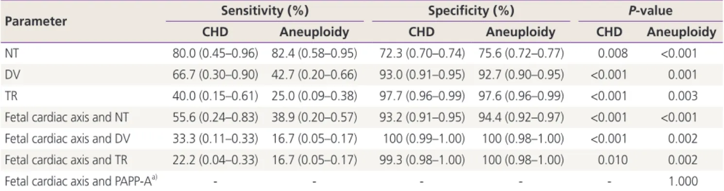
Table 5 shows the efficacy of other variables alone and in combination with the fetal cardiac axis measurement as a screening tool in the first trimester. The sensitivity of NT in screening of CHD and aneuploidy were higher than that of the fetal cardiac axis. DV also had a higher sensitivity than the fetal cardiac axis in CHD and aneuploidy.
Table 3. Frequency of abnormal fetal cardiac axis in each type of aneuploidy
Aneuploidy Number Abnormal cardiac axis (number)
Trisomy 12 1 0
Trisomy 13 3 1
Trisomy 18 3 2
Trisomy 21 3 3
Turner syndrome 7 4
A normal cardiac axis was defined as that angled at 27–67°.
Table 4. The performance of fetal cardiac axis measurement as a screening tool in the first trimester
Parameter Congenital heart
defects Aneuploidy Sensitivity (%) 50.0 (0.21–0.79) 41.2 (0.20–0.64) Specificity (%) 85.6 (0.83–0.88) 86.4 (0.84–0.90) Positive predictive
value (%)
2.8 0.4
Negative predictive
value (%) 99.4 99.9
Odd ratio 5.95 (1.57–22.52) 4.45 (1.49–13.27) Parentheses denote the 95% confidence interval.
Table 5. The performance of each of several independent variables and in combination with fetal cardiac axis measurement as a screen- ing tool in the first trimester
Parameter Sensitivity (%) Specificity (%) P-value
CHD Aneuploidy CHD Aneuploidy CHD Aneuploidy
NT 80.0 (0.45–0.96) 82.4 (0.58–0.95) 72.3 (0.70–0.74) 75.6 (0.72–0.77) 0.008 <0.001 DV 66.7 (0.30–0.90) 42.7 (0.20–0.66) 93.0 (0.91–0.95) 92.7 (0.90–0.95) <0.001 0.001 TR 40.0 (0.15–0.61) 25.0 (0.09–0.38) 97.7 (0.96–0.99) 97.6 (0.96–0.99) <0.001 0.003 Fetal cardiac axis and NT 55.6 (0.24–0.83) 38.9 (0.20–0.57) 93.2 (0.91–0.95) 94.4 (0.92–0.97) <0.001 <0.001 Fetal cardiac axis and DV 33.3 (0.11–0.33) 16.7 (0.05–0.17) 100 (0.99–1.00) 100 (0.98–1.00) <0.001 0.002 Fetal cardiac axis and TR 22.2 (0.04–0.33) 16.7 (0.05–0.17) 99.3 (0.98–1.00) 100 (0.98–1.00) 0.010 0.002
Fetal cardiac axis and PAPP-Aa) - - - 1.000
Parentheses denote the 95% confidence interval. P-values were calculated by student’s t-test or Fisher’s exact test.
CHD, congenital heart defect; NT, nuchal translucency; DV, ductus venosus; TR, tricuspid regurgitation; PAPP-A, pregnancy-associated plasma protein A.
a)Fetal cardiac axis and PAPP-A was used only for screening for aneuploidy.