Address reprint requests to Jung Yeul Kim, MD, PhD
Department of Ophthalmology, Chungnam National University Hospital, #282 Munhwa-ro, Jung-gu, Daejeon 35015, Korea
Tel: 82-42-280-8433, Fax: 82-42-255-3745
Received: 2018. 9. 12 Revised: 2018. 10. 25 Accepted: 2018. 10. 29
Introduction
Macular edema (ME) is one of the most important causes
of decreased visual acuity in retinal vein occlusion [1,2]. It is caused by serous exudation following damage to perifo- veal capillaries distal to the occluded retinal vein. Although
망막분지정맥폐쇄에 의한 황반부종에서 유리체강내 베바시주맙 일회 주사와 연속 주사의 효과 비교
Comparison of Pro Re Nata and Three Loading Injections of Intravitreal Bevacizumab for Macular Edema in Branch Retinal Vein Occlusion
이택훈1,2, 남기엽3,4, 김정열3
Taek Hoon Lee1,2, Ki Yup Nam3,4, Jung Yeul Kim3
1이안과병원, 2충남대학교 대학원 의학과, 3충남대학교 의과대학 안과학교실, 4창원경상대학교병원 안과
1Rhee’s Eye Hospital, Daejeon, Korea
2Graduate School of Medicine, Chungnam National University, Daejeon, Korea
3Department of Ophthalmology, Chungnam National University College of Medicine, Daejeon, Korea
4Department of Ophthalmology, Gyeongsang National University Changwon Hospital, Changwon, Korea
Purpose: To compare pro re nata (PRN) and three scheduled loading injections of intravitreal bevacizumab for macular edema (ME) caused by branch retinal vein occlusion (BRVO).
Methods: We retrospectively analyzed the medical records of 96 patients. We compared a group of patients who underwent treatment with intravitreal bevacizumab PRN with a second group that received three scheduled loading injections at 1 month intervals followed by PRN injections. Changes in visual acuity and central retinal thickness (CRT), and the need for additional injections over a 6 month peri- od were compared.
Results: The improvement in visual acuity and decrease in CRT were greater in the loading injection group, and the decreases in CRT at the 2, 3, and 6 months were statistically significant. The need for additional injections within 3 months after the last injection was signifi- cantly lower in the loading injection group.
Conclusions: Over a 6 month period, the three loading injections seemed to be better than PRN injections for the treatment of ME in BRVO with regard to improvements in visual acuity and ME, reducing the need for additional injections within 3 months after the last injection.
Keywords: Bevacizumab; Intravitreal inejctions; Retinal vein occlusion
it improves spontaneously in some cases, serious damage to blood vessels may provoke a continuous or permanent degenerative change in the retina, making recovery of vi- sual acuity impossible [1,2].Various treatments for ME in retinal vein occlusion have been investigated, and injection of intravitreal anti-vascular endothelial growth factor an- tibodies, such as bevacizumab and ranibizumab, is widely used. Ranibizumab was approved for the treatment of ME in retinal vein occlusion, but due to its high cost, bevacizumab is frequently used in its place as an off-label prescription. Al- though various dosing schedules of bevacizumab have been deemed effective for ME following retinal vein occlusion, an optimal dosing regimen has not been determined. In this study, the authors compared two practical regimens of bev- acizumab dosing for branch retinal vein occlusion (BRVO) patients-pro re nata (PRN, when necessary) injections and three loading injections.
Materials and Methods
We retrospectively analyzed the medical records of 96 pa- tients diagnosed with ME caused by BRVO who were treat- ed with intravitreal injections of bevacizumab (Avastin®, Genentech Inc., South San Francisco, CA, USA) from Sep- tember 2006 to December 2011. Bevacizumab was injected intravitreally when the initial visual acuity was less than or equal to 0.5, or when the central retinal thickness (CRT) was > 300 µm by optical coherence tomography (OCT).
Institutional Review Board (IRB) and ethics committee ap- proval was obtained, and the study adhered to the tenets of the Declaration of Helsinki.
Upon their first visit, all patients received a best-correct- ed visual acuity (BCVA) test, tonometry, slit-lamp exam- ination, fundus examination, fluorescein angiography, and OCT (Stratus OCT3, Carl Zeiss Meditec, Inc., Dublin, CA, USA). Patients with BCVA and OCT results at 1, 2, 3, and 6 months after intravitreal bevacizumab injection were in- cluded in this study. Patients with an initial visual acuity of less than 0.1, follow up period less than 6 months, a history of local laser therapy or steroid injection, an anti-vascular endothelial growth factor antibody (anti-VEGF) injection within 6 months, or a history of vitrectomy, glaucoma, or other causes of decreased visual acuity were excluded from the study. Patients who were did not attend monthly fol-
low-up visits during the 6 months were also excluded.
The subjects were classified into PRN injection and loading injection groups. In the PRN injection group, bevacizumab was injected intravitreally once at diagnosis and additional injections were provided if necessary. In the loading injection group, three injections at monthly intervals were administered and additional injections were provided if necessary. The operator (J.Y.K.) used the PRN injection method in patients seen between September 2006 and July 2009. Thereafter, the three loading injection meth- od was used.
In cases in which ME recurred, visual acuity decreased, or the CRT increased by over 100 μm by OCT, an addition- al injection was administered. Injections were performed after obtaining the approval of patients for the procedure.
The eyelid and the conjunctiva were sterilized with 5%
povidone-iodine solution, and 0.5% proparacaine hydro- chloride (Alcaine®, Alcon, Fort Worth, TX, USA) was administered to induce anesthesia. The eye was widened using an eyelid retractor, and a dose of 1.25 mg in 0.05 mL bevacizumab was injected at the position 4.0 and 3.5 mm away from the limbi of the phakic and pseudophakic eyes, respectively, using a 30-gauge needle.
For analysis, measured visual acuity was converted into logMAR visual acuity and CRT was determined by OCT.
Changes in logMAR visual acuity and decreases in CRT relative to initial measurements were compared between the PRN and loading injection groups at 1, 2, 3, and 6 months after injection. The rates of additional injections needed within 3 months after first injection in the PRN group and third loading injection group (within 3 months after the last injection in each group) were also compared.
A statistical analysis was conducted using IBM SPSS ver.
18.0 (IBM Corp., Armonk, NY, USA). Changes in visual acuity and CRT were compared using independent t-tests, and the proportion of patients that required additional in- jections was compared using a chi-squared test.
Results
The mean age of all 96 subjects was 66.0 (± 9.2) years; 32 were males and 64 were females. The study included 23 patients with diabetes mellitus (DM) and 37 with hypertension. The PRN injection group included 52 subjects, and the remaining
44 subjects were assigned to the loading injection group.
Differences in age, sex, and the prevalence of DM and hy- pertension between the two groups were not significant. The mean logMAR BCVA values at first examination were 0.43 (±
0.25) and 0.45 (± 0.26) in the PRN injection and loading injec- tion groups, respectively. The mean CRT on OCT was 496.1 (± 150.9) and 508.8 (± 123.6) μm, respectively, however, these differences were not statistically significant (Table 1).
The change in visual acuity was investigated by comparing
the acuity during follow-up after intravitreal bevacizumab injection with that measured at the first visit. The changes in visual acuity were -0.24 (± 0.26), -0.24 (± 0.26), -0.25 (± 0.28), and -0.24 (± 0.31) at 1, 2, 3, and 6 months after the first injec- tion, respectively, in the PRN injection group. In the loading injection group, the changes at the same time points were -0.28 (± 0.24), -0.33 (± 0.20), -0.37 (± 0.27), and -0.35 (± 0.34), re- spectively. The loading injection group showed more improve- ment, however, the difference was statistically significant only at the 3- and 6-month follow-up (p = 0.410, 0.348, 0.019, 0.040;
Fig. 1).
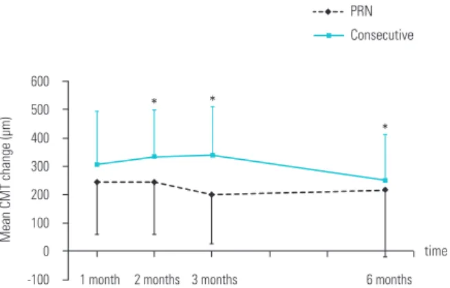
The decreases in the measured CRT during follow-up after the first injection relative to that at the first visit were 202.7 (± 174.6), 203.5 (± 168.7), 182.4 (± 163.1), and 188.6 (± 201.3) μm at 1, 2, 3, and 6 months in the PRN injection group, re- spectively. In the loading injection group, the decreases were 283.1 (± 155.1), 316.3 (± 141.5), 327.9 (± 142.9), and 259.4 (±
132.2) μm, respectively. Decreases in the loading injection group were more marked at all measurement times, and the differences after 2, 3, and 6 months were significant (p = 0.102, 0.001, < 0.001, 0.042; Fig. 2).
The rates of additional injections that were needed within three months after the first injection in the PRN group and third loading injection group were compared; these were 61.5% (32 of 52 eyes) in the PRN injection group and 27.3%
(12 of 44 eyes) in the loading injection group. That is, the proportion of patients without recurrence within 3 months after the last injection was significantly higher in the loading Table 1. Baseline characteristics of two groups
PRN injection (n = 52)
Three consecutive
injections (n = 44)
p-value
Age (years) 65.3 (± 8.2) 66.3 (± 10.1) 0.573*
Sex 0.472†
Male 18 (34.6) 14 (31.8)
Female 34 (65.4) 30 (68.2)
Diabetes mellitus 12 (23.1) 11 (25.0) 0.827†
Hypertension 20 (38.5) 17 (38.6) 0.576†
BCVA at initial visit 0.43 0.45 0.826*
CMT at initial visit (μm) 496.1 508.8 0.649* Values are presented as mean (± standard deviation) or number (%).
PRN = pro re nata; BCVA = best-corrected visual acuity; CMT = central macular thickness.
*Independent t-test; †chi-squared test.
Figure 1. Change in LogMAR BCVA at the monthly follow-up rela- tive to that at the initial visit. Error bars indicate standard deviations.
LogMAR = logarithm of the minimum angle of resolution; BCVA = best-corrected visual acuity; PRN = pro re nata. *p < 0.05, indepen- dent t-test.
0.20
0.00
-0.20
-0.40
-0.60
-0.80
6 months 3 months
2 months 1 month
Mean LogMAR BCVA change
time Consecutive PRN
* *
Figure 2. Reduction in mean CMT at the monthly follow-up relative to that at the initial visit. Error bars indicate standard deviations. CMT
= central macular thickness; PRN = pro re nata. *p < 0.05, indepen- dent t-test.
600 500 400 300 200 100 0
-100 3 months 6 months
* *
*
2 months 1 month
Mean CMT change (µm)
time Consecutive PRN
group (Fig. 3). The rate was significantly lower in the loading injection group (p = 0.001). The mean frequency of injection over 6 months were 2.29 (± 1.13) and 3.43 (± 0.76) times in the PRN and loading injection groups, respectively, and this difference was statistically significant (p < 0.001). No sys- temic or ocular complications was found after intravitreal bevacizumab injection.
Discussion
Retinal vein occlusion is the second most common retinal vascular disease after diabetic retinopathy, and its prevalence has been reported to be 0.3-2% in persons aged over 40 years [3-5].The decrease in visual acuity caused by retinal vein occlusion occurs through the combination of three mecha- nisms [1,2].First, ME develops due to serous exudation in the distal part of the occluded retinal vein. Second, acuity can decrease due to retinal hemorrhage. Third, ischemic damage to the retina caused by vein occlusion impairs capil- laries and may lead to atrophic changes. This lowered visual acuity occasionally improves without any treatment.
However, research into the natural course of retinal vein occlusion has revealed that untreated occlusion is associated with damage to visual acuity and serious ocular complica- tions [6-8].For BRVO, an improvement in acuity of over two
lines compared to that on the first visit was observed in 33- 75% of patients without treatment during follow-up, but an increase to over 20/40 was rare. Untreated central retinal vein occlusion led to gradual decreases in visual acuity over time.
ME is observed in 5-15% of cases of BRVO per year and in most cases of central retinal vein occlusion [7,8]. Untreat- ed ME improves spontaneously in 18-41% of BRVO cases and in 30% of central retinal vein occlusion cases [7,8].
Focal laser treatment, intravitreal triamcinolone acetonide injections, intravitreal anti-VEGF injections, and intravit- real dexamethasone implant injections for the treatment of decreased visual acuity and ME have been investigated. Of these, the intravitreal anti-VEGF antibody injection is used most commonly. Among various protocols of intravitreal anti-VEGF injections, we compared PRN and three loading intravitreal bevacizumab injections.
Previous studies have reported data from 3-6 months of loading or PRN injections. The BRAVO (Ranibizumab for the treatment of ME following BRVO) [9] and CRUISE (Ra- nibizumab for the treatment of ME following central retinal vein occlusion) [10] studies revealed that six intravitreal ran- ibizumab injections at 1 month intervals resulted in signifi- cant improvement in visual acuity and a significant decrease in CRT relative to a placebo after 6 months. A further injec- tion, when necessary, in all groups at the 6 month follow-up resulted in maintenance of the improvement in visual acuity out to 12 months in the ranibizumab group. The placebo group also showed significantly better visual acuity at 12 m- onths compared to 6 months, because they received antibody treatment after 6 months. However, the improvement in visual acuity at 12 months remained significantly greater in the ranibizumab injection group than in the placebo group [11,12]. These studies showed that an intravitreal ranibizumab injection is effective in treating ME in retinal vein occlusion, and that earlier injection produces greater improvement in vi- sual acuity. In addition, Prager et al. [13] reported that three loading treatments with intravitreal bevacizumab at 1 m- onth intervals increased visual acuity and decreased CRT in patients with ME in retinal vein occlusion, which continued for over three months. Demir et al. [14] also reported that three loading injections of bevacizumab led to excellent out- comes in patients with ME caused by BRVO.
The effectiveness of PRN injections also has been report- ed previously. Schaal et al. [15] revealed that an average Figure 3. Optical coherence tomography features for a case in the
loading group. (A) The figure shows the macular edema at initial visit.
(B) Edema resolved 1 month after three loading injections (at 4 m- onths after the first treatment). (C, D) There were no recurrences after loading injections for 3 months (at 5 and 6 months after the first treatment).
C
B
D A
of 2.6 injections of bevacizumab over a mean of 6 months improved visual acuity and reduced CRT significantly in pa- tients with central or BRVO. Ahmadi et al. [16]reported that an average of 2.4 injections of intravitreal bevacizumab over a mean of 356 days was effective in treating ME patients with BRVO.
Thus, both loading and PRN injections of the anti-vascular endothelial growth factor antibodies ranibizumab and bev- acizumab were effective in treating ME caused by retinal vein occlusion. There has been a study that compared the effects of the PRN and three loading injection methods. Ahn et al. [17] compared these methods for BRVO treatment and reported that PRN injection would be better because there were no significant visual or anatomical differences between PRN and the initial dose group, even though the number of injections was similar in both groups.
However, our results differed. The improvement in visual acuity was greater during the entire follow-up period in the loading injection group, and the difference was statistically significant at 3 and 6 months. The decrease in CRT was also greater in the loading injection group and was statistically significant at the 2, 3, and 6 month follow-up evaluations.
These differences from the previous study might have re- sulted from the different rates of ischemic and non-ischemic BRVO or foveal non-perfusion. However, in our study, we were not able to analyze retinal ischemia data.
Another critical point in the course of treatment for ME caused by retinal vein occlusion is the need for additional injections after recurrence. Previous studies of bevacizumab injection have pointed out that the duration of its effect was short and that recurrences were common. Therefore, addi- tional injections are often necessary after the three loading injections and PRN injections to maintain the effects on vi- sual acuity and ME. Ahmadi et al. [16] reported that of 42 p- atients treated with PRN intravitreal bevacizumab injection for ME due to BRVO, only 11 (26.2%) needed a single fur- ther injection, while others (73.8%) needed multiple bevaci- zumab injections, macular laser treatment, or a triamcino- lone injection due to recurrence of edema. Hsu et al. [18] also reported in their study of 30 patients with central retinal vein occlusion that the mean visual acuity was increased for up to two months after one time injection of 1.25 mg/0.05 mL bevacizumab, but declined again without re-injection. Ac- cordingly, this study compared the frequencies with which patients needed a re-injection within 3 months after the first
injection in the PRN group and three loading injection group for recurrence of ME between the two injection methods. As the total follow-up period was 6 months in the current study and 3 months remained after the three loading injections, we investigated the rate of additional injections at an early phase after the initial injection or last loading injection in the PRN and loading injection groups, respectively. The rate in the loading injection group was significantly lower (p = 0.001) than in the PRN group at 27.3% and 61.5%, respectively.
Furthermore, loading injections for 6 months significantly decreased the recurrence of ME compared to PRN injec- tions. However, long-term studies are necessary to obtain more accurate information regarding the rates of recurrence and the necessity for additional injections associated with these two methods.
In conclusion, greater functional and anatomical improve- ment of ME in BRVO resulted from loading rather than PRN injections. The need for additional injections within 3 m- onths after the last injection was significantly lower in the loading injection group, although more injections in total were required. Therefore, loading injections may be more useful for improving ME and preventing recurrence. Howev- er, long-term, large-scale prospective studies are necessary to assess the clinical effects and recurrence rates associated with both injection methods.
Acknwoledgements
Involved in design of study (T.H.L., K.Y.N., J.Y.K.); conduct of study (T.H.L., K.Y.N., J.Y.K.); collection of data (T.H.L., K.Y.N.); analysis (T.H.L., K.Y.N.), and interpretation of data (T.H.L., K.Y.N., J.Y.K.); preparation of manuscript (T.H.L., K.Y.N.) ; and critical review of article (J.Y.K.).
The protocol was approved by the institutional review board of Chungnam National University Hospital, and the study adhered to the tenets of the Declaration of Helsinki.
Intravitreal bevacizumab injection was performed after obtaining the approval of patients for the procedure with in- formed consent.
Conflicts of Interest
The authors declare no conflicts of interest relevant to this article.
References
1. Hayreh SS. Prevalent misconceptions about acute retinal vascu- lar occlusive disorders. Prog Retin Eye Res 2005;24:493-519.
2. Gass JDM. Stereoscopic Atlas of macular diseases: diagnosis and treatment. Vol. 1. 4th ed. St. Louis: Mosby, 1997.
3. Klein R, Klein BE, Moss SE, Meuer SM. The epidemiology of ret- inal vein occlusion: the Beaver Dam Eye Study. Trans Am Oph- thalmol Soc 2000;98:133-41; discussion 141-3.
4. Rogers S, McIntosh RL, Cheung N, et al. The prevalence of reti- nal vein occlusion: pooled data from population studies from the United States, Europe, Asia, and Australia. Ophthalmology 2010;117:313-9.e1.
5. Wong TY, Scott IU. Clinical practice. Retinal-vein occlusion. N Engl J Med 2010;363:2135-44.
6. Laouri M, Chen E, Looman M, Gallagher M. The burden of dis- ease of retinal vein occlusion: review of the literature. Eye (Lond) 2011;25:981-8.
7. Rogers SL, McIntosh RL, Lim L, et al. Natural history of branch retinal vein occlusion: an evidence-based systematic review.
Ophthalmology 2010;117:1094-101.e5.
8. McIntosh RL, Rogers SL, Lim L, et al. Natural history of central retinal vein occlusion: an evidence-based systematic review.
Ophthalmology 2010;117:1113-23.e15.
9. Brown DM, Campochiaro PA, Singh RP, et al. Ranibizumab for macular edema following central retinal vein occlusion: six- month primary end point results of a phase III study. Ophthal- mology 2010;117:1124-23.
10. 1ampochiaro PA, Heier JS, Feiner L, et al. Ranibizumab for mac- ular edema following branch retinal vein occlusion: six-month
primary end point results of a phase III study. Ophthalmology 2010;117:1102-12.
11. Campochiaro PA, Brown DM, Awh CC, et al. Sustained benefits from ranibizumab for macular edema following central retinal vein occlusion: twelve-month outcomes of a phase III study.
Ophthalmology 2011;118:2041-9.
12. Brown DM, Campochiaro PA, Bhisitkul RB, et al. Sustained ben- efits from ranibizumab for macular edema following branch retinal vein occlusion: 12-month outcomes of a phase III study.
Ophthalmology 2011;118:1594-602.
13. Prager F, Michels S, Kriechbaum K, et al. Intravitreal bevacizumab (Avastin) for macular edema secondary to retinal vein occlusion:
12-month results of a prospective clinical trial. Br J Ophthalmol 2009;93:452-6.
14. Demir M, Oba E, Gulkilik G, et al. Intravitreal bevacizumab for macular edema due to branch retinal vein occlusion: 12-month results. Clin Ophthalmol 2011;5:745-9.
15. Schaal KB, Höh AE, Scheuerle A, et al. Bevacizumab for the treat- ment of macular edema secondary to retinal vein occlusion.
Ophthalmologe 2007;104:285-9.
16. Ahmadi AA, Chuo JY, Banashkevich A, et al. The effects of intravit- real bevacizumab on patients with macular edema secondary to branch retinal vein occlusion. Can J Ophthalmol 2009;44:154-9.
17. Ahn SJ, Ahn J, Woo SJ, Park KH. Initial dose of three monthly intravitreal injections versus PRN intravitreal injections of bev- acizumab for macular edema secondary to branch retinal vein occlusion. Biomed Res Int 2013;2013:209735.
18. Hsu J, Kaiser RS, Sivalingam A, et al. Intravitreal bevacizumab (avastin) in central retinal vein occlusion. Retina 2007;27:1013-9.