Received: 2011.12. 1. Revised: 2012. 1.16. Accepted: 2012. 1.20.
Corresponding author: Seok Hyun Kim, MD, PhD Department of Obstetrics and Gynecology, Seoul National University College of Medicine, 101 Daehak-ro, Jongno-gu, Seoul 100-744, Korea
Tel: +82-2-2072-3773 Fax: +82-2-762-3599 E-mail: [email protected]
Th is is an Open Access article distributed under the terms of the Creative Commons Attribution Non-Commercial License (http://creativecommons.org/licenses/
by-nc/3.0/) which permits unrestricted non-commercial use, distribution, and reproduction in any medium, provided the original work is properly cited.
Copyright © 2012. Korean Society of Obstetrics and Gynecology pISSN 2233-5188 · eISSN 2233-5196
Menopause is defi ned as the absence of natural menstruation for more than 1 year and is an important marker of endocrine and re- productive changes in women; this condition refers to permanent loss of fertility. The basic change observed in this reproductive se- nescence is depletion of oocytes in the ovaries, and these changes occur over a wide age range (40 to 60 years) [1] rather than at a defi nite time point. This period is a dynamic transition with respect to clinical and endocrine changes. The Stages of Reproductive Ag- ing Workshop (STRAW) classifi cation system is now widely used for categorizing the menopausal transition period, according to
ANTI-MÜLLERIAN HORMONE, FOLLICLE-STIMULATING HORMONE, ANTRAL FOLLICLE COUNT, AND CLINICAL FINDINGS AS PREDICTIVE MARKERS OF MENOPAUSE IN LATE REPRODUCTIVE-AGED WOMEN
Sun Mie Kim, MD, PhD
1,2, Seok Hyun Kim, MD, PhD
1,3, Jung Ryeol Lee, MD
1,4, Byung Chul Jee, MD, PhD
1,4, Chang Suk Suh, MD, PhD
1,3,4, Joong Yeup Lee, MD
1,5, Ki Chul Kim, MD, PhD
5, Do Yeong Hwang, MD, PhD
5, Jung Gu Kim, MD, PhD
1, Shin Yong Moon, MD, PhD
1,3Department of Obstetrics and Gynecology, 1Seoul National University College of Medicine; 2Seoul National University Hospital Health Care System Gangnam Center;
3Institute of Reproductive Medicine and Population, Medical Research Center, Seoul National University, Seoul; 4Department of Obstetrics and Gynecology, Seoul National University Bundang Hospital, Seongnam; 5Hamchoon Women’s Clinics, Seoul, Korea
Objective
To assess the effi ciency of anti-Müllerian hormone (AMH), follicle-stimulating hormone (FSH), estradiol, antral follicle count (AFC), endometrial thickness (EMT), and clinical fi ndings as predictive markers of menopause in late reproductive-aged Korean women.
Methods
A cohort of 104 women, aged 45 to 55 years in their menopausal transition were selected. The participants were assessed twice (T1 and T2) at a mean interval of 13.1 months. At each time, their menstrual history was determined; pelvic ultrasonography was performed to evaluate AFC and EMT; blood sampling was done. A logistic regression analysis using the SPSS ver. 17.0 was performed, with the outcome measure of menopause at T2.
Results
Of the 104 participants, 33 were postmenopausal based on their menstrual history at T2. Compared with women who stayed in the menopausal transition period, those who became postmenopausal at T2 differed signifi cantly with regard to the following factors at T1: FSH, estradiol, EMT, AFC, days from the last menstrual cycle, and interval between the last 2 cycles. However, AMH levels were not different between the groups. Of all the parameters, a longer number of days from the last menstrual cycle and time interval between the last 2 cycles were signifi cantly associated with the occurrence of menopause.
Conclusion
This study indicates that AMH is not a predictive marker of menopause in late reproductive-aged women over a relatively short timeframe (range, 0.5 to 2.5 years). Time since the last menstruation at T1 was a better predictor of menopause.
Keywords:
Menopausal transition period; Anti-Müllerian hormone; Follicle stimulating hormone; Antral follicle count; Last menstrual period
the study by Soules et al. [2].
While the mean age of menopause is reported worldwide to be 51 years, there are few markers to predict when menopause will occur in individuals, because the reproductive endocrine changes that occur during this transition period are inconsistent and vari- able. An estimation of menopause or the menopausal transition period is important for clinicians for appropriate management of menopausal symptoms and hormone therapy (HT)-related side effects. In addition, the timing of HT initiation might be the main factor determining the cardiovascular protective effect of HT [3-5], thus emphasizing the importance of this prediction.
The level of serum follicle-stimulating hormone (FSH) has tradition- ally been used as a marker of ovarian reserve, but it is limited by large cycle-to-cycle variation, requiring repetitive measurements to be applied clinically. In some cases, it is not as powerful as the last menstrual period for predicting menopause (or the fi nal menstrual period) [6,7].
Recently, anti-Müllerian hormone (AMH), also known as Mülleri- an-inhibiting substance [5], which is a member of the transform- ing growth factor-ß superfamily, has received attention in the reproductive endocrinology fi eld. AMH may be a marker for ovar- ian responsiveness in controlled ovarian-stimulation cycles [8-10];
it is considered to be a reliable ovarian reserve marker because it is produced only from the ovary, and its levels constantly decrease with age [11,12]. Furthermore, AMH is undetectable in women after spontaneous or surgically induced menopause [13]. Many studies have reported AMH levels as a promising predictor for the occurrence of menopausal transition or menopause [14-16].
Ultrasonographically assessed antral follicle count (AFC) and ovar- ian volume have also been suggested as sensitive and specific markers of ovarian aging and menopause [17-19] because the number of total follicles is regarded as a good indicator of the remaining primordial follicle pool size, which decreases with age since sonographic markers refl ect the endocrine status [20-22].
The objectives of this study were to evaluate the usefulness of measuring serum AMH levels for predicting menopause in women who have already entered their menopausal transition period over a relatively short period of 1 to 2 years. In addition, we assessed serum levels of FSH, estradiol, AFC, endometrial thickness (EMT), days from the last menstrual period (LMP), and interval of LMP from the previous cycle as markers for predicting menopause in late reproductive-aged women.
Materials and Methods
1. Study population
A cohort of 104 women, aged 45 to 55 years (mean age 49.7 ± 2.3 years), who were predicted to be in their menopausal transition (previous regular cycles with changes in their menstrua- tion in the previous 6 months) were selected from a group of women who visited Seoul National University Gangnam Center more than twice for an annual routine health check-up between September 2008 and March 2011.
The inclusion criteria were as follows: 1) 45- to 55-year-old pre- menopausal women having an intact uterus and both ovaries;
2) proven fertility: more than 1 live birth; and 3) previous regular menstrual cycles of 21 to 35 days with a change in cycle in the previous 6 months (shortening or lengthening of their cycle for more than 7 days). The exclusion criteria were as follows: 1) his- tory of hysterectomy or ovarian surgery; 2) large-sized myoma, adenomyosis with endometrial distortion, or ovarian tumor exam- ined by ultrasonography; 3) chronic debilitating diseases such as malignancies, thyroid, or renal diseases; 4) use of oral contracep- tive pills; or 5) current smokers.
Participants were assessed twice (T1 and T2) at a mean interval of 13.1 months (range, 6 to 30 months). At each time, their men- strual history (LMP, previous menstrual period) was assessed by a medical interview at the time of their routine gynecologic ex- amination. Pelvic ultrasonography was performed to evaluate AFC and EMT; blood samples were obtained to assess serum levels of AMH, FSH, and estradiol. Reproductive characteristics such as age at menarche and parity were recorded during a medical interview using a structured questionnaire and verbal confi rmation during routine gynecologic examination.
Most of the participants voluntarily paid for their health check- ups; some women were supported by their employer or by their spouses. This study was approved by the Institutional Review Board of Seoul National University Hospital.
2. Methods
1) Classifi cation of menopausal transition status
Menopausal transition status was classified according to the STRAW system. At T1, participants were in either EMT or LMT.
(1) Early menopausal transition (EMT): variable-length cycles with more than 7 days of difference from normal.
(2) LMT: 2 or more skipped menstrual cycles and at least 1 inter-
menstrual interval of 60 days or more.
(3) Menopause: amenorrhea for more than 12 consecutive months.
2) Blood samples and hormonal measurements in serum Blood samples were obtained from the participants after they were subjected to overnight fasting. Samples were obtained from all participants at T1, but from only 62 participants at T2. Serum samples were stored at –20°C until assayed. AMH concentrations were measured from all serum samples by using an enzyme-linked immunosorbent assay (ELISA, Immunotech, Marseille, France).
Intra- and inter-assay coefficients of variation were 12.3% and 14.2%, respectively, with a detection limit of 0.14 ng/mL. Serum FSH and estradiol were assayed using an immunoradiometric as- say [23] and radioimmunoassay [24], respectively (Siemens, Los Angeles, CA, USA). For FSH, the sensitivity of the test was 0.06 IU/
L, with intra- and inter-assay coeffi cients of variation of 3.8% and 5.7%, respectively. For estradiol, the sensitivity of the test was 8 pg/mL, with intra- and inter-assay coeffi cients of variation of 7%
and 8.1%, respectively.
3. Ultrasonographic evaluation
All participants received transvaginal or transrectal ultrasonogra- phy to assess for AFC, EMT, or any pathologic fi ndings in the pelvis by using a 5.5 MHz probe (GE, Seoul, Korea). The examination was performed on the participant’s visiting day for health checkup it was irrespective of menstrual cycle.
4. Statistical analyses
The continuous variables measured in this study were expressed using mean and standard deviation (SD) values. We used Fisher’s exact test to determine if the frequency of menopause was differ- ent between groups with detectable and undetectable AMH levels.
To analyze the change in values between each visit, the paired t-test, Wilcoxon signed-rank test, and McNemar’s test were used.
Pearson’s correlation analysis was used to determine whether a correlation existed between the reproductive characteristics and hormonal levels.
A logistic regression analysis with the outcome measure as oc- currence of menopause at T2 was performed to identify the most predictive marker of menopause. The area under the receiver op-
Table 1. Baseline characteristics of the participants, grouped according to the Stages of Reproductive Aging Workshop scoring system
EMT (n=20) LMT (n=84)
P‐value Mean ± SD Median (min, max) Mean ± SD Median (min, max)
Age, yr 48.9 ± 1.7 49.5 (46, 51) 49.9 ± 2.5 50 (45, 55) 0.119a
Age at menarche 14.3 ± 1.1 14 (12, 16) 14 ± 1.3 14 (11, 18) 0.329a
Parity 2.0 ± 0.5 2 (1, 3) 2.0 ± 0.5 2 (1, 3) 0.929a
BMI (kg/m2) 21.9 ± 1.7 21.9 (18.5, 25) 21.9 ± 2.8 21.8 (16.1, 31.6) 0.774a
FSH, mIU/mL 36.2 ± 32.3 32.5 (2.8, 110) 55.2 ± 44.4 50.7 (1.5, 195) 0.073a
Estradiol, pg/mL 53.9 ± 71.3 20 (3, 218) 44.3 ± 62.9 13.5 (3, 337) 0.264a
EMT, mm 0.6 ± 0.3 0.6 (0.3, 1.2) 0.5 ± 0.3 0.5 (0.1, 1.5) 0.368a
Antral follicle count 2.4 ± 1.7 2.5 (0, 6) 1.9 ± 1.5 2 (0, 7) 0.174a
Days from LMP 24.8 ± 13.0 24 (7, 49) 84.8 ± 78.6 60 (3, 300) <0.001a
Interval between LMP and PMP 31.6 ± 8.2 30 (22, 60) 85.8 ± 77.5 60 (29, 420) <0.001a Menopaused at T2
Yes 1 (5.0%) 32 (38.1%) 0.004b
No 19 (95%) 52 (61.9%)
AMH, ng/mL
<0.14 17 (85.0%) 73 (86.9%) 0.730c
≥0.14 3 (15.0%) 11 (13.1%)
EMT, early menopausal transition; LMT, late menopausal transition; SD, standard deviation; BMI, body mass index; FSH, follicle‐stimulating hormone;
LMP, last menstrual period; PMP, previous menstrual period; AMH, anti‐Müllerian hormone.
aBy Wilcoxon rank sum test.
bBy chi‐square test.
cBy Fisher’s exact test.
erating curve (ROC
AUC) was calculated as a measure of predictive accuracy.
All statistical analyses were performed using the SPSS ver. 17.0 (SPSS Inc., Chicago, IL, USA). A 2-tailed P-value<0.05 was consid- ered statistically signifi cant.
Results
1. Characteristics of the study population at T1 and menopausal status at T2
The mean age of the 104 enrolled participants was 49.7 ± 2.3 years (range, 45 to 55 years). Of these, 20 were in EMT, and 84 were in LMT; 17 of 20 women (85.0%) in EMT and 73 of 84 women (86.9%) in LMT had non-detectable AMH levels. By com- parison, only 1 of 20 women (5.0%) in EMT and 32 of 84 women (38.1%) in LMT were menopausal at T2 ( P = 0.004) (Table 1).
2. Characteristics of the study population at T1 according to menopausal status at T2
When the participants were grouped according to their meno-
pausal status at T2 (non-menopausal women at T2, n = 71, vs.
menopausal women at T2, n = 33), the menopausal group had higher FSH, longer number of days from LMP and interval days between their last 2 cycles, and lower values of estradiol, EMT, and AFC. In contrast, no difference was observed in AMH values between the groups, and most participants had non-detectable levels of AMH (85.9% and 87.9% for the non-menopausal and menopausal groups, respectively) (Table 2).
3. Changes in hormonal, ultrasonographic, and clinical markers at each visit
All participants visited at both T1 and T2; blood samples were ob- tained from all participants at T1, but blood samples for hormonal assays were obtained from only 62 women at T2. When the paired values were compared between the 2 times, levels of estradiol, EMT, and AFC were signifi cantly decreased, and menstrual cycle days became much longer at T2. For AMH levels, 25.9% of the participants that had undetectable levels at T1 had detectable lev- els of AMH at T2 (Table 3).
Table 2. Baseline characteristics of the participants, grouped according to menopausal status at the second visit (T2) Non MP at T2 (n = 71) MP at T2 (n = 33)
P‐value Mean ± SD Median (min, max) Mean ± SD Median (min, max)
Age, yr 49.4 ± 2.1 50 (45, 54) 50.3 ± 2.7 51 (45, 55) 0.054a
Age at menarche 14.1 ± 1.2 14 (12, 18) 13.8 ± 1.3 14 (11, 18) 0.205b
Parity 2.0 ± 0.6 2 (1, 3) 2.0 ± 0.4 2 (1, 3) 0.898b
BMI (kg/m2) 21.8 ± 2.4 21.8 (16.9, 31.6) 22.1 ± 3.1 22.1 (16.1, 30.3) 0.651b
FSH, mIU/mL 41.5 ± 39.2 36.6 (1.5, 195) 73.2 ± 43.0 67.2 (6.1, 167) <0.001b
Estradiol, pg/mL 53.9 ± 67.1 22.5 (3, 337) 25.3 ± 52.2 9 (3, 252) 0.024b
EMT, mm 0.6 ± 0.3 0.6 (0.2, 1.5) 0.4 ± 0.2 0.4 (0.1, 1) <0.001b
Antral follicle count 2.2 ± 1.6 2 (0, 7) 1.4 ± 1.3 1 (0, 4) 0.015b
Days from LMP 47.1 ± 44.9 35 (3, 240) 129.6 ± 93.5 120 (5, 300) <0.001b
Interval between LMP and PMP 55.7 ± 52.5 40 (22, 420) 122.8 ± 91.7 120 (30, 360) <0.001b AMH, ng/mL
<0.14 61 (85.9%) 29 (87.9%) 1.000c
≥0.14 10 (14.1%) 4 (12.1%)
MP, menopause; SD, standard deviation; BMI, body mass index; FSH, follicle‐stimulating hormone; EMT, early menopausal transition; LMP, late meno- pausal transition; PMP, previous menstrual period; AMH, anti‐Müllerian hormone.
aBy t‐test.
bBy Wilcoxon rank sum test.
cBy Fisher’s exact test.
4. Correlation among ovarian reserve markers
The correlation analyses after adjustment for age revealed that FSH levels were negatively correlated with estradiol, EMT, and AFC and were positively correlated with days from LMP. EMT was positively correlated with estradiol levels and was negatively cor- related with prolonged days after the last menstrual cycle and previous menstruation. AFC was also negatively correlated with the number of days since LMP (Table 4).
5. Predictive capacity of ovarian reserve markers and menstrual history assessed at T1 for menopause at T2 The results of univariable logistic regression analysis are presented in Table 5. Among the hormonal and sonographic markers, lower levels of FSH and higher values of estradiol, EMT, and AFC at T1 were associated with a lower probability of menopause at T2.
A longer number of days after LMP and a larger gap between the last 2 cycles were significantly associated with the onset of menopause at T2. Detectable values (>0.14 ng/mL) of AMH at T1
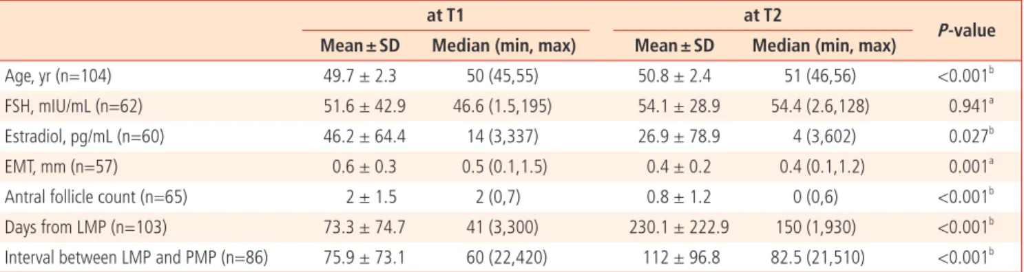
Table 3A. Comparison of hormonal, ultrasonographic and clinical markers at each visitat T1 at T2
P‐value Mean ± SD Median (min, max) Mean ± SD Median (min, max)
Age, yr (n=104) 49.7 ± 2.3 50 (45,55) 50.8 ± 2.4 51 (46,56) <0.001b
FSH, mIU/mL (n=62) 51.6 ± 42.9 46.6 (1.5,195) 54.1 ± 28.9 54.4 (2.6,128) 0.941a
Estradiol, pg/mL (n=60) 46.2 ± 64.4 14 (3,337) 26.9 ± 78.9 4 (3,602) 0.027b
EMT, mm (n=57) 0.6 ± 0.3 0.5 (0.1,1.5) 0.4 ± 0.2 0.4 (0.1,1.2) 0.001a
Antral follicle count (n=65) 2 ± 1.5 2 (0,7) 0.8 ± 1.2 0 (0,6) <0.001b Days from LMP (n=103) 73.3 ± 74.7 41 (3,300) 230.1 ± 222.9 150 (1,930) <0.001b Interval between LMP and PMP (n=86) 75.9 ± 73.1 60 (22,420) 112 ± 96.8 82.5 (21,510) <0.001b SD, standard deviation; FSH, follicle‐stimulating hormone; EMT, early menopausal transition; LMP, late menopausal transition; PMP, previous menstrual period.
aBy paired t‐test.
bBy Wilcoxon signed ranks test.
Table 3B. Comparison of hormonal, ultrasonographic and clinical markers at each visit
AMH, ng/mL (n=62) at T2
P‐value
<0.14 ≥0.14
at T1 <0.14 43 (74.1%) 15 (25.9%) 0.008a
≥0.14 3 (75%) 1 (25%)
AMH, anti‐Müllerian hormone.
aBy Mcnemar test.
Table 4. Correlations between ovarian reserve markers after adjustment for age at the fi rst visit (T1)
FSH Estradiol EMT AFC Days from
LMP Interval between LMP and PMP FSH 1.000
Estradiol ‐0.513a 1.000
EMT ‐0.553a 0.608a 1.000
AFC ‐0.250b 0.117 0.126 1.000
Days from LMP 0.215b ‐0.195 ‐0.215b ‐0.232b 1.000
Interval between LMP and PMP 0.090 ‐0.057 ‐0.229b ‐0.085 0.142 1.000
FSH, follicle‐stimulating hormone; EMT, early menopausal transition; AFC, antral follicle count; LMP, late menopausal transition; PMP, previous menstrual period.
aP < 0.01.
bP < 0.05.
did not show signifi cant correlation with menopause at T2. In the multivariable stepwise logistic analysis, the variables of days since LMP and interval between LMP and the previous menstrual period (PMP) were selected as independent risk factors for predicting menopause in our model (Table 5). When 60 days (2 months) was selected as the cutoff, the sensitivity was 60.1%, and the specifi c- ity was 81.5% for the number of days since LMP; for the interval
between the last 2 menstrual cycles, the sensitivity for predicting menopause at T2 was 64.3%; specifi city was 84.6% with 75 days (2.5 months) as the cutoff (Fig. 1).
Discussion
Our study included women who were more than 45 years of age and were in their late reproductive years with irregular menstrua- tion; serum levels of AMH were undetectable in most cases (86.6%
at T1, 74.2% at T2), and detectable values of AMH were not as- sociated with menopause over a 13-month period. In addition, the mean AMH value did not decrease over time. However, 15 of 58 women (25.9%) with undetectable levels of AMH at T1 showed detectable serum levels of AMH at T2. These results were unex- pected because we had hypothesized a linear decrease in AMH levels over time and that AMH levels at T1 would be a predicting factor of menopause at T2.
A number of recent studies have suggested that AMH is a promis- ing marker for predicting the menopausal transition or menopause [14-16,19]. In contrast to our study, the target population in other studies included young, reproductive-aged women (25 to 46 years old or 40 to 50 years old), and the follow-up interval spanned a longer period (6 to 11 years).
Serum AMH levels have been reported to be constant throughout the natural menstrual cycle in young, reproductive-aged women;
Fig. 1. Receiver operating characteristic (ROC) curves of selected mark- ers after multiple logistic regression analysis for predicting menopause at T2. MCD, menstrual cycle day; LMP, last menstrual period; PMP, previous menstrual period.
Table 5. Predictive capacity of ovarian reserve markers assessed at T1 for menopause at T2
Un-Adjusted OR (95% CI) P‐value ROCAUC (95% CI) Univariate analysis
Age 1.195 (0.995-1.435) 0.057 0.610 (0.475-0.745)
FSH at T1 1.018 (1.007-1.029) 0.001 0.735 (0.629-0.842)
Estradiol at T1 0.989 (0.978-1.001) 0.071 0.646 (0.521-0.772)
EMT at T1 0.015 (0.001- 0.164) 0.001 0.738 (0.619-0.856)
AFC at T1 0.67 (0.484-0.928) 0.016 0.636 (0.509-0.763)
AMH at T1 <0.14 vs ≥0.14 1.189 (0.344-4.111) 0.785 0.502 (0.373-0.631)
MCD at T1 1.017 (1.009-1.024) <0.001 0.785 (0.679-0.890)
Interval between LMP and PMP 1.016 (1.006-1.026) 0.002 0.773 (0.659-0.888)
Multivariable Stepwise logistic analysis Adjusted OR (95% CI)
MCD at T1 1.019 (1.009-1.030) <0.001
Interval between LMP and PMP 1.015 (1.004-1.026) 0.008
OR, odds ratio; CI, confi dence interval; ROCAUC, receiver operating characteristic area under the curve; FSH, follicle‐stimulating hormone; EMT, early menopausal transition; AFC, antral follicle count; AMH, anti‐Müllerian hormone; MCD, menstrual cycle day; LMP, late menopausal transition; PMP, previ- ous menstrual period.
therefore, AMH levels have been regarded as a reliable serum marker of ovarian reserve that can be measured anytime dur- ing the menstrual cycle, at the convenience of clinicians [25-29].
However, Robertson et al. [29] reported recently that AMH levels could change signifi cantly throughout the ovulation cycle in late reproductive-aged women (>45 years old); some women had a 2.8-fold increase, while others had a 4.3-fold decrease in levels in the luteal phase compared with those observed in the follicular phase. Furthermore, 19% of the women had non-detectable levels of AMH during their ovulation cycle based on other hormonal as- says even though the sensitivity of their assay was 10-fold higher than the assay used in our study (0.017 ng/mL). Therefore, the au- thors concluded that AMH levels become less reliable as a marker of ovarian reserve when levels are substantially reduced, as in late reproductive-aged women [29]. Likewise, there have been reports of moderate and reasonable pregnancy rates following extremely low levels of serum AMH in women in vitro fertilization programs [30].
Although we did not monitor intracyclic fl uctuations of AMH levels because we did not measure blood progesterone levels, and blood samples were taken irrespective of the menstrual cycle, we found that 25.8% (all participants with detectable levels of AMH at T2 had lower or undetectable levels of AMH at T1) of participants had higher serum values of AMH after 13 months, which might be due to an unpredictable abrupt change that occurs in women at a late-reproductive age. Elevated basal FSH level, decreased AFC (or ovarian reserve), and the resulting unpredictable ovarian response, are thought to be important markers, but more studies are needed to further understand the underlying ovarian physiology at this age.
As expected, FSH—the classical marker of ovarian aging—and AFC—the well-established sonographic marker of ovarian re- serve—were signifi cantly associated with predicting menopause as a single marker in our study. However, hormonal and sono- graphic markers including FSH and AFC were not signifi cantly as- sociated with development of menopause after multiple stepwise regression analyses. Instead, a longer interval from their LMP and a larger gap between their last 2 cycles in late reproductive-aged women were most strongly and independently associated with occurrence of menopause after 1 year in women who had already entered the late reproductive period in our study.
There were some limitations in this study. First, there is a pos- sibility of recall bias because the information regarding the LMP was retrospectively obtained from the participants. Second, at T2, blood samples were obtained from only 62 of the 104 participants due to a lack of remaining blood samples after the routine blood
examination. A larger study group may be required. Third, since it has been known that the endocrine status of EMT and LMT is different, recruitment of adequate number of participant of each state at T1 (especially subjects in EMT) could have given more detailed information. In addition, the AMH ELISA kit used in this study was not sensitive enough to discriminate differences in very small antral follicle numbers. Only 14 of 104 participants (13.4%) at T1 and 16 of 62 (25.4%) at T2 had detectable levels of AMH.
Accordingly, any statistical analysis that could reveal a quantitative association among other hormonal or sonographic markers with AMH was not possible. Development and application of a more sensitive tool is required for future studies. Furthermore, any study design that investigates the level of AMH to predict menopause would better span longer period to cover reproductively younger women that might have measurable AMH level.
Nonetheless, despite the small study group size, this is the first longitudinal Korean study that evaluates the usefulness of AMH, other hormonal and ultrasonographic markers, and menstrual history as markers for predicting menopause in a relatively short timeframe in late reproductive-aged women. These data can be obtained during a routine gynecologic examination where infor- mation regarding the exact timing of menopause and hormonal status is important to both the clinician and the examinee. Women who have not menstruated for more than 2 months and who are 45 to 55 years old have a high probability of becoming meno- pausal after 1 year without requiring further hormone evaluation.
This study indicates that AMH is not a determining marker for pre- dicting the onset of menopause in late reproductive-aged women in a relatively short timeframe (range, 0.5 to 2.5 years). The tradi- tional ovarian reserve markers FSH and AFC were not independent predictors, but time since the LMP and longer intervals between the last 2 cycles at T1 were more useful predictors of menopause onset.
Serum values of AMH in this age group of women were very low and changed sporadically; hence, a more sensitive assay kit should be used, and ovulation cycle timing should be a consideration.
Acknowledgments
This study was supported by Seoul National University Hospital Research Grant no. 01-2008-107.
Seoul National University Hospital Medical Research Collaborating
Center (SNUH MRCC) has contributed to the statistical analysis of
this paper.
References
1. Treloar AE. Menstrual cyclicity and the pre-menopause. Ma- turitas 1981;3:249-64.
2. Soules MR, Sherman S, Parrott E, Rebar R, Santoro N, Utian W, et al. Executive summary: Stages of Reproductive Aging Work- shop (STRAW). Fertil Steril 2001;76:874-8.
3. Grodstein F, Manson JE, Stampfer MJ. Hormone therapy and coronary heart disease: the role of time since menopause and age at hormone initiation. J Womens Health (Larchmt) 2006;15:35-44.
4. Rossouw JE, Prentice RL, Manson JE, Wu L, Barad D, Barnabei VM, et al. Postmenopausal hormone therapy and risk of car- diovascular disease by age and years since menopause. JAMA 2007;297:1465-77.
5. Salpeter SR, Walsh JM, Greyber E, Ormiston TM, Salpeter EE.
Mortality associated with hormone replacement therapy in younger and older women: a meta-analysis. J Gen Intern Med 2004;19:791-804.
6. Burger HG, Hale GE, Robertson DM, Dennerstein L. A review of hormonal changes during the menopausal transition: focus on fi ndings from the Melbourne Women’s Midlife Health Proj- ect. Hum Reprod Update 2007;13:559-65.
7. Hale GE, Zhao X, Hughes CL, Burger HG, Robertson DM, Fraser IS. Endocrine features of menstrual cycles in middle and late reproductive age and the menopausal transition classified according to the Staging of Reproductive Aging Workshop (STRAW) staging system. J Clin Endocrinol Metab 2007;92:3060-7.
8. Rigon C, Andrisani A, Forzan M, D’Antona D, Bruson A, Cosmi E, et al. Association study of AMH and AMHRII polymorphisms with unexplained infertility. Fertil Steril 2010;94:1244-8.
9. Lee JR, Kim SH, Kim SM, Jee BC, Ku SY, Suh CS, et al. Follicular fluid anti-Mullerian hormone and inhibin B concentrations:
comparison between gonadotropin-releasing hormone (GnRH) agonist and GnRH antagonist cycles. Fertil Steril 2008;89:860-7.
10. Fanchin R, Mendez Lozano DH, Frydman N, Gougeon A, di Clemente N, Frydman R, et al. Anti-Mullerian hormone con- centrations in the follicular fl uid of the preovulatory follicle are predictive of the implantation potential of the ensuing embryo obtained by in vitro fertilization. J Clin Endocrinol Metab 2007;92:1796-802.
11. Shin SY, Lee JR, Noh GW, Kim HJ, Kang WJ, Kim SH, et al.
Analysis of serum levels of anti-Mullerian hormone, inhibin B, insulin-like growth factor-I, insulin-like growth factor binding
protein-3, and follicle-stimulating hormone with respect to age and menopausal status. J Korean Med Sci 2008;23:104-10.
12. Sowers MR, Eyvazzadeh AD, McConnell D, Yosef M, Jannausch ML, Zhang D, et al. Anti-mullerian hormone and inhibin B in the defi nition of ovarian aging and the menopause transition.
J Clin Endocrinol Metab 2008;93:3478-83.
13. La Marca A, De Leo V, Giulini S, Orvieto R, Malmusi S, Giannel- la L, et al. Anti-Mullerian hormone in premenopausal women and after spontaneous or surgically induced menopause. J Soc Gynecol Investig 2005;12:545-8.
14. van Rooij IA, Tonkelaar I, Broekmans FJ, Looman CW, Scheffer GJ, de Jong FH, et al. Anti-mullerian hormone is a promising predictor for the occurrence of the menopausal transition.
Menopause 2004;11:601-6.
15. Tehrani FR, Solaymani-Dodaran M, Azizi F. A single test of antimullerian hormone in late reproductive-aged women is a good predictor of menopause. Menopause 2009;16:797-802.
16. van Disseldorp J, Faddy MJ, Themmen AP, de Jong FH, Peeters PH, van der Schouw YT, et al. Relationship of serum antimul- lerian hormone concentration to age at menopause. J Clin Endocrinol Metab 2008;93:2129-34.
17. Flaws JA, Langenberg P, Babus JK, Hirshfi eld AN, Sharara FI.
Ovarian volume and antral follicle counts as indicators of menopausal status. Menopause 2001;8:175-80.
18. Giacobbe M, Mendes Pinto-Neto A, Simões Costa-Paiva LH, Martinez EZ. The usefulness of ovarian volume, antral follicle count and age as predictors of menopausal status. Climacteric 2004;7:255-60.
19. Broer SL, Eijkemans MJ, Scheffer GJ, van Rooij IA, de Vet A, Themmen AP, et al. Anti-mullerian hormone predicts meno- pause: a long-term follow-up study in normoovulatory wom- en. J Clin Endocrinol Metab 2011;96:2532-9.
20. Flaws JA, Rhodes JC, Langenberg P, Hirshfi eld AN, Kjerulff K, Sharara FI. Ovarian volume and menopausal status. Meno- pause 2000;7:53-61.
21. Scheffer GJ, Broekmans FJ, Looman CW, Blankenstein M, Fauser BC, teJong FH, et al. The number of antral follicles in normal women with proven fertility is the best reflection of reproductive age. Hum Reprod 2003;18:700-6.
22. Yang YS, Hur MH, Kim SY, Young K. Correlation between sono- graphic and endocrine markers of ovarian aging as predictors for late menopausal transition. Menopause 2011;18:138-45.
23. Irmak MK. Self-fertilization in human: having a male embryo without a father. Med Hypotheses 2010;75:448-51.
24. Rashidi BH, Abediasl Z, Tehraninejad ES, Shariat M, Mahdavi
A. Menstrual cycle length in relation to antimullerian hormone and follicle-stimulating hormone. J Reprod Med 2009;54:315-8.
25. Tsepelidis S, Devreker F, Demeestere I, Flahaut A, Gervy C, En- glert Y. Stable serum levels of anti-Mullerian hormone during the menstrual cycle: a prospective study in normo-ovulatory women. Hum Reprod 2007;22:1837-40.
26. La Marca A, Stabile G, Artenisio AC, Volpe A. Serum anti- Mullerian hormone throughout the human menstrual cycle.
Hum Reprod 2006;21:3103-7.
27. La Marca A, Giulini S, Tirelli A, Bertucci E, Marsella T, Xella S, et al. Anti-Mullerian hormone measurement on any day of the menstrual cycle strongly predicts ovarian response in assisted
reproductive technology. Hum Reprod 2007;22:766-71.
28. Hehenkamp WJ, Looman CW, Themmen AP, de Jong FH, Te Velde ER, Broekmans FJ. Anti-Mullerian hormone levels in the spontaneous menstrual cycle do not show substantial fl uctua- tion. J Clin Endocrinol Metab 2006;91:4057-63.
29. Robertson DM, Hale GE, Fraser IS, Hughes CL, Burger HG.
Changes in serum antimullerian hormone levels across the ovulatory menstrual cycle in late reproductive age. Menopause 2011;18:521-4.
30. Weghofer A, Dietrich W, Barad DH, Gleicher N. Live birth chances in women with extremely low-serum anti-Mullerian hormone levels. Hum Reprod 2011;26:1905-9.
폐경이행기 여성에서 폐경 예측지표로서 혈중 anti-Müllerian hormone, follicle-stimulating hormone, 초음파상의 동난포 개수 및 임상적 증상들의 유용성 평가
1서울대학교 의과대학 산부인과학교실, 2서울대학교병원 강남센터, 3서울대학교병원 인구의학연구소, 4분당서ㅎ울대학교병원 산부인과, 5함춘여성클리닉 김선미1,2, 김석현1,3, 이정렬1,4, 지병철1,4, 서창석1,3,4, 이중엽1,5, 김기철5, 황도영5, 김정구1, 문신용1,3
목적
폐경이행기 한국 여성에서 폐경 예측 지표로서 혈중 anti-Müllerian hormone (AMH), follicle-stimulating hormone (FSH), estradiol, 자궁내막 두께, 초음파상의 동난포 개수 및 임상적 증상들의 유용성을 평가하고자 하였다.
연구방법
45-55세 사이의 폐경이행기 여성 104명이 참여하였으며 평균 13.1개월 간격으로 두 번에 걸쳐 방문하였다. 각 방문 시 최근의 월경력을 확인하고 골반 초음파검사를 시행하여 동난포 개수 및 자궁내막 두께를 측정하였으며 호르몬검사를 위한 혈액을 채취하였다. 최종 평가 지표는 두 번째 방문(T2) 시의 폐경 여부였으며 통계적 분석은 SPSS ver. 17.0을 사용하였다.
결과
104명의 참여자 중 두 번째 방문시기에 폐경이 된 여성은 33명이었다. 두 번째 방문 시 폐경된 여성들은 그렇지 않은 여성들과 비교하여 첫 번째 방문 시의 FSH, estradiol, 자궁내막 두께, 동난포 개수, 최종 월경일로부터 경과일 및 최종 월경일과 그 이전 주기와의 기간에 있어 유의한 차이를 보였다. 그러나 AMH 수치는 두 그룹 간 유의한 차이를 보이지 않았다. 모든 인자들 중 최종 월경일로부터 경과일 및 최종 월경일과 그 이전 주기와의 간격이 폐경 발생과 유의한 상관관계를 보인 지표였다.
결론
폐경이행기 여성에서 1년 정도의 짧은 간격을 두고 볼 때 AMH는 폐경을 예측하는 지표가 되지 못하였다. 첫 번째 방문 시 최종 월경일로 부터 경과한 기간이 1년 후 폐경 예측의 더욱 유용한 지표이다.
중심단어: 폐경이행기, 항뮐러관호르몬, 난포자극호르몬, 동난포 개수, 최종 월경일