We successfully treated this with stenting of the left main coronary artery and proximal left anterior descending artery, and this allowed for the definitive surgical correction
6
0
0
전체 글
(2) A. B. Figure 1. The initial ECG showing the tall T wave on the precordial leads (A). The follow up ECG showing the improved tall T wave on the precordial leads (B). The follow up ECG showing the ST segment elevation on the I, aVL and precordial leads (C). ECG: electrocardiogram.. C. A. B. C. D. Figure 2. At the initial phase of contrast injection, the left anterior oblique caudal view (A) and anteroposterior caudal view (B) showed the heterogenous filling defect in the proximal left anterior descending artery. Later, however, the left anterior oblique caudal view (C) and anteroposterior caudal view (D) showed no filling defect in the proximal left anterior descending artery.. 1211.
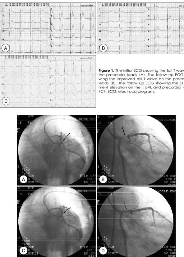
(3) anterior chest radiating toward the back, and this was accompanied by cold sweating, nausea and vomiting. The. 37. There were no specific findings on the physical examination. The results of laboratory tests, including cardiac. blood pressure dropped to 90/60 mmHg with the patient displaying an altered mentality and shallow respiration.. enzyme testing, were within the normal range except for a mild leukocytosis. The initial electrocardiogram (ECG). After endotracheal intubation, he was transferred to our hospital.. showed a tall T wave at the V2-6 precordial lead. There was no elevated ST segment (Figure 1A). Antiplatelet. He was a 20-pack-year smoker without any specific comorbidity. His elder brother died suddenly at the age of. agent and heparin were administered under the impression of acute myocardial infarction. After an hour, follow-up. A. B. C. D. Figure 3. After stenting the proximal left anterior descending artery, IVUS showed malposition of the stent (A). The large false lumen compromising the true lumen and dissection flap (arrow) in the left main coronary artery (B). After left main coronary artery stenting, IVUS showed a residual false lumen (arrow) at the left main coronary artery (C). IVUS showed the membranous structure (arrow) in the ascending aorta near to the left main coronary artery (D). IVUS: intravascular ultrasonography.. A. B. Figure 4. After stenting the proximal left anterior descending artery, the coronary angiogram shows the compromised left main and left circumflex artery flows (arrow) (A). After left main coronary artery stenting, the coronary angiogram shows the recovered blood flow (B).. 1212. Korean Circulation J 2004;34(12):1210-1215.
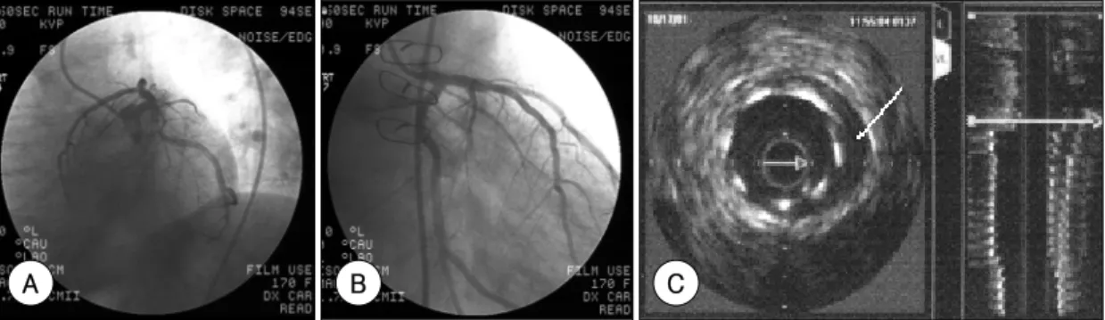
(4) ECG showed a resolution of the tall T wave (Figure 1B), but the chest pain persisted. To differentiate acute myo-. patient was moved to the cardiac catheterization room for primary coronary intervention.. cardial infarction from other possible causes such as aortic dissection, echocardiography and computed tomography. Right coronary angiography (CAG) showed no abnormality except for a mild luminal narrowing at the ostium. (CT) were planned. At that time, he again complained severe chest pain and the follow-up ECG showed ST. of the right coronary artery. Left CAG showed an inhomogenous filling defect, which was probably due to exter-. segment elevation at the I, aVL and precordial leads (Figure 1C). The echocardiography showed hypokine-. nal compression (Figure 2A, B). The arterial lumen appeared to be expanded slightly during contrast injection. sia of the interventricular and the anterior wall of the left ventricle. On the follow up laboratory study, the level of. (Figure 2C, D). During CAG, the coronary artery lumen was compromised again and ventricular fibrillations re-. the cardiac enzymes was elevated (CKMB: 224 ng/mL). So we dia-gnosed the patient as having ST-elevation. quiring DC cardioversion occurred several times. Balloon angioplasty (Scimed, 3.5×20 mm) was done. myocardial infarction because of the typical changes on the ECG and echocardiography. CT was skipped and the. at 6 atmospheres for 30 seconds on the proximal left anterior descending artery. After this, a stent was inserted (3.5×9 mm, 10 atmospheres for 20 seconds). Following this, IVUS was performed to find out the exact pathophysiology. Apposition of stent was poor in the proximal left anterior descending artery (Figure 3A). A large false lumen and dissection flap compressing the true lumen were noted in the left main coronary artery (Figure 3B). With retraction of the guiding catheter to the aorta, we found that the left main coronary artery was compressed. The blood flow was impaired not only in the left anterior descending artery, but also in the left circumflex artery (Figure 4A). The blood flow was restored by an immediate stenting in the left main coronary artery (4.0×13 mm, 12 atmospheres for 20 seconds, Figure 4B). On the followup IVUS, the true lumen was patent, but the false. Figure 5. The computed tomography imaging showing dissection of the ascending aorta and descending aorta. The left main and proximal left anterior descending artery show good patency after stenting.. A. B. lumen still remained (Figure 3C). Membranous structure that seemed to be a dissection flap was seen at the ascending aorta just outside the left main coronary artery (Figure 3D). This lesion extended to the left main coro-. C. Figure 6. Follow up coronary angiogram shows no significant stenosis in the left anterior oblique caudal view (A) and anteroposterior caudal view (B). Follow up IVUS shows the intact stent and the residual false lumen (arrow) (C). IVUS: intravascular ultrasonography.. 1213.
(5) nary artery and the proximal left anterior descending artery. Emergency chest CT showed the ascending aortic. aortic dissection that involved the right coronary artery. They underwent surgical treatment with a aortic graft and. dissection (Figure 5). Upon operating, a large hematoma in the false lumen was found to be compressing the ascen-. coronary artery bypass graft, and 27.3% of these cases displayed postoperative mortality. Neri et al12) studied 34. ding aorta and coronary artery. Glue aortoplasty and aortic valve resuspension were then performed in a timely. cases that were treated with operation for aortic dissection involving the coronary ostia, and they reporting a 20%. fashion. Five days after the operation, the patient was weaned from the mechanical ventilator and he was dis-. rate of in-hospital mortality. Kawada et al13) studied the predictors of postoperative mortality in the 88 cases of. charged without any specific complication. Restenosis was not present in the inserted stent on the follow-up. ascending aortic dissection. They concluded that the preoperative complications from coronary dissection were the. CAG performed at the 19th month after the operation (Figure 6A, B). No intimal proliferation was noted in the. most important predictors of early postoperative mortality. Kawahito et al14) also reviewed the surgical results and. IVUS, but the false lumen in the left main coronary artery and left anterior descending artery persistently remained. the mechanism of malperfusion in a group of 12 patients with coronary malperfusion caused by aortic dissection.. (Figure 6C). The echocardiography showed marked improvement for the left ventricular wall motion and systolic. They reported a 33.3% rate of mortality, and they insisted on the necessity of aggressive coronary revascularization. function.. and early aortic repair to salvage these critically ill patients.. Discussion. Recently, there have been reports of attempts to maintain the coronary artery flow through stent insertion prior. Acute myocardial infarction occurs in 1-2% of patients with dissection of the ascending aorta, and this. to definitive surgical repair. Barabas et al4) have reported a case of emergency left. happens because of compression of the coronary ostia by hematoma or occlusion by an intimal flap.2) The dissec-. main direct stenting that was used as a bridge to surgery in a patient with hemodynamic instability due to occlusion. tion more often affects the right coronary artery than the left, and this explains why these myocardial infarctions. of the left main coronary artery; this occlusion was due to external compression caused by an intamural hema-. tend to be inferior in location.8)9) In case of patients presenting with acute chest pain and. toma stemming from aortic dissection. In addition, Ikari et al,5) Ohara et al,6) and Cardozo et al7) have also reported. showing ST segment elevation on the electrocardiogram, treatment is often done by primary coronary intervention. on treating cases of acute myocardial infarction associated with aortic dissection by stenting the left main coronary. or thrombolytic therapy due to the clinical impression that the patient is only suffering from acute myocardial. artery and/or the other coronary arteries as well. In this case, too, by promptly maintaining adequate. infarction.10) In case of aortic dissection, however, thrombolytic therapy is contraindicated. Our patient was a case. myocardial blood flow, extensive myocardial damage was avoided. Accordingly, this treatment was believed to have. of myocardial infarction resulting from left main coronary artery dissection that was complicated by aortic dissection.. reduced the pre- and post operative complications. Additionally, early stenting was considered to improve the. If thrombolytic therapy had been applied, the condition of the patient might have fatally deteriorated.. patient’s critical state and to have played a role as a bridge to definitive surgical management. This stenting. There have been a few reports on left main coronary artery dissection complicated by aortic dissection. Most. procedure was also believed to have contributed to reducing the patient’s risk of mortality.. of the cases resulted in a high mortality rate due to the low cardiac output from an extensive anterior myocardial in-. In summary, left main coronary artery dissection combined with aortic dissection is a rare but fatal complication. farction. Pego-Fernandes et al11) analyzed 11 patients having. of aortic dissection; this condition has a high mortality rate and it usually requires emergency surgical manage-. 1214. Korean Circulation J 2004;34(12):1210-1215.
(6) ment. We report here on a case of a left main coronary artery dissection complicated by ascending aortic dissection that was treated successfully with left main coronary artery stenting. REFERENCES 1) Kasper DL, Braunwald E, Fauci AS, Hauser SL, Longo DL,. Jameson JL, et al. Harrison’s principles of internal medicine. 16th ed. New York: McGraw-Hill; 2005. p.1483-4. 2) Hirst AE Jr, Johns VJ Jr, Kime SW Jr. Dissecting aneurysms of the aorta: a review of 505 cases. Medicine 1958;37:217-79. 3) Coselli JS. Treatment of acute aortic dissection involving the right coronary artery and aortic valve. J Cardiovasc Surg 1990;31:305-9. 4) Barabas M, Gosselin G, Crepeau J, Petitclerc R, Cartier R, Theroux P. Left main stenting-as a bridge to surgery-for acute type A aortic dissection and anterior myocardial infarction. Catheter Cadiovasc Interv 2000;51:74-7. 5) Ikari Y, Hara K, Tamura T, Hara H, Saeki F, Kikawa I, et al. Intracoronary stenting of a coronary occlusion resulting from an aortic dissection. Cathet Cardiovasc Diagn 1995;36: 160-3. 6) Ohara Y, Hiasa Y, Hosokawa S. Successful treatment in a case of acute aortic aortic dissection complicated with acute myocardial infarction due to occlusion of the left main coronary artery. J Invasive Cardiol 2003;15:660-2.. 7) Cardozo C, Riadh R, Mazen M. Acute myocardial infarction. due to left main compression aortic dissection treated by direct stenting. J Invasive Cardiol 2004;16:89-91. 8) DeSanctis RW, Doroghazi RM, Austen WG, Buckley MJ. Aortic dissection. N Engl J Med 1987;317:1060-7. 9) Pande AK, Gosselin G, Leclerc Y, Leung TK. Aortic dissection complicating coronary angioplasty in cystic medial necrosis. Am Heart J 1996;131:1221-3. 10) Nah DY, Park KU, Kim SH. Acute proximal aortic dissection associated with ST segment elevation on electrocardiography. Korean Circ J 2004;34:795-8. 11) Pego-Fernandes PM, Stolf NA, Hervoso CM, Silva JM, Arteaga E, Jatene AD. Management of aortic dissection that involves the right coronary artery. Cadiovasc Surg 1999;7: 545-8. 12) Neri E, Toscano T, Papalia U, Frati G, Massetti M, Capannini G, et al. Proximal aortic dissection with coronary malperfusion: presentation, management, and outcome. J Thorac Cardiovasc Surg 2001;121:552-60. 13) Kawada T, Okada Y, Alba M, Sekiguchi S, Yamada M, Michihata T, et al. Changing predictors of postoperative mortality in acute type A aortic dissection: is only coronary artery compromise significant? Jpn J Thorac Cardivasc Surg 2001; 49:347-54. 14) Kawahito K, Adachi H, Murata S, Yamaguchi A, Ino T. Coronary malperfusion due to type A aortic dissection: mechanism and surgical management. Ann Thorac Surg 2003;76: 1471-6.. 1215.
(7)
수치

관련 문서
