J O U R N A L O F
Veterinary Science
pISSN 1229-845X, eISSN 1976-555X J. Vet. Sci. (2013), 14(4), 491-494 http://dx.doi.org/10.4142/jvs.2013.14.4.491
Received: 7 Feb. 2013, Revised: 4 Apr. 2013, Accepted: 28 Jun. 2013
Short Communication
*Corresponding author: Tel: +34-927257114; Fax: +34-927257110; E-mail: [email protected]
ⓒ 2013 The Korean Society of Veterinary Science.
This is an Open Access article distributed under the terms of the Creative Commons Attribution Non-Commercial License (http://creativecommons.org/licenses/by-nc/3.0) which permits unrestricted non-commercial use, distribution, and reproduction in any medium, provided the original work is properly cited.
Development of a real-time SYBR Green PCR assay for the rapid detection of Dermatophilus congolensis
Alfredo García*, Remigio Martínez, José Manuel Benitez-Medina, David Risco, Waldo Luis García, Joaquín Rey, Juan Manuel Alonso, Javier Hermoso de Mendoza
Departamento de Sanidad Animal, Facultad de Veterinaria de Cáceres, Universidad de Extremadura, 10003 Cáceres, Spain
Methods such as real time (RT)-PCR have not been developed for the rapid detection and diagnosis of Dermatophilus (D.) congolensis infection. In the present study, a D. congolensis-specific SYBR Green RT-PCR assay was evaluated. The detection limit of the RT-PCR assay was 1 pg of DNA per PCR reaction. No cross-reaction with nucleic acids extracted from Pseudomonas aeruginosa, Mycobacterium tuberculosis, Staphylococcus aureus, or Austwickia chelonae was observed. Finally, the RT-PCR assay was used to evaluate clinical samples collected from naturally infected animals with D. congolensis. The results showed that this assay is a fast and reliable method for diagnosing dermatophilosis.
Keywords: clinical samples, Dermatophilus congolensis, dermatophilosis, diagnosis, real-time PCR
Dermatophilus (D.) congolensis is a filamentous branching actinomycete that causes dermatophilosis, a disease characterized by exudative and proliferative dermatitis that affects a wide range of animals and occasionally humans. Clinical characteristics of the disease include matted hair or wool, scab and crust formation, and sometimes, generalized massive crust formation in chronic cases. Chronic infection tends to result in hair loss and even local loss of the upper skin layers that predisposes the patient to secondary infection by Staphylococcus spp. or Pseudomonas aeruginosa [18].
In some instances, D. congolensis may reach the lymph nodes, resulting in a granulomatous response [5]. In a recent report, this organism was also found to be associated with placentitis, funisitis, and abortion in a horse [14].
Dermatophilosis is an economically important disease of cattle in tropical and sub-tropical regions as well as sheep in areas with high rainfall. This disease causes
significantly reduced meat, milk, and hide production;
failure to thrive in livestock-dependent communities, and increased culling and death [9, 17]. Human infection is rare and most reported cases involve skin infections due to contact with animals. However, D. congolensis has also been associated with infection of the human esophagus [12].
Detection of animals affected by dermatophilosis is frequently difficult and susceptible to confusion with clinically similar diseases such as orf and mange. Direct microscopic examination of stained smears from the lesions or exudates beneath the scabs in acute cases enables visualization of the characteristic branched filaments [15].
However, stained smears taken from dried scabs collected from chronic cases tend to contain low numbers of this pathogen and the characteristic appearance of D.
congonlensis is not so obvious [2]. On the other hand, isolation of the infectious agent by culturing is often difficult due to contamination. For this, special techniques involving filtration, chemotaxis, or selective media are necessary. Immunofluorescence techniques have also proven to be useful and sensitive methods for diagnosing D. congolensis infection. However, laboratories must manufacture their own antisera for diagnostic tests because of the lack of a commercial one [11].
Sensitive and routine laboratory techniques for rapid and accurate dermatophilosis diagnosis are highly desirable.
The aim of this study was to develop a rapid, and sensitive technique for detecting D. congolensis in cultures and clinical samples. For this investigation, a specific SYBR Green real-time (RT)-PCR assay for detecting D.
congolensis DNA was developed.
Twenty D. congolensis isolates were used for this study.
The host species and geographic origin are listed in Table
1. The isolates were grown in 20 mL of 2% (w/v) brain
492 Alfredo García et al.
Table 1. Available data of the strains used for assay development and assessment of primer specificities
Strain Host species Origin
HDC (DSM 43037) HA2 (NCTC 5175) BC87
HD201 HWAN HATI HISL HEQ
BE3 (ATCC 14637) OT8
BG45 (IMTV M6445) OG1
OTS1 OPOA BF13 (5.11 A) HA11 (3.86) B556 POF OT10 ORL
Austwickia chelonae Pseudomonas aeruginosa Staphylococcus aureus (ATCC 29213)
Mycobacterium tuberculosis (H37Rv)
Horse Horse Cattle Horse Horse Horse Horse Horse Cattle Sheep Cattle Sheep Sheep Sheep Cattle Horse Cattle Sheep Sheep Sheep Tortoise Sheep
UK UK Cameroon Germany Spain Spain Spain Spain Zimbabwe Spain Guadeloupe Spain Spain Spain Nigeria UK Spain Spain Spain Spain Australia Spain Reference strain Reference strain
heart infusion (Oxoid, UK) supplemented with 1.7% (w/v) neutralized soya peptone (Oxoid) in glass McCartney bottles (Fisher, UK) at 37
oC in air for 48 h. A dense growth of hyphae and filaments with a clear supernatant were typically observed in the cultures.
Genomic DNA was extracted from the cultures using a previously described method [8]. The final DNA pellet was suspended in 25 μL of ultrapure water (Life technologies, Spain) and stored at -20
oC until further use.
Concentration of the genomic DNA was calculated based on the absorbance measured at 260 nm with a Synergy HT Multi-Mode Microplate Reader (BioTek, USA) using Gen 5TM 182 V1.09 data analysis software (BioTek). Genomic DNA samples with 260/280 ratios between 1.7 and 1.9 were considered to be pure and of high quality.
To determine the detection limit of the RT-PCR assay, D.
congolensis DNA from a reference strain (DSM 43037) (kindly provided by Dr. Alex Morrow, Centre for Tropical Veterinary Medicine, UK) was used. In order to estimate the analytical specificity of the D. congolensis-specific RT-PCR assay, DNA from Pseudomonas aeruginosa,
Mycobacterium tuberculosis, Staphylococcus aureus, and Austwickia chelonae (formerly Dermatophilus chelonae) [6] from in vitro cultures (Table 1) were also used. Primers were designed with the help of an online primer design program, Primer3 [13], based on the nucleotide sequence of the agac gene (GenBank accession no. AJ496026.1) encoding an alkaline ceramidase protein [4]. The resulting primer set named DermaF 5´-TGGCAGCTCTGATGA GTACCACAA-3´ (Tm 60.1
oC) and DermaR 5´-AATGTG CCGGGAACGGAAATCAAC-3´ (Tm 60.3ºC) amplified a 127-bp fragment between the locus positions 930 and 1065. To evaluate their potential annealing specificity, the primer sequences were analyzed with the BLAST search homology database [1].
Purified DNA templates were amplified in an iCycler iQ real-time detection system (BioRad, USA). Briefly, a 25- μL PCR mixture was prepared containing 1 μL of DNA, 12.5 μL SensiMix SYBR and fluorescein (Quantance, UK), and 200 nM of each primer.
Amplification was carried out with one cycle at 50
oC for 2 min and one cycle at 95
oC for 2 min followed by 35 cycles of 95
oC for 30 sec, 60
oC for 30 sec, and 72
oC for 1 min. The plate reading was taken after extension at 72
oC and a melting curve was obtained using temperatures between 65
oC and 95
oC that increased at a rate of 0.5
oC/s.
Duplicate positive and negative controls were included in all tests and at least two separate experiments were performed. Negative controls contained all components of the reaction mixture except for template DNA. The PCR products were analyzed with ethidium bromide-stained 2% agarose gels (Pronadisa, Spain).
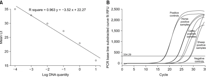
Genomic DNA from the reference D. congolensis strain (DSM 43037) was serial diluted 10-fold (from 10 ng to 1.10
-4ng) and used to test the sensitivity of the RT-PCR assay. The resultant data were used to generate a standard curve by plotting threshold cycle (Ct) values against the logarithmic values of the starting DNA quantities.
Amplification efficiency and coefficient of determination (R
2) were automatically calculated using the Statistical Package for Social Sciences 15.0 for Windows (SPSS, USA).
In order to test the efficacy of the RT-PCR assay, eight tissue specimens (skin scabs) were collected. Six of the samples (two from cattle, two from horses, and two from sheep) were obtained from dermatophilosis naturally infected animals diagnosed by direct smears and cultures.
Additionally, two samples from non-infected cattle
(negative according to direct smears and cultures) were used
as the negative control. DNA extraction was carried out
using an UltraClean Tissue DNA kit (MoBio, USA)
according to the manufacturer’s instructions. All samples
were stored at -20
oC until use and were allowed to thaw at
room temperature before testing. RT-PCR was performed as
described above except that the volume of DNA template
Real-time PCR technology for diagnosing Dermatophilus congolensis 493
Fig. 1. (A) The linear standard curve obtained for the dilution series of D. congolensis DNA ranging from 10 ng to 1.10
-4ng. (B) Real time (RT)-PCR results for the clinical samples.
per reaction was 5 μL. Positive controls with D. congolensis DNA were also routinely included. All experiments were repeated at least twice for reproducibility.
All 20 D. congolensis strains tested produced positive results while no positive signal was detected for DNA templates from other bacteria. Specificity of the primers and the absence of nonspecific products were also observed based on the analysis of the melting curve. It was discovered that the melting temperature (Tm) was 84.5
oC
± 0.5 for all laboratory strains and D. congolensis-positive skin samples.
In addition, PCR amplicons were also analyzed on 2%
agarose gels. A fragment of the expected length (127 bp) for D. congolensis was observed. Moreover, no nonspecific PCR products were detected.
A standard curve based on the dilutions of purified D.
congolensis DNA showed a linear relationship between the initial amounts of DNA template and the Ct values (R
2= 0.96), indicating that the test was highly precise (Fig. 1).
Additionally, the minimum level of detection was 1 pg of DNA per PCR reaction. As expected, we obtained positive results for all the infected samples tested with an increase of fluorescence and consequently low Ct values. The assay was also tested with DNA extracted from non-D.
congolensis-infected cattle scabs as negative controls. No cross-reaction with skin DNA was observed, thus providing a clear distinction between positive and negative samples (Fig. 1).
In most laboratories, dermatophilosis is routinely diagnosed using conventional methods such as direct smear and isolation of the causative agent. However, there is a consensus among experts that skin samples submitted to the laboratory for D. congolensis isolation are usually contaminated with different species of bacteria [3]. This can made culturing unsuccessful due to the faster growth of
commensal skin bacteria that inhibits the growth of D.
congolensis.
Although there are some conventional PCR assays for detecting D. congolensis [7,16], they require gel electrophoresis to confirm the presence of PCR products, which makes them laborious and time-consuming processes. The present study is the first to describe the use of RT-PCR to detect D. congolensis DNA in both cultures and clinical animal samples. Nowadays, RT-PCR assays are becoming widely used as a major diagnostic tool since they can quantify and detect a pathogen more rapidly than conventional PCR. In addition, the risk of contamination is considerably reduced with RT-PCR compared to conventional PCR [10]. For all these reasons, conventional PCR assays are now gradually being replaced with more convenient and rapid RT-PCR assays. Furthermore, SYBR Green is one of the most inexpensive fluorescence dyes used for RT-PCR, which helps limit the cost of testing large numbers of samples. In conclusion, the real-time PCR assay developed in this study is a fast and reliable method for detecting DNA from D. congolensis in clinical samples.
Acknowledgments
Alfredo García received a research contract from the INIA-CCAA (Instituto Nacional de Investigaciones Agrarias – Comunidades Autónomas) program (ref.
DR07-0027).
References
1. Altschul SF, Gish W, Miller W, Myers EW, Lipman DJ.
Basic local alignment search tool. J Mol Biol 1990, 215, 403-410.
2. Awad WS, Nadra-Elwgoud MIA, El-Sayed AA.
494 Alfredo García et al.