Transrenal and Translumbar Hemodialysis Accesses:
Good Alternatives for Exhausted Central Vascular Access
Jin Young Kim1, Ji Hoon Shin1, Sang Hwan Lee2
1Department of Radiology and Research Institute of Radiology, Asan Medical Center, University of Ulsan College of Medicine, Seoul, Korea
2Department of Radiology, H Plus Yangji Hospital, Seoul, Korea
INTRODUCTION
Hemodialysis (HD) is the most common method for patients who start renal replacement therapy (RRT) and is typically performed by inserting permanent catheter via central venous routes such as jugular veins, even in the process of waiting for maturation of arteriovenous fistula (AVF) or arteriovenous graft (AVG). Exhaustion of the central venous routes is considered as a significant cause for morbidity and mortality in HD patients and the frequency of that situation is increasing
[1,2]. When the traditional central venous access sites are no longer available, alternative access routes such as translumbar, transrenal, transhepatic, or transfemoral, become necessary [3].
However, current standard has not been established for which alternative routes are more stable and usable for longer periods in conditions with exhausted central access [1].
The purpose of this study was to evaluate the outcome of transrenal and translumbar catheter insertion for HD in patients with central venous access failure, based on experience in two medical centers.
Received: Oct 10, 2019, Revised: Nov 7, 2019, Accepted: Nov 14, 2019 Corresponding Author : Ji Hoon Shin
Department of Radiology and Research Institute of Radiology, Asan Medical Center, University of Ulsan College of Medicine, 88 Olympic-ro 43-gil, Songpa-gu, Seoul 05505, Korea
Tel: 82-2-3010-4400, Fax: 82-2-476-0090, E-mail: [email protected]
Background: Transrenal or translumbar access for insertion of permanent hemodialysis (HD) catheters is considered as one of several options for alternative routes in patients with exhausted central vascular access.
Material and Method: The authors retrospectively reviewed all five patients with impaired central venous condition for HD, who underwent transrenal or translumbar permanent catheter insertion between January 2014 and December 2018 at the two medical centers.
Result: All five patients were found to have occlusion of central vein or malfunction of catheter due to long-term use of permanent HD catheter, leading to access different routes, three in transrenal and two in translumbar approaches.
Though transhepatic and transfemoral routes were chosen as an alternative for HD in three patients, they were eventually transferred to transrenal, translumbar routes for reasons such as short patency or infection. The mean patency of the alternative catheters was about 118.4 (range, 22-165) days. There was no complication after application of transrenal or translumbar permanent catheter in all patient. After a considerable period of time using the alternative catheters, all patients were transferred to subsequent renal replacement therapy such as arteriovenous fistula or kidney transplantation.
Conclusion: Placement of transrenal or translumbar permanent catheter to perform HD is relatively safe and tolerable technique. When central venous access is impaired, these options should be considered.
Key Words: Hemodialysis, Transrenal, Translumbar
This is an Open Access article distributed under the terms of the Creative Commons Attribution Non-Commercial License (http://creativecommons.org/licenses/by-nc/4.0) which permits unrestricted non-commercial use, distribution, and reproduction in any medium, provided the original work is properly cited.
Copyright © The Korean Society for Dialysis Access | eISSN: 2635-8603
MATERIALS AND METHODS
1. Patients
The authors performed a retrospective analysis of the medi- cal records in five patients who were suffering from impaired central venous access and converted to alternative routes of transrenal or translumbar approaches between January 2014 and December 2018 at the two medical centers. Impaired central venous access was defined as a condition in which catheter insertion was not possible with any central veins due to thrombosis or stenosis.
We reviewed their medical records to collect information about demographic data and clinical characteristics. Each patient’s RRT, vascular access history and its radiologic studies were also reviewed. Specific attention was paid to the location, duration of using previous central venous catheters, patency of transrenal or translumbar catheters and their complications.
Institutional review board approval was obtained for this retrospective analysis.
2. Catheter Insertion Technique
All procedures were performed with sufficient explanation and consent from the patients.
1) Transrenal Catheter Insertion (Figure 1)
A 21-gauge Chiba needle was advanced into mid- or inferior renal parenchyma under ultrasound guidance. The ideal target was segmental interpolar veins. Once confirmed the intraluminal
location in a renal vein with contrast injection, 0.018-inch hair wire was passed into the inferior vena cava (IVC), and it was exchanged for a 6Fr coaxial transitional sheath (i.e. Neff set:
Cook, Bloomington, IN, USA) and a 150-cm long, stiff 0.035- inch guidewire was inserted into the IVC.
Then, 5-Fr long sheath (Terumo Medical Corporation, Somerset, New Jersey, USA) was inserted over the stiff 0.035- inch guidewire, and another stiff 0.035-inch guidewire was inserted through the same lumen of the sheath. One of two inserted guidewires was kept as a safety guidewire in case of tract loss during HD catheter insertion. Then, a subcutaneous tunnel was created, and HD catheter was inserted into it through a peel-away sheath following tract dilatation. The tip of the peel- away sheath was intentionally bent to be advanced smoothly into the IVC.
3. Translumbar Catheter Insertion (Figure 2)
The access to the IVC was decided after thorough pre- procedural review of CT images. With the patient in prone position, a 21-gauge Chiba needle was advanced medial and cephalad from the skin entry point. The guidewire, pig tail catheter or balloon catheter in the IVC were used as a target for percutaneous access if possible. After confirming needle placement in the IVC by contrast injection, a 0.018-inch guidewire was passed into the IVC and exchanged for a coaxial transitional sheath.
Then, a 150-cm long, stiff 0.035-inch guidewire was then inserted into the IVC using a coaxial transitional sheath in the routine fashion. Tunneling of the catheter was performed
A B C D
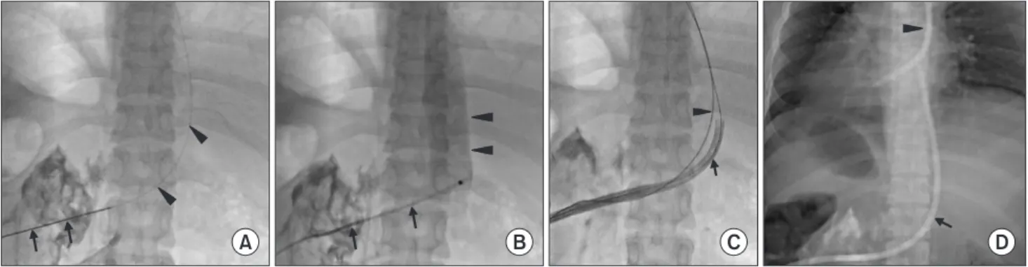
Fig. 1. Transrenal inferior vena cava (IVC) access in a 31-year-old female patient with exhausted central venous access. (A) The left renal vein was punctured successfully with a 21-gauge needle (arrows) after making several attempts. A 0.018-inch guidewire (arrowheads) was inserted into the IVC through the needle. (B) After exchanged for a coaxial transitional sheath (arrows), small amount of contrast media was injected for confirmation the lumen of IVC (arrowheads). (C) Double guide wire technique was performed. A catheter was inserted via one guidewire (arrow) and another wire (arrowhead) was kept as a safety guidewire in case of tract loss. (D) A transrenal permanent HD catheter (arrow) was inserted with the tip located in the upper IVC. A central venous permanent catheter (arrowhead) previously inserted with malfunction is visible.
between the vein entry point and the exit site near the mid- axillary line. Then the tract was dilated serially, and a peel-away sheath was inserted within it. The tip of permanent catheter was in the mid- to low right atrium.
RESULTS
Demographics and clinical characteristics of the study population are in Table 1. Mean age of the five study patients was 56 years old (range, 31-83). Three patients had diabetes mellitus for the causes of end stage renal disease (ESRD) and the others had unknown etiology of it. Type of initial RRT included kidney transplantation (KT) (Patient No. 1, 2) and AVF (No. 3, 4, 5), and mean treatment period was 10 years (range, 3-16).
Due to graft rejection or malfunction of the fistula, it had
been converted to HD through permanent catheter insertion.
After failure of the initial RRT, HD was performed with a permanent catheter through the central veins for an average of 21.8 months (range, 10-48). One patient (No. 1) had long- term chemotherapy for the right ureter cancer using ports via central veins. She needed transrenal permanent catheter because of chemotherapy-induced graft rejection and central venous impairment. Three patients (No. 3, 4, 5) underwent the permanent catheter placement via transhepatic or transfemoral access for the alternative routes before transrenal or translumbar approach. It was failed to maintain HD properly because of vulnerability of infection and malfunction, resulting in the search of the other alternative routes.
The transrenal (n=3) and translumbar (n=2) access were all technically successful. In transrenal access, the tract was lost due
A B C D
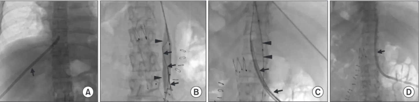
Fig. 2. Translumbar inferior vena cava (IVC) access in a 48-year-old female patient with exhausted central venous access. (A) Insertion of transhepatic catheter (arrow) was performed in advance of translumbar catheter placement. Note the aortic stent graft which was inserted for the inadvertent aortic puncture during IVC cannulation. (B) The inflated 16-mm diameter balloon catheter (arrowheads) which was inserted through the transhepatic route was punctured using a 21-gauge Chiba needle, and an inserted hair wire (arrows) in the Neff set could be confirmed to be in the lumen of the balloon, and which indicates successful IVC cannulation. (C) Serial dilation (arrows) was performed after exchanging with a stiff 0.035-inch guidewire. Note the deflated balloon catheter (arrowheads) and guidewire via the transhepatic route along the IVC course. (D) The final spot radiograph with the patient in the prone position shows the successfully inserted HD catheter (arrow) with the tip near the right atrium.
Table 1. Patient characteristics and clinical outcome
No./Sex/
Age Causes of ESRD
Duration of initial (years)RRT
Duration of CVC (months)
Alternative permanent catheter
Subse-
quent RRT Remark
Kinds Duration
(days) Complication
1/F/64 DM 13 48 Transrenal 22 AVF Chemotherapy for ureter
cancer
2/F/31 DM 3 23 Transrenal 145 AVF
3/M/83 DM 3 18 Transrenal 145 AVF
4/M/54 Unknown 15 10 Translumbar 115 KT
5/F/48 Unknown 16 10 Translumbar 165 Aorta puncture AVF Aorta puncture was
managed with stent graft ESRD, end stage renal disease; RRT, renal replacement therapy; CVC, central venous catheter; AVF, arteriovenous fistula; KT, kidney transplantation; DM, diabetes mellitus.
to the shortage of the peel away sheath, about 13 cm in length, in one patient (No. 1); the catheter inserted through the peel- away sheath did not enter the renal vein after the guidewire and dilator of the sheath have been removed. However, with a safety guidewire, the tract was reused, and the catheter could enter the renal vein by further advancing the peel-away sheath at the second attempt. In two patients with translumbar access, the 16 mm diameter balloon catheter inserted through transhepatic route (Fig. 1, No. 5) or 18 mm diameter balloon catheter inserted through transfemoral route (No. 4) was used as a puncture target.
In both transrenal and translumbar accesses, the distal portion of the peel-away sheath was bent to be smoothly advanced into the IVC. The duration of use of alternative transrenal or translumbar permanent catheters averaged 118.4 days (range, 22-165 days).
There was one procedure-related complication in one patient (No. 5)- the aorta was inadvertently punctured and a 6Fr Neff sheath was inserted. After noticing it was into the aorta, stent graft was placed with simultaneous removal of the Neff sheath uneventfully.
After using alternative HD catheters, four patients have been transferred to AVF and one patient underwent KT at the last follow-up. Catheter removal was the same as that of catheter removal at the jugular vein, and there were no compilations related to catheter removal.
DISCUSSION
To maintain durable vascular access is essential to the patients with ESRD for the long-term use of HD. Using AVF or AVG is regarded better method because the risk of infection is lower than using HD catheters [4]. However, permanent HD catheter is the only reasonable way to HD when malfunction of the fistula or graft occurs or maturation of them after surgery is necessary [5,6].
Repeated and prolonged venous access can lead to impairment such as vascular stenosis, thrombosis or infection, which results in the progressive loss of vascular access and significant morbidity and mortality [5,7]. Typical examples include patients with superior vena cava obstruction, or patients with unilateral obstruction of a brachiocephalic vein in whom access using the contralateral side is contraindicated. When traditional venous access routes such as jugular and subclavian veins are exhausted, alternative access routes, i.e. hepatic vein, translumbar infra- and suprarenal IVC, renal vein, neck collaterals, brachiocephalic, cephalic, hemiazygos and azygos veins have been considered for performing HD [4,6-8]. The
procedures using the alternative routes are considerably more complicated than the common central venous access procedures [9].
The venous anatomy, history of prior access sites and physical examination of the patients are important to choose both the best alternative route as well as the device. Histories about the prior use of ports, tunneled catheters, temporary HD catheters, pacemakers, and IVC filters are all particularly important [9].
The presence of underlying coagulopathy should be noted and disorders related with bleeding and thrombosis must be checked in advance [5]. Current imaging studies such as ultrasound, computed tomography (CT) or diagnostic venography should be taken to obtain the accurate venous anatomy [3]. The review of CT images before the procedure can reduce the risk of complications such as renal vein or aortic injury, damage to the ipsilateral renal parenchyma, and bowel injury especially in patients with limited ultrasonic windows [5,10].
Translumbar catheterization is a technically challenging interventional procedure. IVC could be accessed by targeting a marker from a guide wire or a pigtail catheter placed in the IVC from the femoral venous approach under C arm fluoroscopy [11]. In one patient in this study, blind cannulation resulted in inadvertent aortic puncture with management with a stent graft.
Successful IVC cannulation was possible by puncturing the inflated balloon catheter in the IVC under fluoroscopic guidance which was inserted through transhepatic or transfemoral routes.
Transrenal venous access can be considered an another good alternative when conventional access options are impaired [5].
There are a few case reports about the patency of the transrenal catheters which functioned for six months [8] or two years [6]. Renal parenchymal injury related with the procedure is not a significant issue because the native kidneys are hypo- or nonfunctional in ESRD. Based on the anatomy of renal hilum, however, the possibility of renal arterial injury is always present in the procedure. Thus, embolization is necessary in the event of bleeding with arterial injury [5,8]. Double guidewire insertion technique, that is keeping another guidewire as a safety guidewire in cases of tract loss, seems quite necessary especially when long peel-away sheath is not available. The length of the used peel-away sheath in this study was 13cm, and which can be short for patients where skin entry point is away from puncturable renal vein.
The patency of the transrenal or translumbar permanent catheters, used as an alternative route in this study, averaged 118.4 days (range, 22-165). Actual patency will be more than this because the catheters were removed for conversion to other
types of RRT, such as AVF or KT, rather than dysfunction or complications. Furthermore, the average patency duration is longer about two months than that reported in previous article by Liu et al [7].
The most common reported complications of transrenal or translumbar catheter were catheter-related thrombosis and infection and other minor things included fibrin sheath formation or decreased blood flow [12]. During the use of them in all five patients in this study, no thrombosis or infection occurred.
All the patients of this study have been successfully transferred to AVF or KT as a subsequent RRT after using alternative permanent catheters for more stable HD. It could be suggested that transrenal or translumbar catheters can provide intermediate process as a bridge to a new permanent or more effective access for patients who have exhausted traditional central access for HD [7].
This study has several limitations related with retrospective nature and small numbers of patients and their catheters. Risk factors of stenosis or infection in the transrenal or translumbar catheter could not be determined due to small patient number and short follow-up period.
In conclusion, placement of HD catheters using transrenal or translumbar route is relatively safe, feasible and tolerable method when central venous access is exhausted, and their catheters can be maintained for a reasonable period of time.
REFERENCES
1. Gameiro J, Fonseca JA, Jorge S, et al. Management of end-stage vascular access failure patients: a retrospective analysis. Portuguese Journal of Nephrology & Hypertension.
2018; 32: 324-30.
2. Pereira M, Lopez N, Godinho I, et al. Life-saving vascular access in vascular capital exhaustion: single center experience in intra-atrial catheters for hemodialysis. Brazilian Journal of Nephrology. 2017; 39: 36-41.
3. Falk A. Use of the Brachiocephalic Vein for Placement of Tunneled Hemodialysis Catheters. American Journal of Roentgenology. 2006; 187: 773-7.
4. Power A, Singh S, Ashby D, et al. Translumbar central venous catheters for long-term haemodialysis. Nephrol Dial Transplant. 2010; 25: 1588-95.
5. Shin JH. Hemodialysis Access Salvage Techniques in Patients with Exhausted Access. Journal of Korean Dialysis Access. 2018; 1: 21-7.
6. Law WP, Cheung CY, Chan HW, et al. Hemodialysis catheter insertion using transrenal approach. Hemodial Int. 2015; 19:
E14-6.
7. Liu F, Bennett S, Arrigain S, et al. Patency and Complications of Translumbar Dialysis Catheters. Seminars in Dialysis.
2015; 28: E41-E7.
8. Murthy R, Arbabzadeh M, Lund G, et al. Percutaneous transrenal hemodialysis catheter insertion. J Vasc Interv Radiol. 2002; 13: 1043-6.
9. Denny DF Jr. Venous access salvage techniques. Tech Vasc Interv Radiol. 2011; 14: 225-32.
10. Thakor AS, Chung J, Patel R, et al. The use of cone-beam CT in assisting percutaneous translumbar catheter placement into the inferior vena cava. Clinical Radiology. 2015; 70: 21-4.
11. Yaacob Y, Zakaria R, Mohammad Z, et al. The vanishing veins: difficult venous access in a patient requiring translumbar, transhepatic, and transcollateral central catheter insertion. The Malaysian Journal of Medical Sciences : MJMS. 2011; 18: 98-102.
12. Rahman S, Kuban JD. Dialysis Catheter Placement in Patients With Exhausted Access. Techniques in Vascular and Interventional Radiology. 2017; 20: 65-74.