Renal Dysfunction in Patients with Chronic Liver Disease
Jay Wook Lee, M.D.
Division of Nephrology, Department of Internal Medicine, Chung-Ang University College of Medicine, Seoul, Korea
Renal dysfunction in patients with chronic liver disease encompasses a clinical spectrum of hyponatremia, ascites, and hepatorenal syndrome. Clinical observation has suggested that patients with cirrhosis have hyper- dynamic circulation, and recent studies strongly suggest that peripheral arterial vasodilatation and subsequent development of hyperdynamic circulation are responsible for disturbances in renal function. Arterial vaso- dilatation predominantly occurs in the splanchnic vascular bed, and seems to precede an increase in blood flow in the splanchnic circulation. Nitric oxide plays a central role in progressive vasodilatation, as evidenced by in vivo and in vitro studies. Renal dysfunction negatively affects the prognosis of patients with cirrhosis, as hyponatremia, ascites, and azotemia are associated with increased rate of complications and mortality.
Recent advances in understanding the pathophysiology of renal dysfunction have enabled clinicians to devel- op new diagnostic criteria and therapeutic recommendations. Hepatorenal syndrome is regarded as a poten- tially reversible disorder, as systemic vasoconstrictors with concomitant albumin administration are emerging as a promising management option, especially in terms of providing bridging therapy for patients awaiting liver transplantation.
Electrolytes Blood Press 7:42-50, 2009ㆍdoi: 10.5049/EBP.2009.7.2.42 Key Words: liver cirrhosis; hepatorenal syndrome; hyponatremia
1)
Introduction
Renal dysfunction in chronic liver disease is charac- terized by impaired natriuresis, decreased free water clear- ance, and decreased glomerular filtration rate (GFR).
Hyponatremia, ascites, and hepatorenal syndrome (HRS) represent the clinical consequences of disturbances in renal function. Optimal management of renal dysfunction in cir- rhosis is extremely important in that renal dysfunction fre- quently complicates the clinical course of advanced liver disease and is invariably associated with poor clinical outcomes. Hyponatremia is present in about 50% of pa-
Received November 2, 2009. Accepted November 23, 2009.
Corresponding author: Jay Wook Lee, M.D.
Department of Internal Medicine, Chung-Ang University College of Medicine Yong-San Hospital, 65-207, Hangangro-3-ga, Yongsan-gu, Seoul, 140-757, Korea
Tel : +82-2-748-9841, Fax : +82-2-790-2068 E-mail: [email protected]
tients with cirrhosis and is associated with increased rate of other complications such as gastrointestinal bleeding, spontaneous bacterial peritonitis, and hepatic encephalop- athy1). The presence of ascites predicts poor clinical out- come in cirrhotic patients, as shown by the 3-year survival rate for patients with ascites at 50%2). Progressive liver failure and superimposition of precipitating events culmi- nate in the development of HRS, a state of severe intrarenal vasoconstriction and reduced GFR without intrinsic renal damage. Survival of patients with liver disease continues to be affected by the presence of renal dysfunction, even after they underwent liver transplantation3).
Renal dysfunction in cirrhosis is a clinical consequence of peripheral arterial vasodilatation and hyperdynamic cir- culation caused by portal hypertension. Clinical ob- servations and recent experimental studies have shed light on the pathogenesis of hyperdynamic circulation in chronic
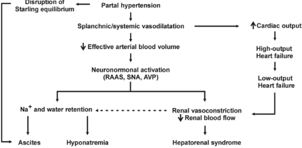
Fig. 1. Pathogenesis of renal dysfunction in chronic liver disease. RAAS, renin-angiotensin- aldosterone system; SNS, sympathetic nervous system; AVP, arginine vasopressin.
liver disease. Better understanding of the pathophysiology enabled clinicians to introduce effective therapies for renal dysfunction once considered irreversible or medically in- tractable, and led to the proposal of new concepts and diag- nostic criteria for HRS4).
Pathophysiology of renal dysfunction in chronic liver disease
1. Peripheral arterial vasodilatation and hyperdy- namic circulation in chronic liver disease Portal hypertension in cirrhosis is one of the best exam- ples of hyperdynamic circulation, which results from a combination of increased cardiac output and dilated periph- eral vascular bed5). Investigators have been aware of the importance of primary vasodilatation in cirrhosis for a long time, and bedside observation made by astute clinicians describes the typical features of hyperdynamic circulation in patients with cirrhosis, including increased pulse pres- sure, warm extremities, and capillary pulsations in the nail bed. Based on these findings, Kowalski and Abelmann were the first to demonstrate an increase in cardiac output and a decrease in peripheral vascular resistance in a patient with alcoholic cirrhosis6). A subsequent study corroborated this finding7). In addition, a host of studies using Doppler ultrasonography to assess blood flow in various organs in patients with cirrhosis have demonstrated (1) the state of hyperdynamic circulation in chronic liver disease, (2) pre- dominant vasodilatation in the splanchnic vessels, and (3)
relative vasoconstriction in other organs such as the kid- neys8, 9).
Splanchnic and systemic vasodilatation in the wake of portal hypertension creates a state of relative hypovolemia, which activates sodium-conserving mechanisms and leads to an increase in plasma volume. Most of the increase in plasma volume is used to fill up the increased splanchnic vascular compartment. Meanwhile, portal blood flow in- creases in the face of increased intrahepatic vascular resist- ance, as portosystemic collaterals partially decompress the portal vein and provides conduits for pooled splanchnic blood. The importance of increased blood flow in the portal vein was shown in a previous study, which reports that an increase in portal venous flow is a major contributing factor in maintaining and aggravating portal hypertension in conditions of increased intrahepatic resistance10).
2. Alterations in renal function
Splanchnic and systemic vasodilatation leads to renal vasoconstriction and impaired renal function (Fig. 1).
Relative hypovolemia activates the sympathetic nervous system and renin-angiotensin-aldosterone system, and in- creases nonosmotic secretion of arginine vasopressin (AVP), resulting in sodium and water retention and devel- opment of ascites and hyponatremia. Renal hypoperfusion and cardiac dysfunction combine to provoke severe intra- renal vasoconstriction leading to HRS.
The clinical course of renal impairment in cirrhosis is
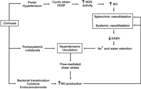
Fig. 2. Pathogenesis of hyperdynamic circulation in cirrhosis. VEGF, vascular endothelial growth factor; NOS, nitric oxide synthase; NO, nitric oxide; EABV, effective arterial blood volume.
further complicated by progressive cardiac dysfunction. As a result of peripheral vasodilatation and retention of so- dium and water, cardiac output increases in the early phase of chronic liver disease. However, this increase in cardiac output fails to meet the need of the body and results in high-output cardiac failure. As peripheral vasodilatation continues to progress, myocardial contractility and cardiac output begin to decrease and low-output cardiac failure en- sues11). Reduced cardiac output decreases renal perfusion and may precipitate the development of HRS, as renal hy- poperfusion seems to trigger severe intrarenal vaso- constriction12).
3. Pathogenesis of splanchnic arterial vasodilatation In vivo and in vitro studies have investigated the mecha- nisms of splanchnic vasodilatation in chronic liver disease by using experimental animal models for cirrhosis. Major findings from these studies are as follows: (1) the source of vascular hyporeactivity to vasoconstricting stimuli lies in the vascular endothelium13); (2) endothelial and neuronal isoforms of nitric oxide synthase (eNOS and nNOS, re- spectively) are upregulated in the splanchnic circulation14); (3) cirrhotic animals are remarkably sensitive to the effect of nonspecific inhibition of nitric oxide (NO) synthesis as compared with normal controls15); (4) and inhibition of NO
synthesis almost completely normalize major hemody- namic abnormalities and renal function16, 17). Vascular hy- poresponsiveness and increased NO production were ob- served in major systemic arteries as well as in splanchnic arteries18, 19).
Nitric oxide is believed to be a key player in patho- genesis of splanchnic and systemic vasodilatation in chron- ic liver disease. Portal hypertension activates eNOS and nNOS in the splanchnic circulation through a myriad of putative mechanisms, including increased shear stress on the mesenteric arterial wall, increased expression of vas- cular endothelial growth factor (VEGF) in the splanchnic microcirculation, overproduction of inflammatory cyto- kines (e.g., tumor necrosis factor), and bacterial trans- location (Fig. 2). Upregulation of NO synthases is not like- ly to be induced by increased mesenteric blood flow, as the time-course of NO production has shown that over- production of NO occurs before the blood flow increases in the superior mesenteric artery20). Rather, other signals located upstream of NO, such as cyclic stress in the mesen- teric arterial wall or increased expression of VEGF, seem to be involved in the enhanced eNOS activity. Additional studies lend support to the role of VEGF in splanchnic vasodilatation by showing that inhibition of VEGF receptor effectively attenuates splanchnic vasodilatation21, 22).
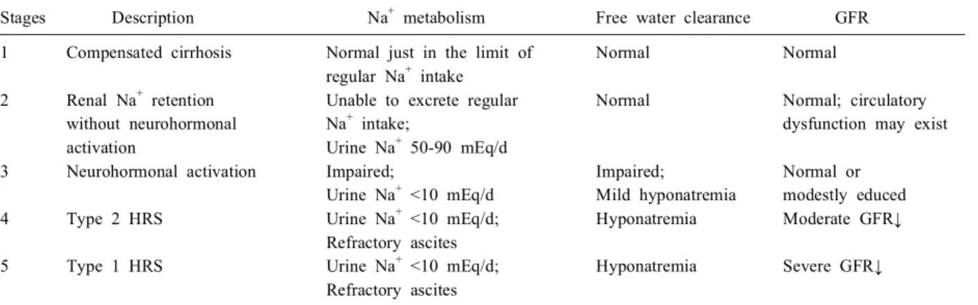
Table 1. Stages of Renal Dysfunction in Chronic Liver Disease
Stages Description Na+ metabolism Free water clearance GFR
1 Compensated cirrhosis Normal just in the limit of Normal Normal
regular Na+ intake
2 Renal Na+ retention Unable to excrete regular Normal Normal; circulatory
without neurohormonal Na+ intake; dysfunction may exist
activation Urine Na+ 50-90 mEq/d
3 Neurohormonal activation Impaired; Impaired; Normal or
Urine Na+ <10 mEq/d Mild hyponatremia modestly educed
4 Type 2 HRS Urine Na+ <10 mEq/d; Hyponatremia Moderate GFR↓
Refractory ascites
5 Type 1 HRS Urine Na+ <10 mEq/d; Hyponatremia Severe GFR↓
Refractory ascites GFR, glomerular filtration rate; HRS, hepatorenal syndrome.
Increased NO production leads to splanchnic and sys- temic arterial vasodilatation, which, combined with in- creased effective arterial blood volume, increases blood flow and augments NO production, as an increase in blood flow is a well-known stimulus for NO synthases (Fig. 2).
As mentioned before, portosystemic shunts have a sig- nificant role in maintaining or aggravating hyperdynamic circulation, and increased portal blood flow can enhance the production of NO in the splanchnic circulation. Circu- lating hormones (e.g., endocannabinoids), gastrointestinal hormones (e.g., glucagon), and proinflammatory cytokines (e.g., tumor necrosis factor-alpha) induced by bacterial translocation can contribute to increased NO produc- tion23-25).
There are NO-independent mechanisms involved in hy- perdynamic circulation in cirrhosis, as knockout of eNOS did not completely prevent the development of hyper- dynamic circulation in portal hypertension26). Studies on impaired reactivity of the endothelium to vasoconstrictors in cirrhotic animals have reported reduction in phosphor- ylation of the myosin light chain (MLC) of the vascular smooth muscle cell (VSMC). Contractile agonists usually stimulate MLC phosphorylation via the activation of MLC kinase or the inhibition of MLC phosphatase. Phosphory- lated MLC in turn activates actin-myosin ATPase, thereby crosslinking actin-myosin to induce smooth muscle con- traction. Vasoconstrictors such as epinephrine and vaso- pressin bind to their respective receptors on the surface of the VSMC and activate phospholipase C (PLC), which
produces inositol triphosphate (IP3) and diacylglycerol (DAG). IP3 mobilizes calcium from the sarcoplasmic retic- ulum, and DAG activates protein kinase C which is in- volved in the increased activity of MLC kinase. In addi- tion, receptor activation leads to increased activity of rho kinase, which is believed to inhibit the activity of MLC phosphatase. As a result, increased MLC phosphorylation and increased intracellular calcium concentration leads to smooth muscle contraction.
Reported alterations in PLC and rho kinase signaling in cirrhotic animals include decreased activation of PLC;
subsequently diminished production of IP3 and DAG; re- duced activity of DAG-dependent protein kinase C; and reduced rhoA activity27-29).
Clinical manifestations
Hyponatremia, ascites, and HRS are among the principal clinical consequences of the progressive arterial vaso- dilatation and hyperdynamic circulation in cirrhosis.
Impaired sodium excretion and free water clearance result in the development of hyponatremia and ascites, and pro- gressive decrease in GFR results in the development of HRS.
Progression of renal dysfunction in cirrhosis can be div- ided into five stages (Table 1)30). In the early phase of liver dysfunction (stage 1), patients can excrete daily so- dium intake, though just in the range of normal daily so- dium intake, and show normal GFR and absence of ascites.
In the next phase of renal dysfunction (stage 2), impaired
natriuresis takes place without overt activation of neuro- hormonal systems, as patients cannot mount an adequate natriuretic response to exogenous sodium load while con- centrations of plasma catecholamine, aldosterone or AVP remain within the normal range. Increased expression of renal sodium transporters in the distal tubule seems to be involved in this phenomenon31). Subtle unidentified circu- latory dysfunction also may play a role. As liver function deteriorates (stage 3), overt activation of neurohormonal systems occurs, and ascites and hyponatremia develop. In the final stages of cirrhosis, severe vasodilatation and renal vasoconstriction lead to refractory ascites and HRS. Type 2 HRS (stage 4) is considered to be a mild, slowly pro- gressive form of renal failure, and type 1 HRS (stage 5) is characterized by rapidly worsening renal function, re- fractory ascites, and severe sodium retention.
Hyponatremia is associated in a graded fashion with oth- er serious complications of liver cirrhosis. As compared with patients with mild hyponatremia (plasma sodium 131-135 mEq/L), patients with moderate-to-severe hypona- tremia (plasma sodium ≤130 mEq/L) have significantly higher risk of developing hepatic encephalopathy, sponta- neous bacterial peritonitis, and gastrointestinal bleeding1). The most common precipitating factor for hyponatremia is inappropriate use of diuretics, especially for patients with ascites but without peripheral edema. As the peritoneum has limited capacity for mobilizing ascitic fluid (~500 mL/day), excessive diuresis can reduce intravascular vol- ume and renal free water clearance, leading to development of hyponatremia.
Ascites develops as a result of sodium retention by the kidney and disruption of the Starling equilibrium in the splanchnic circulation. As mentioned earlier, activation of the sympathetic nervous system, renin-angiotensin-aldo- sterone axis, and nonosmotic AVP secretion leads to so- dium and water retention by the kidney. Increased hydro- static pressure and decreased oncotic pressure in the capil- lary of the bowel, which occur as a result of portal hyper- tension, increases hepatic lymph production.
HRS is caused by severe renal vasoconstriction which occurs in patients with advanced liver disease and circu-
latory dysfunction. By definition, HRS is not associated with intrinsic renal disease or nephrotoxic injury, as kid- neys from patients with HRS function perfectly when transplanted to other patients. HRS may occur sponta- neously, but is frequently precipitated by bacterial in- fections such as spontaneous bacterial peritonitis (SBP), gastrointestinal bleeding, or inadequate albumin replace- ment after therapeutic paracentesis. These precipitating events abruptly reduce renal perfusion, thereby tipping the balance toward vasoconstriction between intrarenal vaso- dilators such as prostaglandins and vasoconstrictors. Imbal- ance between vasoactive mediators within the kidney fur- ther diminishes renal blood flow and causes HRS.
The diagnosis of HRS is based upon documentation of progressive renal failure and exclusion of intrinsic struc- tural renal damage and other systemic illness affecting re- nal function. Type 1 HRS is a severe, rapidly progressive type of renal failure (i.e. doubling of serum creatinine to a level greater than 2.5 mg/dL in less than 2 weeks), while type 2 HRS represents moderate, slowly progressive renal failure (i.e. serum creatinine 1.5-2.5 mg/dL). Since the first proposal of diagnostic criteria in 199432), new concepts have emerged from experimental and clinical studies: (1) peripheral arterial vasodilatation predominantly occurs in the splanchnic vascular bed; (2) cardiac output in patients with HRS is insufficient for the patient’s needs; (3) devel- opment of type 1 HRS is often triggered by other super- imposing factors, most commonly spontaneous bacterial peritonitis or gastrointestinal bleeding; (4) renal function can be improved by medical treatment in patients with HRS4). As compared with the diagnostic criteria in 1994, new diagnostic criteria proposed in 2007 (1) eliminated the additional criteria as they are not essential for diag- nosis; (2) recommended that plasma volume expansion should be performed with albumin rather than saline; (3) excluded creatinine clearance as it is impractical and diffi- cult to interpret in chronic liver disease; (4) suggested that renal failure in the setting of ongoing bacterial infection should be considered as HRS, emphasizing that effective therapy should be started before the resolution of bacterial infection4).
Management 1. Hyponatremia
Restriction of free water intake is the mainstay of treat- ment for hyponatremia. Severe symptomatic hyponatremia should be managed with hypertonic saline infusion.
However, both treatments are not aimed to address the un- derlying pathophysiology of water retention, the non- osmotic AVP secretion.
Pharmacologic inhibition of AVP action is emerging as a valuable addition to the armamentarium for management of hypervolemic or euvolemic hyponatremia. Binding to V2 receptor on the basolateral surface of the collecting tub- ular epithelial cells, V2 receptor antagonists (vaptans) in- hibit the action of AVP and increase free water clearance.
Several studies on cirrhotic patients with ascites and hypo- natremia showed that V2 receptor antagonists increased uri- nary excretion of free water and plasma sodium concen- tration and were not associated with increased rates of seri- ous adverse events33-35). A multi-center, randomized, place- bo-controlled trial on 110 patients with ascites and hypona- tremia reported that satavaptan improved the control of as- cites, moderately increased plasma sodium concentration, and was not associated with significant alterations in plas- ma and urine electrolyte concentrations33). Patients who start on V2 receptor antagonists may require admission, as clinical trials with tolvaptan showed that a small fraction of patients (~2%) had early-stage increases in plasma so- dium concentration higher than the acceptable range35).
2. Ascites
To counterbalance sodium retention in cirrhosis, neg- ative sodium balance should be achieved by reducing daily sodium intake to less than 2 g/d and by starting diuretic therapy. Sodium restriction should be maintained through- out the entire course of ascites management. Mineralocorti- coid antagonists are the first-line diuretics in management of ascites, as secondary hyperaldosteronism has been con- sidered important in the pathogenesis of ascites. Loop diu- retics and thiazide may be added if natriuresis is insuffi- cient. As excessive natriuresis can greatly reduce intra-
vascular volume and precipitate acute renal failure, caution should be exercised especially for patients with ascites in the absence of peripheral edema. Refractory ascites should be managed with therapeutic paracentesis with albumin re- placement, construction of peritoneovenous shunt, or trans- hepatic intrajugular portosystemic shunt (TIPS).
3. Hepatorenal syndrome
An ideal treatment for HRS should be aimed at improv- ing renal perfusion and glomerular filtration rate. It should achieve (1) reduction of serum creatinine to less than 1.5 mg/dL, (2) prolongation of survival, and (3) avoidance of serious adverse effects. Despite remarkable advances in the understanding of the pathophysiology of HRS, no ideal medical therapy has been discovered so far.
Liver transplantation is the only treatment proven to pro- long survival in patients with HRS. It is important, how- ever, to provide adequate management aimed at reversal of azotemia, because preoperative azotemia negatively af- fects the prognosis of the patients who underwent liver transplantation, and because effective treatment for HRS can earn valuable time for patients who await liver trans- plantation3).
Given the central role of splanchnic vasodilatation and hyperdynamic circulation in the development of renal dys- function, new medical therapies are designed to reduce in- tense arterial vasodilatation and to expand the effective cir- culating volume. Systemic vasoconstrictor therapies, com- bined with albumin infusion, were found to be effective in patients with HRS in reversing renal dysfunction, and the survival of the patients who recovered from their renal dysfunction was significantly improved36-39). The available vasoconstrictors proven to be effective in clinical trials are vasopressin analogues (e.g., terlipressin, ornipressin)36, 37), midodrine plus octreotide38), and norepinephrine39). Com- plications such as myocardial infarction, cerebrovascular disease or hypertension may preclude the use of vaso- constrictors. Concomitant volume expansion with intra- venous albumin is essential, as vasoconstrictor therapy without albumin produced little effect in reversing azote- mia40). An international group of experts issued treatment
Table 2. A Summary of Treatment and Prevention of Hepatorenal Syndrome Treatment of hepatorenal syndrome
Liver transplantation
Systemic vasopressors with concomitant albumin infusion
Terlipressin: 0.5 mg IV q4-6 h; raise the dose up to 1 mg/4 h (maximum: 2 mg/4 h) Midodrine with octreotide
Midodrine: 2.5-7.5 mg PO three times a day (maximum: 12.5 mg three times a day) Octreotide: 100 mcg SC three times a day (maximum: 200 mcg three times a day) Norepinephrine: 0.5-3 mg/h IV
Albumin: 20-40 g/day after 1 g/kg on the first day of vasoconstrictor administration Transjugular intrahepatic portosystemic shunt (TIPS)
Renal replacement therapies (only for transplant candidates) Prevention of hepatorenal syndrome
Avoidance of volume depletion
Aggressive management of gastrointestinal bleeding and spontaneous bacterial peritonitis Administration of norfloxacin
IV, intravenous; PO, per os; SC, subcutaneous.
recommendations for the management of type 1 HRS4). TIPS may be considered as a therapeutic option for HRS, especially when the risk of hepatic encephalopathy is low. Unfortunately, most patients with HRS are too ill to undergo TIPS. The Model for End-stage Liver Disease (MELD) score, a scoring system developed to predict the survival of patients with chronic liver disease and to priori- tize organ allocation to patients in more critical condition, can be used to predict the survival of patients who under- went TIPS. A computational model suggested that TIPS should not be recommended for patients with a MELD score >18, as their median survival would be less than 3 months41).
Renal replacement therapies should be considered when patients are awaiting a liver transplant or when there is the possibility of improvement in liver function. In one retrospective study, 30 percent of patients who required dialysis survived to liver transplantation42). Patients with an acute and potentially reversible liver dysfunction may benefit from renal replacement therapy until they recover their liver function. Management of HRS is summarized in Table 2.
4. Prevention of HRS
Two studies have evaluated the efficacy of preventive measures for HRS. Sort et al. reported that intravenous albumin infusion combined with antibiotics for patients
with SBP was associated with reduced rate of impaired renal function and increased survival, as compared with antibiotics alone43). Intravascular volume expansion might have been helpful to reverse the progression of renal dys- function in patients with SBP, as bacterial translocation and increased inflammatory cytokines might augment nitric oxide production through the putative tetrahydrobiopterin synthesis pathway.
A randomized trial reported significant benefits with the administration of norfloxacin at 400 mg/day to patients with cirrhosis who met the following criteria: (a) ascites fluid total protein <1.5 g/dL and either of the following:
(b) a Child-Pugh score ≥9 points and serum bilirubin ≥ 3 mg/dL; or (c) serum creatinine ≥1.2 mg/dL, blood urea nitrogen ≥25 mg/dL, or serum sodium ≤130 mEq/L44). Norfloxacin was associated with decreased one-year proba- bility of SBP and HRS, and improved 3-month survival.
Acknowledgements
The author is grateful to the Korean Society of Electro- lyte Metabolism and the Korean Society of Nephrology for granting an opportunity to present and publish this review.
References
1) Angeli P, Wong F, Watson H, Gines P: Hyponatremia in cirrhosis: Results of a patient population survey. Hepatology 44:1535-1542, 2006
2) Arroyo V, Gines P, Planas R, Panes J, Rodes J: Management of patients with cirrhosis and ascites. Semin Liver Dis 6:353-369, 1986
3) Gonwa TA, McBride MA, Anderson K, Mai ML, Wadei H, Ahsan N: Continued influence of preoperative renal func- tion on outcome of orthotopic liver transplant (OLTX) in the US: where will MELD lead us? Am J Transplant 6:
2651-2659, 2006
4) Salerno F, Gerbes A, Gines P, Wong F, Arroyo V:
Diagnosis, prevention and treatment of hepatorenal syn- drome in cirrhosis. Gut 56:1310-1318, 2007
5) Wiest R: Splanchnic and systemic vasodilation: the ex- perimental models. J Clin Gastroenterol 41(Suppl 3):S272- 287, 2007
6) Kowalski HJ, Abelmann WH: The cardiac output at rest in Laennec's cirrhosis. J Clin Invest 32:1025-1033, 1953 7) Murray JF, Dawson AM, Sherlock S: Circulatory changes
in chronic liver disease. Am J Med 24:358-367, 1958 8) Rivolta R, Maggi A, Cazzaniga M, et al.: Reduction of renal
cortical blood flow assessed by Doppler in cirrhotic patients with refractory ascites. Hepatology 28:1235-1240, 1998 9) Sacerdoti D, Bolognesi M, Merkel C, Angeli P, Gatta A:
Renal vasoconstriction in cirrhosis evaluated by duplex Doppler ultrasonography. Hepatology 17:219-224, 1993 10) Sikuler E, Groszmann RJ: Interaction of flow and resistance
in maintenance of portal hypertension in a rat model. Am J Physiol 250:G205-212, 1986
11) Ruiz-del-Arbol L, Monescillo A, Arocena C, et al.: Circula- tory function and hepatorenal syndrome in cirrhosis.
Hepatology 42:439-447, 2005
12) Arroyo V, Colmenero J: Ascites and hepatorenal syndrome in cirrhosis: pathophysiological basis of therapy and current management. J Hepatol 38(Suppl 1):S69-89, 2003 13) Atucha NM, Shah V, Garcia-Cardena G, Sessa WE,
Groszmann RJ: Role of endothelium in the abnormal re- sponse of mesenteric vessels in rats with portal hypertension and liver cirrhosis. Gastroenterology 111:1627-1632, 1996 14) Niederberger M, Gines P, Martin PY, et al.: Comparison
of vascular nitric oxide production and systemic hemody- namics in cirrhosis versus prehepatic portal hypertension in rats. Hepatology 24:947-951, 1996
15) Claria J, Jimenez W, Ros J, et al.: Pathogenesis of arterial hypotension in cirrhotic rats with ascites: role of endogenous nitric oxide. Hepatology 15:343-349, 1992
16) Pizcueta MP, Pique JM, Bosch J, Whittle BJ, Moncada S:
Effects of inhibiting nitric oxide biosynthesis on the sys- temic and splanchnic circulation of rats with portal hyper- tension. Br J Pharmacol 105:184-190, 1992
17) Niederberger M, Martin PY, Gines P, et al.: Normalization of nitric oxide production corrects arterial vasodilation and hyperdynamic circulation in cirrhotic rats. Gastroenterology 109:1624-1630, 1995
18) Claria J, Jimenez W, Ros J, et al.: Increased nitric oxide-de- pendent vasorelaxation in aortic rings of cirrhotic rats with ascites. Hepatology 20:1615-1621, 1994
19) Gadano AC, Sogni P, Yang S, et al.: Endothelial cal- cium-calmodulin dependent nitric oxide synthase in the in vitro vascular hyporeactivity of portal hypertensive rats. J Hepatol 26:678-686, 1997
20) Colombato LA, Albillos A, Groszmann RJ: Temporal rela- tionship of peripheral vasodilatation, plasma volume ex- pansion and the hyperdynamic circulatory state in portal-hy- pertensive rats. Hepatology 15:323-328, 1992
21) Wiest R, Shah V, Sessa WC, Groszmann RJ: NO over- production by eNOS precedes hyperdynamic splanchnic cir- culation in portal hypertensive rats. Am J Physiol 276:
G1043-1051, 1999
22) Abraldes JG, Iwakiri Y, Loureiro-Silva M, Haq O, Sessa WC, Groszmann RJ: Mild increases in portal pressure upre- gulate vascular endothelial growth factor and endothelial ni- tric oxide synthase in the intestinal microcirculatory bed, leading to a hyperdynamic state. Am J Physiol Gastrointest Liver Physiol 290:G980-987, 2006
23) Batkai S, Jarai Z, Wagner JA, et al.: Endocannabinoids act- ing at vascular CB1 receptors mediate the vasodilated state in advanced liver cirrhosis. Nat Med 7:827-832, 2001 24) Wiest R, Das S, Cadelina G, Garcia-Tsao G, Milstien S,
Groszmann RJ: Bacterial translocation in cirrhotic rats stim- ulates eNOS-derived NO production and impairs mesenteric vascular contractility. J Clin Invest 104:1223-1233, 1999 25) Lopez-Talavera JC, Merrill WW, Groszmann RJ: Tumor ne- crosis factor alpha: a major contributor to the hyperdynamic circulation in prehepatic portal-hypertensive rats. Gastroen- terology 108:761-767, 1995
26) Iwakiri Y, Cadelina G, Sessa WC, Groszmann RJ: Mice with targeted deletion of eNOS develop hyperdynamic cir- culation associated with portal hypertension. Am J Physiol Gastrointest Liver Physiol 283:G1074-1081, 2002 27) Trombino C, Tazi KA, Gadano A, Moreau R, Lebrec D:
Protein kinase C alterations in aortic vascular smooth mus- cle cells from rats with cirrhosis. J Hepatol 28:670-676, 1998
28) Tazi KA, Moreau R, Heller J, Poirel O, Lebrec D: Changes in protein kinase C isoforms in association with vascular hyporeactivity in cirrhotic rat aortas. Gastroenterology 119:201-210, 2000
29) Hennenberg M, Biecker E, Trebicka J, et al.: Defective RhoA/Rho-kinase signaling contributes to vascular hypo- contractility and vasodilation in cirrhotic rats. Gastroenter- ology 130:838-854, 2006
30) Moller S, Henriksen JH, Bendtsen F: Pathogenetic back- ground for treatment of ascites and hepatorenal syndrome.
Hepatol Int 2:416-428, 2008
31) Angeli P, Gatta A, Caregaro L, et al.: Tubular site of renal sodium retention in ascitic liver cirrhosis evaluated by lith- ium clearance. Eur J Clin Invest 20:111-117, 1990 32) Arroyo V, Gines P, Gerbes AL, et al.: Definition and diag-
nostic criteria of refractory ascites and hepatorenal syn- drome in cirrhosis. International Ascites Club. Hepatology 23:164-176, 1996
33) Gines P, Wong F, Watson H, Milutinovic S, del Arbol LR, Olteanu D: Effects of satavaptan, a selective vasopressin V(2) receptor antagonist, on ascites and serum sodium in cirrhosis with hyponatremia: a randomized trial. Hepatology 48:204-213, 2008
34) Gerbes AL, Gulberg V, Gines P, et al.: Therapy of hypona- tremia in cirrhosis with a vasopressin receptor antagonist:
a randomized double-blind multicenter trial. Gastroenterol- ogy 124:933-939, 2003
35) Schrier RW, Gross P, Gheorghiade M, et al.: Tolvaptan, a selective oral vasopressin V2-receptor antagonist, for hyponatremia. N Engl J Med 355:2099-2112, 2006 36) Solanki P, Chawla A, Garg R, Gupta R, Jain M, Sarin SK:
Beneficial effects of terlipressin in hepatorenal syndrome:
a prospective, randomized placebo-controlled clinical trial.
J Gastroenterol Hepatol 18:152-156, 2003
37) Sanyal AJ, Boyer T, Garcia-Tsao G, et al.: A randomized, prospective, double-blind, placebo-controlled trial of terli- pressin for type 1 hepatorenal syndrome. Gastroenterology 134:1360-1368, 2008
38) Angeli P, Volpin R, Gerunda G, et al.: Reversal of type 1 hepatorenal syndrome with the administration of mido- drine and octreotide. Hepatology 29:1690-1697, 1999
39) Duvoux C, Zanditenas D, Hezode C, et al.: Effects of nora- drenalin and albumin in patients with type I hepatorenal syn- drome: a pilot study. Hepatology 36:374-380, 2002 40) Ortega R, Gines P, Uriz J, et al.: Terlipressin therapy with
and without albumin for patients with hepatorenal syn- drome: results of a prospective, nonrandomized study.
Hepatology 36:941-948, 2002
41) Malinchoc M, Kamath PS, Gordon FD, Peine CJ, Rank J, ter Borg PC: A model to predict poor survival in patients undergoing transjugular intrahepatic portosystemic shunts.
Hepatology 31:864-871, 2000
42) Wong LP, Blackley MP, Andreoni KA, Chin H, Falk RJ, Klemmer PJ: Survival of liver transplant candidates with acute renal failure receiving renal replacement therapy.
Kidney Int 68:362-370, 2005
43) Sort P, Navasa M, Arroyo V, et al.: Effect of intravenous albumin on renal impairment and mortality in patients with cirrhosis and spontaneous bacterial peritonitis. N Engl J Med 341:403-409, 1999
44) Fernandez J, Navasa M, Planas R, et al.: Primary prophy- laxis of spontaneous bacterial peritonitis delays hepatorenal syndrome and improves survival in cirrhosis. Gastroenter- ology 133:818-824, 2007