□ Case Report □
300
Testicular Sertoli Cell Tumor in an Adult
Seongik Bang, Sang Don Lee
From the Department of Urology, Pusan National University Yangsan Hospital, Pusan University College of Medicine, Busan, Korea
Sertoli cell tumors of the testis are very rare and are usually benign. Here we report a case of a Sertoli cell tumor of the testis in a 46-year-old man.
His chief complaint was a painless, palpable testicular mass that he had for 4 years. Serum levels of tumor markers were within normal limits.
Testicular ultrasonography showed a 1.5 cm sized well-demarcated non- homogeneous echogenic mass in the left testis. Chest x-ray and abdomi- nopelvic CT showed no metastasis. Radical orchiectomy was performed.
Histopathology showed a Sertoli cell tumor with no evidence of malig- nancy. (Korean J Urol 2009;50:300-302)
Key Words: Sertoli cell tumor, Testis
Korean Journal of Urology Vol. 50 No. 3: 300-302, March 2009 DOI: 10.4111/kju.2009.50.3.300
Received:September 10, 2008 Accepted:September 29, 2008 Correspondence to: Sang Don Lee
Department of Urology, Pusan National University Yangsan Hospital, Beomo-ri, Mulgeum-eup, Yangsan 626-770, Korea
TEL: 055-360-2134 FAX: 055-360-2931 E-mail: lsd@pusan.ac.kr
Ⓒ The Korean Urological Association, 2009
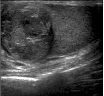
Fig. 1. Testicular ultrasonogram showing a 1.5x1.5 cm well-de- fined heterogeneous echogenic mass.
Sertoli cell tumors are very rare and account for only 1 per- cent of all testicular neoplasms. Ten percent of these tumors metastasize and are considered malignant. They can be found at any age, but most occur in the age range of 30 to 40 years.
The most common clinical finding is a painless scrotal mass, and one-third are associated with gynecomastia and hormonal imbalance. Treatment of benign Sertoli cell tumors is radical orchiectomy. If pelvic lymphatic metastasis is present, pelvic lymph node dissection is usually done, but the effect of radia- tion and chemotherapy is questionable.1 A review of the Korean literature showed 2 cases of malignant Sertoli cell tumors and 1 case of a benign tumor in Korea.2-4 Here we report the case of a 46-year-old man with a Sertoli cell tumor of the testis and review the literature.
CASE REPORT
A 46-year-old man visited the urology clinic with a com- plaint of a left scrotal mass. He had discovered a hard mass in his left scrotum 4 years previously but he had not visited the urology clinic because the mass was asymptomatic.
Physical examination revealed a 1.5 cm, well-defined, hard, painless mass. There was no sign of gynecomastia or other feminization. There was no scrotal trauma or other medical problems on his past history.
Serum α-fetoprotein, β-human chorionic gonadotropin, and
lactate dehydrogenase were 4.26 IU/ml, 1 mIU/ml, and 397 IU/l, respectively, which were within the normal limits. Serum luteinizing hormone (LH) was slightly elevated (6.22 mIU/ml), but serum follicle stimulating hormone (FSH), prolactin, testos- terone, and estradiol were normal. Ultrasonography revealed a 1.5x1.5 cm, well-defined mass with irregular echogenicity in the left testis (Fig. 1).
We planned to perform either a radical orchiectomy or or-
Seongik Bang and Sang Don Lee, et al:Testicular Sertoli Cell Tumor in an Adult 301
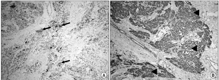
Fig. 3. Immunohistochemical staining of specimens showing that tumor cells were positive for cytokeratin (arrow)(A) and vimentin (arrow head)(B) (x100).
Fig. 2. Microscopic findings of the specimen showing tubular and focal solid growth of the tumor cell (arrow head) surrounded by stromal elements (H&E, x200).
gan-sparing surgery on the basis of the frozen biopsy results.
On gross examination, the mass was 1.7x1.3 cm in size with a white-yellow color and a hard nodular appearance. There was no gross hemorrhage or necrosis in the mass of testis. The specimen was examined quickly by using frozen section analysis. It was proven to be a Sertoli cell tumor, and the pa- tient underwent a radical orchiectomy. Microscopically, the mass was confined in the testicle and there was no invasion to the tunica albuginea or epididymis. The testicular tumor showed a cord of cells with abundant eosinophilic cytoplasm, had prominent centrally located nucleoli, and was surrounded by stromal cells. There was no evidence of malignancy, such
as morphological abnormality, necrosis, mitosis, vascular in- vasion, or solid structure (Fig. 2). Immunohistochemical stain- ing was positive for vimentin and cytokeratin and focally pos- itive for S-100 but negative for C-kit and SMA (Fig. 3).
Chest X-ray and abdominopelvic CT scan revealed no evi- dence of lymphatic or distant metastasis. The patient continues with his follow-up 6 months postoperatively, and no recurrence has been observed.
DISCUSSION
Sertoli cell tumors are very rare and account for only 1 per- cent of all testicular neoplasms. These tumors can occur at any age, including in newborns, but the peak incidence of Sertoli cell tumors is in the 30s and 40s. The etiology of testicular cancer is unknown. Although most Sertoli cell tumors develop in normal testis, there are some reports of these tumors in un- descended testis.5
The most common clinical feature of Sertoli tumors is a
“slow growing painless testicular mass.” About one-third of Sertoli cell tumors are associated with gynecomastia derived by estrogen production, but it is not clear that estrogen is produced by Sertoli cells or stromal cells.1 Forty percent of benign Sertoli cell tumors are associated with genetic syndromes such as Carney syndrome and Peutz-Jeghers syndrome; these phenom- enon are more common in bilateral or multiple tumors.6 In the present case, the patient was 46 years old and presented with a painless testicular mass without hormonal abnormalities or
302 Korean Journal of Urology vol. 50 300-302, March 2009
gynecomastia.
Most Sertoli cell tumors fall into the “not otherwise speci- fied”, or NOS, category, but subsets are represented by 2 var- iants: the sclerosing Sertoli cell tumor (SSCT) and the large cell calcifying Sertoli cell tumor (LCCSCT). These 2 variants are determined by histologic features of Sertoli cell-like epi- thelial components and stromal components. LCCSCTs are characterized histologically by large tumor cells, abundant eosi- nophilic cytoplasm, tubular and trabecular differentiation, and extensive calcified debris. SSCTs are characterized histologi- cally by solid and hollow, simple and anastomosing tubules;
large irregular aggregates; and thin cords of Sertoli cells in a prominent collagenous background.7 Immunohistochemical stain- ing may add some differential diagnostic information from oth- er testicular tumors. Sertoli cell tumors are usually positive for cytokeratin and vimentin and weakly positive for S-100.5 Our patient was diagnosed as having a Sertoli cell tumor NOS be- cause the tumor was positive for cytokeratin and vimentin and weakly positive for S-100 and there was no calcified lesion or prominent collagenous background.
A large tumor size, irregular margin of tumor, invasion to adjacent tissue, lymphatic or vascular invasion, and mitotic fig- ure usually indicate a malignant Sertoli cell tumor, but meta- stasis is essential to prove a clinically malignant tumor.1,5 Most Sertoli cell tumors are benign, but 10 percent of Sertoli cell tumors present with distant metastasis, and late metastasis has been reported during follow-up after radical orchiectomy.
Young et al,5 in their mean 3.8 year follow-up of 16 patients with Sertoli cell tumors after radical orchiectomy, reported that 3 of 12 patients who had no metastasis at presentation were found to have late metastasis. The histologic findings of these 3 patients showed microvascular invasion in 2, nuclear pleo- morphism in 2, and necrosis in 1 patient. Eight of 9 patients who had a benign clinical course presented with a tumor less than 5 cm, but 4 of 7 patients who had metastasis presented with a tumor of more than 5 cm; data on tumor size were not available for 3 patients. Metastasis was found in the retro- peritoneal lymph nodes in 6 patients, in the lung in 4, in the inguinal lymph node in 3, in the rib and supraclavicular lymph node in 2, and in the vertebra and liver in 1 patient.
The prognosis of a benign Sertoli cell tumor is good, but the prognosis of malignant Sertoli cell tumors has not been
established. Metastatic Sertoli cell tumors have a poor prog- nosis; Godec8 reported a mean 15 month survival period after diagnosis of metastasis in 9 patients.
Although metastasis is the only evidence of malignancy in Sertoli cell tumors, there are some reports of late metastasis.
The latest metastasis reported in the literature occurred 10 years after radical orchiectomy.9 Kolon et al10 suggested that a mini- mal 5 years of follow-up is necessary because of tumor characteristics.
In our case, we think that the prognosis of our patient will be good because of the small size of the mass, the lack of any histologic abnormalities indicating malignancy, and no evidence of metastasis. But close follow-up will be necessary for detect- ing late metastasis.
REFERENCES
1. Richie JP, Steele GS. Neoplasms of the testis. In: Wein AJ, Kavoussi LR, Novick AC, Partin AW, Peters CA, editors.
Campbell's urology. 9th ed. Philadelphia: Saunders; 2007;893- 935
2. Jeong CS, Seo IY, Rim JS. Malignant Sertoli cell tumor of testis. Korean J Urol 2003;44:1064-6
3. Kim YH. Malignant Sertoli cell tumor of the testis in adult.
J Soonchunhyang Med Coll 2004;10:1373-6
4. Jung SI, Min KD, Kwon DD, Oh BR, Ryu SB, Park YI, et al. Sertoli cell tumor of the testis in a young child. Korean J Urol 2001;42:675-7
5. Young RH, Koelliker DD, Scully RE. Sertoli cell tumors of the testis, not otherwise specified: a clinicopathologic analysis of 60 cases. Am J Surg Pathol 1998;22:709-21
6. Kratzer SS, Ulbright TM, Talerman A, Srigley JR, Roth LM, Wahle GR, et al. Large cell calcifying Sertoli cell tumor of the testis: contrasting features of six malignant and six benign tumors and a review of the literature. Am J Surg Pathol 1997;21:1271-80
7. Zukerberg LR, Young RH, Scully RE. Sclerosing Sertoli cell tumor of the testis. A report of 10 cases. Am J Surg Pathol 1991;15:829-34
8. Godec CJ. Malignant Sertoli cell tumor of testicle. Urology 1985;26:185-8
9. Comperat E, Tissier F, Vieillefond A. Late metastasis after a testicular Sertoli cell tumor. Ann Pathol 2004;24:45-6 10. Kolon TF, Hochman HI. Malignant Sertoli cell tumor in a
prepubescent boy. J Urol 1997;158:608-9