□
증례보고
□472
Embryonal Carcinoma of a Pexed Testis
Hyuk Soo Chang, Kwang Sae Kim
From the Department of Urology, Keimyung University School of Medicine, Daegu, Korea
A 24-year-old patient who had a history of right-side orchidopexy 16 years ago presented with right testicular pain. A hypoechoic mass was seen on the ultrasonography. Inguinal orchiectomy of the right testis was performed. On microscopic examination, the specimen was identical as an embryonal carcinoma. (Korean J Urol 2008;49:472-474)
Key Words: Testicular neoplasms; Testis, undescended
대한비뇨기과학회지 제 49 권 제 5 호 2008
계명대학교 의과대학 비뇨기과학교실 장혁수ㆍ김광세
접수일자:2008년 3월 10일 채택일자:2008년 4월 10일 교신저자: Hyuk Soo Chang
Department of Urology, Keimyung University School of Medicine, 194, Dongsan- dong, Jung-gu, Daegu 700-712, Korea TEL: 053-250-7023 FAX: 053-250-7643 E-mail: [email protected]
Fig. 2. The gross findings show an ill-demarcated, pale tan to white, solid mass with foci of necrosis.
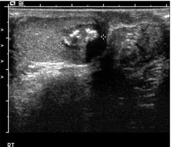
Fig. 1. Ultrasonography of the right testis showed a hypoechoic mass with focal areas of increased echogenicity.
Testicular cancer is the most common urological cancer among young men. The incidence of testicular cancer has increased steadily since the last century. But cancer of pexed testis is relevantly rare disease. We report a case of testicular cancer of 24-year old men who received orchidopexy at age of 8 years.
CASE REPORT
A 24-year-old man who had right orchidopexy at age of 8 years presented with right testicular pain for 3 weeks. On physical examination, both testes were located in scrotum and a nodular tender mass was palpated in right epididymal area.
Epididymitis was suspected and the patient received antibiotic medication.
Ultrasonography showed 2 cm sized round shaped hypoechoic mass with focal areas of increased echogenicity in right testis.
The mass showed focal bulging contour which suggestive of tunica albugineal invasion (Fig. 1). Alpha-fetoprotein and beta-human chorionic gonadotropin were 3.13 ng/ml and 0.928 mIU/ml, respectively.
Right testis was explored through inguinal approach and found to have a hard irregular mass on upper pole area. The testis was removed with high ligation of spermatic vessels.
Cut surface of the specimen showed an ill-demarcated, pale tan to white and solid mass, measuring 1.2x1.0x1.2 cm in size (Fig. 2).
Hyuk Soo Chang and Kwang Sae Kim:Embryonal Carcinoma of Pexed Testis 473
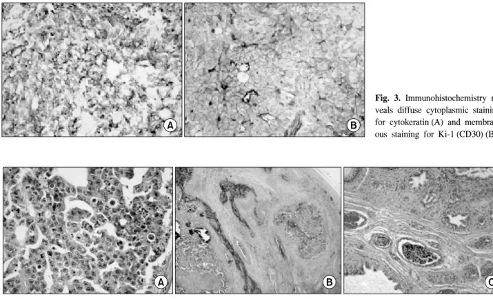
Fig. 3. Immunohistochemistry re- veals diffuse cytoplasmic staining for cytokeratin (A) and membran- ous staining for Ki-1 (CD30) (B).
Fig. 4. (A) The tumor cells have pleomorphic and vesicular nuclei with large nucleoli (H&E, x400). (B) The tunica albuginea is involved by scattered clusters of tumor cells (H&E, x40). (C) Vascular invasion was present in the epididymis.
On microscopic examination, the mass consisted of solid sheets or glandular pattern of undifferentiated cells with marked coagulative necrosis. There were typical, large, irregular and vesicular nuclei with prominent macronucleoli. There was no evidence of other germ tumor components in this specimen.
Immunohistochemistry revealed diffuse strong cytoplasmic staining for cytokeratin and membranous staining for Ki-1 (CD30) (Fig. 3). These microscopic and immunohistochemical findings were identical to the embryonal carcinoma. Tunica albuginea was involved by scattered clusters of tumor cells and foci of vascular invasions. There were scattered foci of vascular invasion in epididymis (Fig. 4). Spermatic cord was not in- volved by tumor.
An abdominal computed tomography showed no evidence of tumor metastasis.
This patient still alive in good heath for 2 years after opera- tion.
DISCUSSION
Undescended testis is associated with a 3-10-fold increase in
the risk of testicular cancer than normal testis.1 Approximately 10% of testicular cancers arise from an undescended testis, and 7 to 10% of patients with testicular cancer have a prior history of cryptorchidism.2,3
The association between cryptorchidism and testicular cancer might be explained by 2 theories; the common cause theory which related to a common hereditary or environmental factor and the position theory that an abnormal location of testis in suboptimal microenvironment and macroenvironment to develop cancer.4 There are disputes on reducing the increased cancer risk of orchidopexy. Prener et al5 reported that there was no decrease in the risk for testicular cancer associated with undescended testis after correction in early childhood. Pike et al6 reported that there was no correlation between the age at orchidopexy and the risk of subsequent malignancy. But orchidopexy had benefit because the resultant scrotal position is optimal for the early detection of malignant change.7 Recently, Walsh et al8 reported a beneficial effect of early orchidopexy for testicular cancer with systematic review and meta-analysis of the literature. They concluded that prepubertal (before age 10 to 11 years) orchidopexy may decrease the risk
474 대한비뇨기과학회지:제 49 권 제 5 호 2008
of testicular cancer, thus early surgical intervention is indicated in children with cryptorchidism.
The most common tumor that develops from a undescended testis is seminoma. But in pexed testis nonseminomatous tumors are predominant. Jones et al9 reported 34 cases of testi- cular cancers associated with undescended testis. Nonsemino- matous tumors were predominant in patients who had prior orchidopexy (78%, 7/9), and seminomas were predominant in patients with uncorrected undescended testis (64%, 16/25). The number of cases which stages III and IV are 4 (44%) in prior orchidopexy group and 4 (16%) in uncorrected group. Because the higher proportion of advanced nonseminomas in prior orchidopexy group, 4 (44%) in prior orchidopexy group and 6
(24%) in uncorrected group were died. The relationship between early orchidopexy and more favourable disease stage is not statistically significant. And the age at orchidopexy and pathological type had no correlation.
In this case, our patient received orchidopexy at age of 8 years, and showed embryonal carcinoma with favourable stage.
He is still in good health, but intensive follow up may be needed.
REFERENCES
1. Garner MJ, Turner MC, Ghadirian P, Krewski D. Epidemiology of testicular cancer: an overview. Int J Cancer 2005;116:331-9 2. Whitaker RH. Management of the undescended testis. Br J
Hosp Med 1970;4:25
3. Abratt RP, Reddi VB, Saremboch LA. Testicular cancer and cryptorchidism. Br J Urol 1992;70:656-9
4. Husmann DA. Cryptorchidism and its relationship to testicular neoplasia and microlithiasis. Urology 2005;66:424-6 5. Prener A, Engholm G, Jensen OM. Genital anomalies and risk
for testicular cancer in Danish men. Epidemiology 1996;7:14-9 6. Pike MC, Chilvers C, Peckham MJ. Effect of age at orchi- dopexy on risk of testicular cancer. Lancet 1986;1:1246-8 7. Martin DC. Malignancy in the cryptorchid testes. Urol Clin
North Am 1982;9:371-6
8. Walsh TJ, Dall'Era MA, Croughan MS, Carroll PR, Turek PJ.
Prepubertal orchiopexy for cryptorchidism may be associated with lower risk of testicular cancer. J Urol 2007;178:1440-6 9. Jones BJ, Thornhill JA, O'Donnell B, Kelly DG, Walsh A,
Fennelly JJ, et al. Influence of prior orchiopexy on stage and prognosis of testicular cancer. Eur Urol 1991;19:201-3