Infection &
Chemotherapy
전이성 유방암 환자에서 발생한 Mycobacterium abscessus 에 의한 파종성 피부 및 연부조직 감염 1예
권지은
1 손준혁
1 이윤빈
1 임주현
1 최윤진
1 한미연
1 전윤경
2 임석아
1 박완범
1서울대학교 의과대학 내과학교실1, 병리학교실2
Case Report
Submitted: August 13, 2011 Revised: September 15, 2011 Accepted: September 30, 2011
Correspondence to Wan Beom Park, M.D.
Department of Internal Medicine, Seoul National University College of Medicine, 101 Deahang-no, Jongno-gu, Seoul 110-744, Korea
Tel: +82-2-2072-3596, Fax: +82-2-762-9662 E-mail: [email protected]
http://dx.doi.org/10.3947/ic.2012.44.3.201 Infect Chemother 2012;44(3):201-204 pISSN 2093-2340 eISSN 2092-6448
www.icjournal.org
A Case of Disseminated Skin and Soft Tissue Infec- tion due to Mycobacterium abscessus with Metastatic Breast Cancer
Mycobacterium abscessus is a rapidly growing species of environmental myco- bacteria commonly found in soil, dust, and water throughout the world. In immuno- competent patients, M. abscessus usually causes localized infection of skin and soft tissue in association with a traumatic or surgical wound. Although rare, it may cause disseminated systemic infection in patients with HIV, diabetes, or medically induced immunosuppression. Here we report a case of a 53-year-old female patient with disseminated skin and soft tissue infection due to M. abscessus who presented with multiple skin lesions on the trunk, back and four extremities. The patient had undergone salvage chemotherapy, modified radical mastectomy, and palliative chemotherapy for metastatic breast cancer. Granulomatous inflam mation and acid-fast bacilli were found on skin biopsy. M. abscessus was identified via mycobacterial culture and PCR-restriction fragment length polymorphism analysis.
The patient responded well to clarithromycin, cefoxitin and amikacin therapy, and was subsequently discharged on oral antimicrobial therapy. Non-tuberculous mycobacterial (NTM) infection is a rare cause of skin and soft tissue infection, and a very high index of suspicion is required to initiate an evaluation for NTM. In metastatic cancer patients with multiple skin lesions, skin infection due to NTM must be differentiated not only from cutaneous metastasis but also from bacterial or fungal infection.
Key Words: Mycobacterium abscessus, Atypical mycobacterium infection, Dis- seminated, Soft tissue infections, Neoplasm metastasis
Ji Eun Kwon1, Jun Hyuk Son1, Yun Bin Lee1, Joo Hyun Lim1, Yoon Jin Choi1, Miyeun Han1, Yoon Kyung Jeon2, Seock-Ah Im1, and Wan Beom Park1
Departments of 1Internal Medicine, and 2Pathology, Seoul National University Hospital, Seoul, Korea
서론
Mycobacterium abscessus는 과거에는 M. chelonae subspecies abscessus로 불리 던 균주로 DNA 염기서열 분석결과 M. chelonae와는 다른 것으로 밝혀져 M. abscessus
This is an Open Access article distributed under the terms of the Creative Commons Attribution Non-Commercial License (http://creativecommons.
org/licenses/by-nc/3.0) which permits unrestricted non-commercial use, distribution, and reproduction in any medium, provided the original work is properly cited.
Copyright © 2012 by The Korean Society of Infectious Diseases | Korean Society for Chemotherapy
202
JE Kwon, et al. • A Case of Disseminated Skin Infection due to M. abscessus www.icjournal.org로 명명되었다. M. abscessus는 M. chelonae, M. fortuitum과 함께 Runyon 분류 중 제 4군 신속발육 비결핵항산균으로 분류된다[1, 2].
이 균주는 토양이나 물 등의 환경에 광범위하게 분포하며, 드물게 피부 의 작은 외상을 통해 감염을 일으킬 수 있고[3], 때로는 면역저하 환자 에서 중증의 감염증도 일으킬 수 있다[4, 5]. 국내에 이 균에 의한 피부 감염 증례는 종종 있었으나[6-10], 파종성으로 발생한 피부, 연부조직 감염에 대한 보고는 없었다.
저자들은 전이성 유방암에 대해 구제항암화학요법과 수술적치료, 이후 고식적 항암치료를 진행하던 여자 환자에서 다발성 피부결절로 발현한 M. abscessus에 의한 파종성 피부 및 연부 조직감염을 경험하 였기에 문헌고찰과 함께 보고하는 바이다.
증례
53세 여성이 내원 20일전 처음 발견된 피부의 다발성 결절에 대한 검 사와 전이성 유방암에 대한 재평가를 위하여 내원하였다.
내원 9개월 전, 자가 촉진으로 왼쪽 가슴의 결절을 발견하여 외부병 원에서 간전이를 동반한 유방암으로 진단받은 환자는 docetaxel과 epirubicin을 이용한 구제항암화학요법을 7회 시행 받고 부분관해 상 태였다. 이후 chemoport 삽입시술과 8번 째 주기의 구제항암화학요법 을 받았고 내원 3개월 전, 8번째 항암주기 제8일 째에 호중구 감소열로 입원하여 항균제로 치료받았다. 하지만 발열이 지속되고 chemoport 삽입부위에 발적, 부종, 농집이 관찰되어 chemoport 제거시술과 추가 적인 항균제 치료를 받은 후 chemoport 주위 감염은 호전되었다. 내 원 2개월 전, 변형근치유방절제술 및 림프절곽청술을 받았으며, 수술 직후 상처는 깨끗하였다.
내원 20일 전, 양하지 피부에 다발성 결절이 발견되었으며, 내원 16 일 전, 수술상처를 따라 국소 농양이 발견되어 국소 절개와 배농을 하 였고, 내원 14일 전 시행한 흉부 컴퓨터단층촬영, 양전자방출단층촬영 결과 폐에 전이성 병변과 전이성 흉수가 의심이 되어 추후 검사와 치료 를 위해 내원하였다. 환자는 수술 후 내원까지 지속적으로 경구용 1세 대 세파로스포린을 복용하고 있었다.
환자는 상태 평가를 위해 입원하였고, 당시 혈압은 117/78 mmHg, 심박수는 74/min, 호흡수는 20/min, 체온은 36.6℃ 였다. 혈액 검사에 서 백혈구 7,530/mm3 (호중구 71.8%, 림프구 16.3%), 혈색소 11.7 g/
dL, 혈소판 309,000/mm3, 혈중요소질소 9 mg/dL, 크레아티닌 0.61 mg/dL였으며, C-반응성 단백은 2.39 mg/dL로 약간 증가되어 있었다.
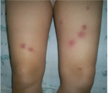
피부의 다발성 병변은 양하지에서 다양한 크기의 붉은색의 결절로 관 찰되었다(Fig. 1).
말초 혈액, 객담, 액와림프절의 절제생검 검체, 그리고 새로 발생한 흉수에 대해서 그람염색과 일반미생물 배양검사를 시행하였다. 아울 러 전이성 흉수를 감별하기 위해 세포진검사를 시행하였으며 수술절 개선을 따라 소량의 체액저류가 관찰되어 세침흡인으로 검체를 얻은 후, 그람염색과 일반미생물 배양검사를 시행하였다. 동시에 피부의 다 발성 결절 중에 우측 넙다리에서 한 군데, 좌측 전경골 부위 한 군데에
서 펀치생검을 시행하여, 병리검사, 그람염색, 일반미생물 배양검사, 항 산균 염색 및 결핵균 배양검사, 결핵균에 대한 중합효소연쇄반응 검사 를 시행하였다.
임상적으로 파종성 감염을 시사할 만한 증상이나 징후가 관찰되지는 않았으나, 미생물 검사결과가 보고되기 전까지 ampicillin/sulbactam을 단독으로 사용하며 경과관찰을 하였고, 그 동안에도 피부의 다발성 병 변은 양하지 뿐만 아니라 양상지, 복부와 등에 걸쳐 느린 속도로 생겼 다가 사라지기를 반복하였다. 다발성의 피부 병변은 붉은색을 띠는 결 절로 일부 병변은 촉지할 때 통증을 동반하였으나, 일부는 통증을 동 반하지 않았으며, 시간이 지나면 갈색으로 변하며 가피를 형성하였고, 농포나 수포로 변화하지는 않았다.
말초 혈액, 객담, 액와림프절의 절제생검 검체 및 흉수를 이용한 미 생물검사 시행 결과, 그람염색은 모두 음성이었고 일반미생물 배양검 사에서도 배양된 균은 없었다. 그러나 수술절개선 부위 체액을 접종 한 혈액배양배지에서 잘 염색이 되지 않는 그람양성 간균이 관찰되었 고 이를 혈액한천배지에 접종하였을 때 7일만에 흰색의 작은 점상 집 락이 자라, 추가로 시행한 Ziehl-Neelsen 염색에서 붉은 색의 간균 이 관찰되어 신속발육 비결핵항산균을 의심하였다. 또한 펀치생검한 피부병변 조직에서 비건락성 육아종(Fig. 2A)이 관찰되었고 Ziehl- Neelsen 염색에서 붉은 색의 간균(Fig. 2B)이 확인되었다. 내원 10 일 째, clari thromycin, cefoxitin, amikacin의 병합 요법으로 비결 핵항산균에 대한 치료를 시작하였다. 혈액배양배지에서 자라 혈액한 천배지에 접종하였던 균주는 중합효소연쇄반응-제한절편길이다형 성 검사를 이용한 최종 동정 결과 M. abscessus로 확인되었다[11].
Clinical and Laboratory Standards Institute (CLSI)의 기준에 따 른 액체배지 미량희석법(broth microdilution method)을 이용한 항 생제 감수성 검사 결과, amikacin과 clarithromycin에는 감수성을, cefoxitin, doxycycline에는 중간내성을, imipenem, ciprofloxacin,
Figure 1.Multiple erythematous nodular lesions secondary to M. abscessus infection were observed on both lower extremities.
http://dx.doi.org/10.3947/ic.2012.44.3.1 • Infect Chemother 2012;44(3):201-204
203
www.icjournal.org
sulfamethoxazole에는 내성을 나타냈다. 내원 40일 째, 환자는 새로 운 피부병변의 발생이 점차 줄어드는 상태로 경구 clarithromycin과 levofloxacin으로 변경하여 퇴원하였고 현재 외래 관찰 중이다.
고찰
신속발육 비결핵항산균은 외상을 통한 접종 이후에 발생하는 피부 및 연부조직 감염, 인공심장판막이나 인공관절 삽입 등 이물질을 삽입 하는 수술과 관련된 감염, 그리고 만성 폐감염 등 다양한 종류의 감염 을 일으키는 것으로 알려져 있다[12]. 이러한 감염증은 환자의 기저 면 역상태에 따라 그 임상양상, 경과와 예후가 달라지며, 면역력이 정상인 상태에서는 느린 임상경과를 보이거나, 국소적 감염증으로 나타나는 반면에, 면역저하 상태에서는 빠른 임상경과를 보이거나, 균혈증과 같 은 치명적인 감염증으로 나타날 수도 있다[5, 13].
최근 M. abscessus의 임상보고가 증가하는 경향을 보이고 있다. 이 는, 후천성면역결핍증후군 환자가 증가하고 스테로이드나 면역억제제 를 사용하는 환자가 늘고 있을 뿐 아니라[4], 비결핵항산균에 대한 관 심이 증대되어 면역저하 환자를 대상으로 선별 검사를 하는 경우가 많 아졌으며 이 균에 대한 진단법 또한 발전했기 때문이다.
M. abscessus 감염증의 진단은 생검 조직에서 이 균을 배양하여 동 정하는 것이 가장 확실하지만[14], 그 외에도 중합효소연쇄반응법이나 염기서열분석 등의 방법도 이용된다[15]. M. abscessus에 의한 병변의 조직검사 소견은 급성 화농성 병변에서 전형적인 육아종성 병변에 이 르기까지 다양하게 나타날 수 있는데, 진피와 피하지방층에 다형핵백 혈구가 모여있는 농양과 뚜렷하지 않은 경미한 육아종이 공존하는 이 상성(biphasic) 염증 반응이 특징적인 것으로 알려져 있다. 항산성 염 색에 붉은 색의 간균이 관찰되고 이러한 이상성 염증반응이 나타날 경 우에는 반드시 비정형항산균의 감염을 의심해야 한다[16]. 본 증례의 조직검사에서는 이상성 염증 반응은 뚜렷하지 않았으나, 경미한 육아 종의 형성과 항산균이 관찰되었다.
치료는 항균제 치료와 수술적 치료가 있는데, 감염부위의 절제나 배
농이 국소감염의 치료로 적절할 수 있다. 또한, 인공관절 감염과 같이 삽입된 기구와 연관된 감염의 경우, 삽입된 기구의 제거 및 괴사 부위 의 변연절제술이 필요할 수 있다[17]. 피부, 연부 조직 감염증을 일으키 는 M. fortuitum complex나 M. abscessus와 같은 신속 성장형 항산 균은 macrolide를 근간으로 한 항균제 치료가 추천되며[18], 본 증례 도 clarithromycin을 포함한 경험적 항생제로 치료를 시작하였다.
신속발육 비결핵항산균의 경우, 종간에 다양한 항생제 감수성 결과 를 보이며, 같은 종이더라도 분리된 균주에 따라 항생제 감수성 결과가 상이할 수 있다[3]. 그러므로 우선 알려진 경험적 항생제로 치료를 시 작하되, 반드시 최종 동정되는 균주를 확인하고, 항생제 감수성 검사 를 시행하여 그 결과에 따라 항생제의 조정이 필요하겠다. 다만, 본 증 례에서 분리된 균주는 항생제 감수성 검사 결과 ciprofloxacin에 내성 이었으나 항생제 감수성을 보이는 적절한 경구 항생제가 없는 상황에 서 clarithromycin를 단독으로 사용할 경우 내성발현의 가능성을 고 려하여 levofloxacin을 함께 사용하였다.
본 증례는 악성 종양 환자에서 백혈구증다증이나 발열 등 급성 감염 의 증거가 미약한 상태로 다발성 피부병변이 지속될 때 악성병변 뿐만 아니라 비결핵항산균 감염을 반드시 감별진단에 포함해야 한다는 것 을 보여준다. 따라서, 악성병변과의 감별을 위해 조직검사를 시행하는 경우 병리검사와 함께 결핵균을 포함한 미생물 배양검사를 함께 시행 해야 하며, 미생물학적 확진과 적절한 항균제의 선택을 위해 생검조직 의 결핵균 배양검사가 중요하겠다[19].
References
1. Timpe A, Runyon EH. The relationship of atypical acid-fast bacteria to human disease: a preliminary report. J Lab Clin Med 1954;44:202-9.
2. Kusunoki S, Ezaki T. Proposal of Mycobacterium peregrinum sp. nov., nom. rev., and elevation of Mycobacterium chelonae subsp. abscessus (Kubica et al.) to species status: Myco bac-
A B
Figure 2.(A) Hematoxylin and eosin stain reveals granulomatous inflammation in subcutaneous tissue (×200). (B) Ziehl-Neelsen (Acid Fast) stain reveals a few acid-fast bacilli (×1,000).
204
JE Kwon, et al. • A Case of Disseminated Skin Infection due to M. abscessus www.icjournal.orgterium abscessus comb. nov. Int J Syst Bacteriol 1992;42: 240-5.
3. Wolinsky E. Mycobacterial diseases other than tuberculosis.
Clin Infect Dis 1992;15:1-10.
4. Wagner D, Young LS. Nontuberculous mycobacterial infec- tions: a clinical review. Infection 2004;32:257-70.
5. Landau W, Feczko J, Kaplan RL. Radiometric detection of mycobacteria in routine blood cultures. J Clin Microbiol 1980;
12:477–8.
6. Lee SH, Kim KY, Hong SP, Kim MJ, Yang MH, Seou JT. A Mycobacteirum chelonae subsp. abscessus wound infection after percutaneous endoscopic gastrostomy. Korean J Med 1997;53:842-6.
7. Kim YS, Hong IC, Kim CK, Kim SW, Kim S, Peck KR, Kim BJ, Kook YH, Song JH. A case of skin and soft tissue infection caused by Mycobacterium abscessus. Korean J Infect Dis 2000;32:64-8.
8. Cho JH, Kim MY, Park YM, Kim HO. A case of cutaneous infection due to Mycobacterium abscessus. Korean J Der- matol 2004;42:512-5.
9. Choi YL, Lee KJ, Lee DY, Lee ES. A case of skin infection caused by Mycobacterium abscessus. Korean J Dermatol 2005;43:852-5.
10. Km JH, Choe WH, Kang JO, Choi TY. Five cases of Myco- bacterium abscessus. Korean J Clin Microbiol 2004;7:84-9.
11. Park CM, Heo SR, Park KU, Song J, Lee JH, Lee CT, Kim EC.
Isolation of nontuberculous mycobacteria using polymerase chain reaction-restriction fragment length polymorphism.
Korean J Lab Med 2006;26:161-7.
12. Wallace RJ Jr, Swenson JM, Silcox VA, Good RC, Tschen JA, Stone MS. Spectrum of disease due to rapidly growing mycobacteria. Rev Infect Dis 1983;5:657-79.
13. Sastry V, Brennan PJ. Cutaneous infections with rapidly growing mycobacteria. Clin Dermatol 1995;13:266-71.
14. Villanueva A, Calderon RV, Vargas BA, Ruiz F, Aguero S, Zhang Y, Brown BA, Wallace RJ Jr. Report on an outbreak of postinjection abscesses due to Mycobacterium abscessus, including management with surgery and clarithromycin therapy and comparison of strains by random amplified polymorphic DNA polymerase chain reaction. Clin Infect Dis 1997;24:1147-53.
15. Henry MT, Inamdar L, O'Riordain D, Schweiger M, Watson JP.
Nontuberculous mycobacteria in non-HIV patients: epide- miology, treatment and response. Eur Respir J 2004;23:741-6.
16. Bartralot R, Pujol RM, García-Patos V, Sitjas D, Martín- Casabona N, Coll P, Alomar A, Castells A. Cutaneous infec- tions due to nontuberculous mycobacteria: histopathological review of 28 cases. Comparative study between lesions ob- served in immunosuppressed patients and normal hosts. J Cutan Pathol 2000;27:124-9.
17. Ingram CW, Tanner DC, Durack DT, Kernodle GW Jr, Corey GR. Disseminated infection with rapidly growing myco- bacteria. Clin Infect Dis 1993;16:463-71.
18. Griffith DE, Aksamit T, Brown-Elliott BA, Catanzaro A, Daley C, Gordin F, Holland SM, Horsburgh R, Huitt G, Iademarco MF, Iseman M, Olivier K, Ruoss S, von Reyn CF, Wallace RJ Jr, Winthrop K; ATS Mycobacterial Diseases Subcommittee;
American Thoracic Society; Infectious Disease Society of America. An official ATS/IDSA statement: diagnosis, treat- ment, and prevention of nontuberculous mycobacterial diseases. Am J Respir Crit Care Med 2007;175: 367-416.
19. Lee S, Yun NR, Kim KH, Jeon JH, Kim EC, Chung DH, Park WB, Oh MD. Discrepancy between histology and culture in filamentous fungal infections. Med Mycol 2010;48:886-8.