pISSN 1598-9992 eISSN 2233-6869
CASE REPORT
제왕절개 출산 후 발생한 특발성 중증 급성 췌장염 1예
김 정, 최진호, 신방섭, 남준열, 강은애, 김주성, 황진혁1, 김재환1
서울대학교 의과대학 서울대학교병원 내과학교실 및 간연구소, 서울대학교 의과대학 분당서울대학교병원 내과학교실1
A Case of Idiopathic Severe Acute Pancreatitis following Cesarean Section Delivery
Jung Kim, Jin Ho Choi, Bang Sup Shin, Joon Yeul Nam, Eun Ae Kang, Joo Seong Kim, Jin Hyeok Hwang1, and Jaihwan Kim1
Department of Internal Medicine and Liver Research Institute, Seoul National University Hospital, Seoul National University College of Medicine, Seoul, Department of Internal Medicine, Seoul National University Bundang Hospital, Seoul National University College of Medicine, Seongnam1, Korea
Acute pancreatitis rarely occurs in the postpartum period. Furthermore, there are very few reports of it after cesarean section delivery. A 35-year-old woman presented with dyspnea and abdominal distension on the third day after cesarean section delivery. Under a suspicion of acute pancreatitis, she was initially managed with conservative treatment. However, she developed intra-abdominal fluid collections and gastric bleeding, which were managed with percutaneous drainage, endoscopic hemostasis, and angiographic embolization. She was discharged with good clinical recovery. Postpartum pancreatitis, especially after cesarean section, is rare; however, its management is not different from that for usual pancreatitis. (Korean J Gastroenterol 2016;68:161-165) Key Words: Pancreatitis; Postpartum period; Cesarean section; Gastric varix; Stomach ulcer
Received March 25, 2016. Revised June 27, 2016. Accepted July 15, 2016.
CC This is an open access article distributed under the terms of the Creative Commons Attribution Non-Commercial License (http://creativecommons.org/licenses/
by-nc/4.0) which permits unrestricted non-commercial use, distribution, and reproduction in any medium, provided the original work is properly cited.
Copyright © 2016. Korean Society of Gastroenterology.
교신저자: 김재환, 13620, 성남시 분당구 구미로 173번길 82, 분당서울대학교병원 내과
Correspondence to: Jaihwan Kim, Department of Internal Medicine, Seoul National University Bundang Hospital, 82 Gumi-ro 173beon-gil, Bundang-gu, Seongnam 13620, Korea. Tel: +82-31-787-7075, Fax: +82-31-787-4290, E-mail: [email protected]
Financial support: None. Conflict of interest: None.
INTRODUCTION
Acute pancreatitis is a sterile inflammation that pro- gresses to development of systemic inflammatory response syndrome and multisystem organ failure.1 Knowledge of the etiology and pathogenesis of acute pancreatitis has sig- nificantly increased; however, there are several aspects that have not been fully described. There are some common causes of acute pancreatitis such as gallstones, alcohol consumption, and endoscopic retrograde cholangiopancreatography.2,3
Compared to the number of reports related to these com- mon causes, there are few reports on acute pancreatitis as- sociated with pregnancy. However, the incidence of acute pancreatitis in pregnant populations is similar to that in
non-pregnant populations;1,3-7 moreover, its causes in preg- nant and non-pregnant subjects are similar.4,6 Ramin and Ramsey4 observed an incidence of acute pancreatitis during pregnancy of 19% in the first trimester, 26% in the second tri- mester, 53% in the third trimester, and 2% in the postpartum period. Therefore, acute pancreatitis rarely occurs in the postpartum period. Furthermore, there are few reports about acute pancreatitis occurring after cesarean section delivery.
Herein, we report a case of acute pancreatitis following ce- sarean section delivery.
CASE REPORT
The subject, a 35-year-old woman, presented to the emer-
Fig. 1. CT reveals pancreatic swelling and abdominal fluid collection. (A) On axial CT scan, diffuse pancreatic swelling and slightly reduced parenchymal attenuation are seen, but no indication of pancreatic necrosis or duct dilatation. (B) On coronal CT scan, the peri-pancreatic area, retroperitoneal space, mesentery, and transverse mesocolon fluid collections are seen. In the omentum, mesentery, and peritoneum, diffuse infiltration is seen. (C) On axial CT scan, a small stone is seen in the gallbladder.
managed with ritodrine, and nifedipine due to preterm labor.
Three days previously, she underwent full-term emergency cesarean section. Although there was bleeding after the cesar- ean section without hypovolemic shock, she did not need a transfusion. Uterine bleeding improved after uterine massage and administration of a drug to cause uterine contraction. One day after her cesarean section, she had dyspnea and ab- dominal distension.
She had been healthy previously and had not consumed alcohol. On review of systems, she complained of dyspnea and abdominal distension without abdominal pain. On phys- ical examination, her abdomen was distended without ten- derness, and breath sounds were decreased in both lower lung fields.
On admission to the emergency department, she was con- scious and oriented. Her vital signs were as follows: blood pressure, 163/82 mmHg; heart rate 139 beats/minute; res- piratory rate 46 breaths/minute, and body temperature 38.3oC. Her arterial blood gas had a pH of 7.511, a PaO2 of 53.0 mmHg, a PaCO2 of 24.9 mmHg, and a HCO3 concen- tration of 19.9 mmol/L.
Further investigation revealed a total leukocyte count of 34.7×10³/L (4.0-10.0×10³/L), hemoglobin level of 15.1 g/dL (12-16 g/dL), platelet count of 253×103/L (130-400×
103/L), BUN concentration of 12 mg/dL (10-26 mg/dL), cre-
CRP level of 15.10 mg/dL (0-0.5 mg/dL), total bilirubin level of 1.4 mg/dL (0.2-1.2 mg/dL), albumin level of 2.0 g/dL (3.3-5.2 g/dL), AST level of 38 IU/L (0-40 IU/L), ALT level of 9 IU/L (0-40 IU/L), ALP level of 68 IU/L (30-115 IU/L), and a GGT level of 6 IU/L (8-35 IU/L). An initial chest X-ray showed bilateral pleural effusion and pulmonary edema. Contrast- enhanced CT of the abdomen confirmed pancreatitis and re- vealed a diffuse swollen pancreas with peri-pancreatic and retro-peritoneal fluid collection. A gallstone was observed, but there was no evidence of pancreatic duct dilatation or ne- crosis (Fig. 1). She developed respiratory distress and was given a 6 L O2 facial mask. Pleural fluid was removed by two chest percutaneous drainage (PCD) catheters (right and left) over a period of six days. She was conservatively managed with intravenous broad-spectrum antibiotics, parenteral nu- trition, and other supportive care in the intensive care unit.
One week after admission, her total leukocyte count and c-re- active protein level began to decline. She had daily fever of more than 38oC, but the fever peak declined. She had ab- dominal distension without any other symptoms.
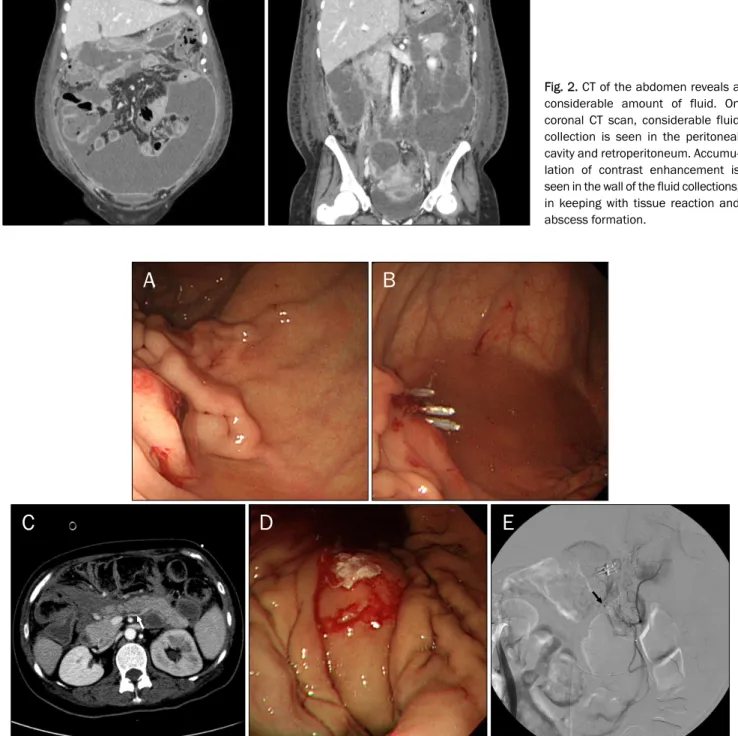
Two weeks after admission, her abdominal distension had worsened. Follow-up CT revealed a considerable amount of fluid in the peritoneal cavity and retroperitoneum (Fig. 2).
Three abdominal PCD tubes were placed for intra-abdominal collections. We checked the tubes and changed tube posi-
Fig. 2. CT of the abdomen reveals a considerable amount of fluid. On coronal CT scan, considerable fluid collection is seen in the peritoneal cavity and retroperitoneum. Accumu- lation of contrast enhancement is seen in the wall of the fluid collections, in keeping with tissue reaction and abscess formation.
Fig. 3. (A) An exposed vessel is present in the high body greater curvature anterior wall side. (B) Successfully installed hemoclips are shown.
(C) Splenic vein obliteration (white arrow) is suspected on CT scan. (D) Histoacryl injection to the gastric varix is seen on the posterior wall side of the high body greater curvature. (E) Locations of posterior gastric artery pseudoaneurysm (black arrow) and left gastric artery angiographic embolization sites are shown.
tions repeatedly over a period of 22 days. A fever of more than 38oC continued throughout the three weeks following admission. Intravenous antibiotics were provided for one month followed by oral antibiotics for one week. The patient
was discharged after a 31-day stay in hospital. She exhibited a good clinical recovery during her visit two weeks after discharge.
However, three weeks after that discharge she presented
the emergency department with gastric variceal bleeding caused by splenic vein obliteration (Fig. 3C). After undergoing endoscopic sclerosing treatment (Fig. 3D), she was dis- charged. Two weeks later, she presented to the emergency department with melena, and underwent angiographic em- bolization (Fig. 3E). Six months after the melena treatment she reported no further bleeding.
DISCUSSION
The incidence of acute pancreatitis in pregnancy differs among the studies reported.3 Eddy et al.6 observed an in- cidence of acute pancreatitis of 101 among 305,101 preg- nancies (1 in 3,021) in the United States from 1992 to 2001.
Ramin and Ramsey4 found that 43 of 147,197 pregnant women (1 in 3,333) had acute pancreatitis, but noted only one case of postpartum pancreatitis in the United States from 1983 to 1993. In that case, the cause of the pancreatitis was gallstone. Hernandez et al.7 reported that the incidence of acute pancreatitis in pregnancy was 21 in 93,440 (1 in 4,449) in the United States from 1996 to 2006, whereas Xu et al.5 showed that it was 36 in 34,292 (1 in 953) in China from 1991 to 2014. The incidence of acute pancreatitis in these pregnant populations are similar to those in general and non-pregnant populations.1,3-7 Compared to the in- cidence of pancreatitis during pregnancy, postpartum acute pancreatitis is rare.4 Previous to this case report, few cases of acute pancreatitis following cesarean section have been reported.8-11 During pregnancy, increased estrogen and pro- gesterone levels induce gallbladder smooth muscle relaxa- tion and biliary stasis, which increases the volume of the gall- bladder and decreases the flow of bile and ejection fraction.3 Thus, gallstone is the most common cause of pancreatitis in pregnant populations.3,4
The pathogenesis of acute pancreatitis is not fully understood. Opie hypothesized that gallstones impacted in the common channel (junction between the common bile duct and the pancreatic duct) obstruct the outflow of bile, provoke bile reflux into the pancreatic duct and induce pancreatitis.12 Pancreatic duct obstruction can activate a cascade of diges- tive enzymes leading to autodigestive injury which stimulates
high pancreatic ductal pressure, and the observation that non-infected bile does not cause pancreatic injury.12
White et al.14 reported that the incidence of postoperative pancreatitis is higher in biliary surgery, gastrectomy, splenec- tomy, and aortic graft surgery, but its cause is not known. They presumed that trauma to the pancreatic tissue and ducts or impairment of the blood supply precipitate pancreatitis.
In the present case, it seems less likely that gallstone pres- ence is the cause of the subject’s pancreatitis, despite the presence of a gallstone, because liver function tests revealed nearly normal levels, except for mild hyperbilirubinemia (total bilirubin 1.4 mg/dL) and there was no bile duct dilatation.
Although we could not exclude passed stone or microlithiasis as a cause of pancreatitis, we thought the possibility was low.
In addition, there was bleeding after cesarean section with- out hypovolemic shock. We could not exclude ischemia as a cause of pancreatitis. However, it is unclear whether the is- chemia induces pancreatitis.
Alcohol is not considered a likely cause of the subject’s pancreatitis because she denied alcohol use. Although hy- perlipidemia, hyperparathyroidism, trauma, medication, and fatty liver are less common causes of acute pancreatitis in pregnancy, there was no candidate cause of acute pan- creatitis in this subject. However, a triglyceride check was missed at the time of admission, but the triglyceride level was 221 mg/dL (normal 0-150 mg/dL) one week after admi- ssion. Although a mild to moderate elevation in triglyceride levels (175-870 mg/dL) and a rapid decrease within 72 hours of presentation are common in the early phase of acute pancreatitis of any etiology,15,16 we could not exclude trigly- ceride as a possible cause of the subject’s pancreatitis, as we did not check its level on admission.
Treatment of postpartum acute pancreatitis is not differ- ent from that for usual acute pancreatitis. Conservative man- agement consisting of bowel rest, intravenous fluid hydra- tion, and analgesia is recommended.3,17 In the present case, PCD for multiple intra-abdominal fluid collection, endoscopic hemoclipping and angiographic embolization were neces- sary after initially implementing a conservative management approach.
In summary, we report a case of postpartum idiopathic
acute pancreatitis following cesarean section. Postpartum pancreatitis, especially after cesarean section, is rare, but its management is not different from that for usual acute pancreatitis.
REFERENCES
1. Granger J, Remick D. Acute pancreatitis: models, markers, and mediators. Shock 2005;24 Suppl 1:45-51.
2. Wang GJ, Gao CF, Wei D, Wang C, Ding SQ. Acute pancreatitis: eti- ology and common pathogenesis. World J Gastroenterol 2009;15:1427-1430.
3. Papadakis EP, Sarigianni M, Mikhailidis DP, Mamopoulos A, Karagiannis V. Acute pancreatitis in pregnancy: an overview. Eur J Obstet Gynecol Reprod Biol 2011;159:261-266.
4. Ramin KD, Ramsey PS. Disease of the gallbladder and pancreas in pregnancy. Obstet Gynecol Clin North Am 2001;28:571-580.
5. Xu Q, Wang S, Zhang Z. A 23-year, single-center, retrospective analysis of 36 cases of acute pancreatitis in pregnancy. Int J Gynaecol Obstet 2015;130:123-126.
6. Eddy JJ, Gideonsen MD, Song JY, Grobman WA, O'Halloran P.
Pancreatitis in pregnancy. Obstet Gynecol 2008;112:1075-1081.
7. Hernandez A, Petrov MS, Brooks DC, Banks PA, Ashley SW, Tavakkolizadeh A. Acute pancreatitis and pregnancy: a 10-year sin- gle center experience. J Gastrointest Surg 2007;11:1623-1627.
8. Redechová S, Féderová L, Hammerová L, Filkászová A, Horváthová D, Redecha M. Thrombotic microangiopathy in pregnancy com-
plicated by acute hemorrhagic-necrotic pancreatitis during early puerperium. Ceska Gynekol 2014;79:190-192.
9. Jeon SY, Jung E, Seol HJ, Hur YJ. Development of Purtscher-like retinopathy after pre-eclampsia combined with acute pancreatitis. Obstet Gynecol Sci 2013;56:261-264.
10. Raheem M, Lala A, Abukhalil I. An aggressive presentation of postpartum acute necrotizing pancreatitis. J Obstet Gynaecol 2004;24:703-704.
11. Guth A, Ekoundzola JR, Beauséjour B, Nassar J, Colin JP. Acute pancreatitis in the puerperium. Diagnostic problems following cesarean section. J Gynecol Obstet Biol Reprod (Paris) 1985;14:753-756.
12. Vonlaufen A, Wilson JS, Apte MV. Molecular mechanisms of pan- creatitis: current opinion. J Gastroenterol Hepatol 2008;23:
1339-1348.
13. Lankisch PG, Apte M, Banks PA. Acute pancreatitis. Lancet 2015;386:85-96.
14. White MT, Morgan A, Hopton D. Postoperative pancreatitis. A study of seventy cases. Am J Surg 1970;120:132-137.
15. Gan SI, Edwards AL, Symonds CJ, Beck PL. Hypertriglyceridemia- induced pancreatitis: a case-based review. World J Gastroenterol 2006;12:7197-7202.
16. Dominguez-Muñoz JE, Malfertheiner P, Ditschuneit HH, et al.
Hyperlipidemia in acute pancreatitis. Relationship with etiology, onset, and severity of the disease. Int J Pancreatol 1991;
10:261-267.
17. Pitchumoni CS, Yegneswaran B. Acute pancreatitis in pregnancy. World J Gastroenterol 2009;15:5641-5646.