I. Introduction
Dental implants do not decay and do not have dental pulps that may give indications of symptoms or disease. But, the mere pres- ence of an implant in site is not acceptable as the single criterion for “success,” because potentially critical peri-implant pathology is not considered. Therefore, detailed criteria for what constitutes success are necessary.
Albrektsson et al1) described the criteria necessary for implant success: Dental implants should be immobile, and radiographs of implants should not demonstrate any evi- dence of peri-implant radiolucencies. Bone loss should not exceed 0.2mm annually after the first year, and there should be an absence of pain, infection, paresthesia, an other implant- related neuropathies.
Whereas earlier studies included mostly radiographic assessments of bone height changes and implant mobility to characterize the peri-implant conditions1-7), later reports also emphasized the importance of the use of modified periodontal indices2, 4, 8-14).
For the natural dentition, examinations such as for bleeding on probing, probing depth have been found to be important periodontal parameters, so their usefulness in the assess- ment of implants has been reported previously by many researchers3, 4, 9, 15). The interface of implant surface and connective tissues was different from natural teeth due to the absence of periodontal ligament. It may, therefore, be expected that periodontal para- meters around natural teeth and around implant fixtures are not the same. From a histological point of view, the per-implant marginal cuff shows a different structure than natural teeth. Fibers inserting directly on the surface of the implants, as they do in the cementum of root, are not found. Therefore, the question may be raised as to whether periodontal parameters are reliable criteria for monitoring implant and whether there is a biological similarity between implants and teeth16). However, similar to the periodontal evaluation, modified clinical indices are also applied for the assessment of peri-implant tis- sues.
대한치주과학회지 : Vol. 28, No. 1, 1998
Interrelationship between periodontal parameters for the evaluation of clinically stable dental implants
Dong Hwan Kim·Joon-Bong Park·Man-Sup Lee·Young-Hyuk Kwon, Yeek Herr Department of Periodontics, College of Dentistry, Kyung Hee University
Plaque is the primary etiologic factor in long-term peri-implant tissue destruction as with the natural dentition. It would therefore seem appropriate to monitor oral hygiene through plaque indices or some other related method17, 18).
Bleeding from the junctional epithelium of the implant has been considered an early symptom of peri-implantitis. Sulcus bleeding in well-controlled natural dentition has been reported to be less than 10%19). It was reported that the higher sulcus bleeding index was recorded from the healthy implant sites compared with the heathy natural dentition in the dog20). These authors also revealed that the resistance offered by the gingiva to prob- ing was greater than that offered by the peri-implant mucosa. The slightly higher sul- cus bleeding index indicated also suggested that the juctional epithelium around implants might be more fragile than that of natural teeth.
Pocket probing depth is a commonly accepted measurement in monitoring peri- odontitis patients and is often applied in implants. In stable implants the pocket prob- ing depth ranges between 1.3 and 3.8 mm15,
21-23). Increased pocket depth may be correlat- ed with a higher degree of inflammation of the peri-implant mucosa15, 24). Unsuccessful implant sites were characterized by probing depths 6mm or greater, suppuration, bone loss, and microbiota consisting primarily of Gram negative anaerobic rods9, 24).
Implant mobility is regarded not only as an important indicator of implant success or fail- ure, but also as an important clinical parame- ter that warrants continual assessment during the maintenance period1, 2, 6, 25). To date, peri-
implant bone loss and implant mobility have gained wide acceptance as measures of osseointegration and therefore as criteria for implant success26, 27). An electronic device, the Periotest (Siemens, Bensheim, Germany), which has been reported to be able to mea- sure the damping characteristics of the peri- odontium in a very reproducible way, could permit the objective discrimination between an implant that has a close bone apposition and one that is fibrously encapsulated28, 29).
It is considered that the long-term success of dental implant treatment depends on peri- odical maintenance and precise follow-up examinations, among other factors. Presently, a few long-term controlled follow-up studies on the use of periodontal parameters for implants in patients have been reported2, 4, 10,
11, 25). In this study, we measured periodontal parameters for evaluating dental implants in a periodical recall program.
This study focused on 5 modified periodon- tal parameters applied for implant that would allow for objective evaluation of clinically sta- ble dental implants. The goal of this study was to define the interrelationship between these periodontal parameters in peri-implant tissues.
II. Materials and Methods
Between January 1991 and April 1996, a total of 178 of implants, 51 Bra°emark(Nobel Biocare, Go..
eborg, Sweden) dental implants, 109 IMZ(Interpore International, Irvine, CA, USA) titanium plasma flame-sprayed type implant, and 18 ITI(Bonefit Straumann, Waldenburg, Switzerland) solid screw type implants, were placed in 43 patients(18 males
and 25 females) with a mean age of 44.7 years(range of 26 to 65 years). The patient selection from the patient pool included candi- dates with a good oral hygiene status and without systemic and/or local contra-indica- tions. All patients had at least a 1-year fol- low-up of prosthesis function(mean 670 days ranging from 373 to 2265 days), and there was no evidence of continual peri-implant radiolucency.
The details of preoperative patient educa- tion, surgical technique, and postoperative care were performed according to the manual of each dental implant. The patients were recalled every 3 months for prophylaxis. After removal of the superstructure, the modified periodontal parameters of each implant were taken. The clinical status of all 178 implants showed no detectable mobility or sign of peri- implant infection. All measurements were per- formed by one periodontist(DHK) using iden- tical methods on all patients.
1) The Modified Plaque Index(M-PI)3) was determined on all 4 surfaces(buccal, lin- gual, mesial, distal) of each implant. : score 0=no detection of plaque, score 1
=plaque only recognized by running a probe across the smooth marginal surface of the implant. Implants covered by plasma spray in this area always score 1, score 2=Plaque can be seen by the naked eye, score 3=Abundance of soft matter.
2) The Modified Sulcus Bleeding Index (M-SBI)3) was assessed at the same surfaces by checking for the occurrence of bleeding from the implant sulcus in response to pressure applied using a Plastic Perio Probe(Implant Innovations
West Palm Beach, FL, USA). : score 0
=no bleeding when a periodontal probe is passed along the gingival margin adja- cent to the implant, score 1=isolated bleeding spots visible, score 2=Blood forms a confluent red line on margin, score 3=heavy or profuse bleeding.
3) Keratinized Mucosa Index(KMI)13) was assessed at the same surfaces. : score 0
=no keratinized mucosa, score 1=1mm or less of keratinized mucosa present, score 2=between 1 and 2mm of kera- tinized mucosa, score 3=greater than 2mm of keratinized mucosa.
4) Probing Depth(PD)10) was assessed at the same surfaces to the nearest mm using a Plastic Perio Probe(Implant Innovations West Palm Beach, FL, USA), attention being given to main- taining parallelism between the probe tip and the long axis of the implant. The PD was determined by measuring the distance from the gingival margin to the bottom of the pocket.
5) Periotest Values(PTVs) was measured with the Periotest device(Siemens, Bensheim, Germany) as used by Teerlinck et al29). The tip of the hand- piece was applied perpendicularly to the surface of the abutment, at 3 mm from the implant shoulder and with the patient seated in a vertical position. The measurements were repeated until the same score was obtained 3 times.
Data Analysis
ANOVA using the SAS system with General Linear Models procedure was used to analyze the clinical parameters. Frequency
distribution graphs were constructed of all periodontal parameters. Correlation analysis between variables was done using the Pearson Correlation Coefficient. Student t-test was used to compare of PTVs between maxilla and mandible.
III. Results
1. Peri-implant parametersThe measured parameters were analyzed in terms of overall distribution. The maximum, minimum and mean values for each parame- ter were summarized in Table 1.
(1) Modified Plaque Index(M-PI)
The patients have demonstrated excellent
oral hygiene. Approximately 80% of the sites showed a M-PI of≤1. The overall mean value and standard deviation of M-PI was 0.83±0.82, indicating good oral hygiene prac- tices(Fig. 1, Table 1).
(2) Modified Sulcus Bleeding Index(M-SBI) The M-SBI 0 was assessed in 33% of the evaluated sites. The overall mean value and standard deviation of M-SBI was 1.04±
0.86(Fig. 2, Table 1).
(3) Keratinized Mucosa Index(KMI)
The peri-implant tissue has demonstrated adequate keratinized tissue. The KMI 3 was assessed in 72% of the evaluated sites. The overall mean value and standard deviation of M-SBI was 2.47±0.95(Fig. 3, Table 1).
Fig 1 Distribution of Modified Plaque Index Fig 2 Distribution of Modified Sulcus Bleeding Index
40
30
20
10
0 0 1 2 3
%
M-PI
40
30
20
10
0 0 1 2 3
%
M-PI
Parameters Sites Mean Min Max
M-PI 712 0.83±0.82 0 3
M-SBI 712 1.04±0.86 0 3
KMI 712 2.47±0.95 0 3
PD 712 3.12±1.14 1 7
PTVS 178 -0.66±4.28 -7 9
Table 1 Results of 178 implants
M-SBI
(4) Probing Depth(PD)
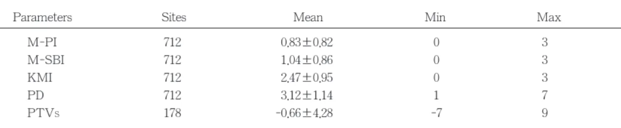
The PD ranged between 1 and 7 mm. The overall mean value and standard deviation of PD was 3.12±1.14 mm. In 88 % of total probed sites, PD were 4 mm or less(Fig. 4, Table 1).
(5) Periotest Values(PTVs)
None of the implants exhibited mobility on manual examination. The Periotest measure- ments produced values ranging from -7 to +9. Over 60% of the implants showed PTV of±0. The overall mean value and standard deviation of PTVs was -0.66±4.28, indicating clinically stable implants(Fig. 5, Table 1).
2. Interrelationship between peri-implant parameters
It was summarized in Fig. 6-13.
M-SBI showed a significant increase in higher M-PI(R2=0.170, P<0.01, Fig. 6).
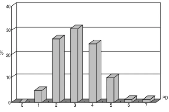
Therefore, the presence of supragingival plaque seemed to affect the peri-implant tis- sue health. Peri-implant sulcus bleeding also correlated with deeper pocket depth. The presence of subgingival plaque may have been the reason for the positive correlation (R2=0.170, P<0.01, Fig. 7). Peri-implant sul- cus bleeding was inversely correlated with the width of keratinized mucosa(R2=-0.140, P<0.01, Fig. 8).
80
60
40
20
0 0 1 2 3
%
M-PI
40
30
20
10
0 0 1 2 3 4 5 6 7 PD
%
Fig 3 Distribution of Keratinized Mucosa Index Fig 4 Distribution of Probing Depth
Fig 5 Distribution of Periotest Values(PTVS)
Fig 6 Relationship between Modified Plaque Index and Modified Sulcus Bleeding Index (R2= 0.170)
15
12
9
6
3
0 -7 -6 -5 -4 -3 -2 -1 0 1 2 3 4 5 6 7 8 9 PTV
%
3
2
1
00 1 2 3 M-PI
M-SBI
KMI
The correlation analysis performed on the present data showed no link between M-SBI and PTVs(P>0.05, Fig. 9). Furthermore, there was no correlation between PD and PTVs (P>0.05, Fig. 10). These results may be due to absence of periodontal ligament around an
implant body.
There was an inverse correlation between the diameter of implant fixture and PTVs (R2=-0.120, P<0.01, Fig. 11). The length of implant fixture also inversely correlated with PTVs(R2=-0.154, P<0.01, Fig. 12).
3
2
1
00 2 4 6 8 PD
M-SBI
3
2
1
0
0 1 2 3 KMI
M-SBI
12 8 4 0 -4
-80 1 2 3 M-SBI
PTVs
12 8 4 0 -4
-80 1 2 3 4 5Diameter
PTVs
12 8 4 0 -4
-80 2 4 6 8 10 12 14 16 18Length
PTVs
1 2 8 4 0 -4
-80 1 2 3 4 5 6 7 8 PD
PTVs
Fig 7 Relationship between Pocket Depth and Modified Sulcus Bleeding Index(R2=0.170)
Fig 8 Relationship between Keratinized Mucosa Index and Modified Sulcus Bleeding Index(R2=-0.140)
Fig 9 Relationship between Modified Sulcus Bleeding Index and Periotest Values(PTVS)(R2=-0.001)
Fig 11 Relationship between diameter of fixture and Periotest Values(PTVS)(R2= -0.120)
Fig 12 Relationship between length of fixture and Periotest Values(PTVS)(R2=-0.154).
Fig 10 Relationship between Probing Depth and Periotest Values(PTVS)(R2= -0.004)
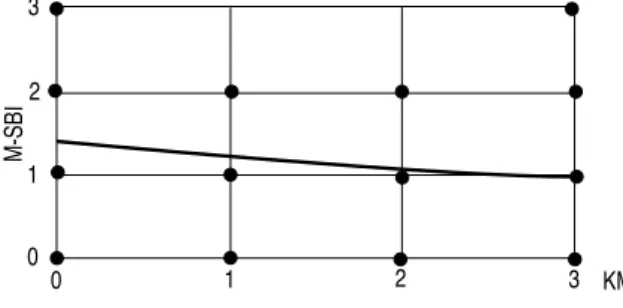
The mean PTVs in maxilla was +2.14, and the mean PTVs in mandible was -1.38. In the present study, Periotest values(PTVs) demonstrated significantly high values in the maxilla compared to the mandible, which may be due to difference of bone quality(Fig. 13).
IV. Discussion
Periodontal parameters were frequently used as criteria for the peri-implant tissues24, 30). Increased indices for plaque accumulation, marginal inflammation and increased probing depths values are associated with the devel- opment of peri-implant lesions in experimental animals31-33).
Modified plaque and sulcus bleeding indices can be used to evaluate the standard of oral hygiene and degree of inflammation in peri- implant mucosa34). A modified plaque index in the present study was well controlled, showing mean values of 0.83, and similar results were reported in other studies3, 4, 5).
High plaque index scores are positive corre- lated with high gingival index scores3, 15, 23). In animal studies, it could be demonstrated that
ligature-induced peri-implantitis including bone loss, could be caused by placing ligatures around the implant35, 36). A correlation between higher bone loss around implants and increased plaque formation was also found in humans5). The bone loss occurred slowly and to a lesser extent in the implants compared with the natural teeth controls.
A sulcus bleeding index has been routinely used as a clinical parameter for assessment of the peri-implant tissue health. Since this para- meter is an important sign of periodontal dis- ease, its use may be of great value in assess- ing peri-implant health. The most common sulcus bleeding gingival index used for implants is suggested by Mombelli et al9). But, implant success is not so related to gin- gival health as in the natural tooth. The inflammation may be limited to above the bone, because there is less fibrous tissue between the implant and bone interface.
An analysis of the clinical data obtained from successful implant throughout this obser- vation period indicated that healthy peri- implant soft tissues with little bleeding ten- dency were found. There were no remarkable signs of gingival inflammation in any patient.
The mean M-SBI values obtained was 1.04, comparable to results from other reported studies3, 4, 15). The excellent home care per- formed by the patients was certainly the major contributing factor for the health of the peri-implant soft tissues.
Poor oral hygiene is the primary cause of sulcus bleeding on probing. When the sulcus depth is less than 5 mm and the bleeding index increases, chlorhexidine often is indicat- ed, along with other professional and home care methods. However, bleeding on probing
6 4 2 0 - 2 - 4
- 6 Maxilla Mandible
Fig 13 Comparison of Periotest Values(PTVS) between maxilla and mandible
PTVs
with sulcus depths in excess of 5 to 6 mm is more common and may require reentry surgery. Radiographic bone loss and increased pocket depth have been correlated with sulcus bleeding37).
More often, bleeding sites were found with either increased plaque accumulation or increased probing depths in natural dentition.
Absence of bleeding on probing has been demonstrated to be a reliable parameter to indicate healthy periodontal tissues37). Bleeding on probing has been also reported as a typical sign of peri-implant infection9, 38), but it has been shown that bleeding on probing may be influenced by probing pressure37). Therefore, it is still open to debate that absence of bleed- ing on probing should be used as a goal for successful maintenance. However, the results of the present study seem to indicate that low bleeding prevalences were associated with maintenance of peri-implant tissues.
In recent years the discussion has been focused on the necessity of the presence of keratinized mucosa around dental implants. It has repeatedly been postulated that the establishment of a circumferential sealing effect by a dense connective tissue collar at the site of implant penetration into the conta- minated environment of the oral cavity was a prerequisite for long-term success of the implants25). The absence of keratinized mucosa usually represents mobility of the peri-implant soft tissue collar. In some studies the width of the surrounding attached mucosa was inversely correlated to pocket depth23), but in other studies no correlation could be found between the parameters35, 39, 40).
In some studies, the role of attached mucosa at the peri-implant cuff appears to be
of minor importance for the peri-implant health as long as good hygiene is practiced and achievable. Experimental research has demonstrated that keratinized mucosa is not a prerequisite for the maintenance of healthy peri-implant tissues, and its lack does not appear to result in an increased risk of loss of attachment41, 42). In clinical studies, it seems to indicate that healthy peri-implant tissues may be maintained even in the absence of kera- tinized mucosa. The sufficient width of kera- tinized mucosa generally did not result in more favorable results for the peri-implant parameters than a narrow zone or absence of keratinized mucosa13, 14, 43).
However, nonkeratinized mucosa was postu- lated to be less resistant to mechanical irrita- tion such as tooth brushing. The mobility of the peri-implant mucosa might affect cleaning procedures and may render these tissues more susceptible to plaque-induced inflammation.
Many clinicians still believe that an implant penetrating through attached keratinized mucosa is easier to maintain and more resis- tant to mechanical stress23, 30).
In the present study, mean value of kera- tinized mucosa index was 2.47, and the width of the keratinized mucosa was inversely corre- lated to sulcus bleeding on probing. Despite similar amounts of plaque accumulation, implants placed in keratinized mucosa showed significantly more decrease in modified sulcus bleeding index. A possible explanation of this increased susceptibility to plaque-induced peri- implant mucositis in the absence of kera- tinized mucosa may be the lack of tight tis- sue adaptation providing the necessary seal for a functionally optimal epithelial attach- ment.
In conclusion, the present study has indicat- ed that the presence of a keratinized mucosal tissue collar around dental implants play a role for the longevity of the implant by providing a soft tissue seal that can cope with the bac- terial invasion.
Probing depth is one of the fundamental parameters of periodontal examination. Mean probing depth in the present study was 3.12 mm, similar to the finding of a previous report2, 4, 8, 9, 10, 12, 13). Compared to the healthy natural dentition, the mean probing depth of the peri-implant mucosa in the present study was slightly higher. Probing depth depends totally on the degree of penetrability of the tissue by the probe. It is suggested, therefore, that peri-implant mucosa is more penetrable than that around natural teeth.
In stable implants the pocket probing depth ranges between 1.3 and 3.8 mm15, 21-23), and several studies indicated that in clinically suc- cessful implants, a probe penetration of about 3 mm was observed2, 4, 8, 9, 10, 12, 13). Partially edentulous patients have consistently greater probing depths around implants than around teeth15).
An increasing probing depth is more of diagnostic criterion because it usually signifies bone loss, except in the case of gingival hyperplasia or hypertrophy. In failing implants, deepened pockets are always found, but absolute pocket depth is not always indicative of implant failure24, 30, 44). The thickness of the mucoperiosteal flap at abutment surgery often influences the future pocket depth2). A tissue thickness of 5mm results with an initial 5mm implant sulcus, unless gingivoplasty or flap thinning is performed. However, implants' sul- cus depths of 6 mm or more provide an
environment favorable to gram-negative microorganisms and gingival inflammation, which favor loss of bone44). There is a direct relationship between probing depth and the effect on subgingival microflora. Therefore, the tissue thickness and implant sulcus depth should be reduced to an ideal 3 mm or less sulcular depth when esthetics are not a pri- mary concern. Gingivoplasty or flap thinning to reduce pocket depths may be performed at the initial surgery, the uncovery surgery after initial healing, or before the final prosthetic impression.
A more accurate assessment of pocket probing depth may be obtainable after removal of the restoration that prevents probe insertion parallel to the long axis of the implant30). It is also recommended that sulcu- lar probing around metal implants be accom- plished with available periodontal probes of similar metal or plastic. This will help prevent scratching and electrochemical interaction between dissimilar metals, which could be detrimental to the biocompatibility of the implant17).
The location of the probe tip subgingivally depends on the pressure used, the presence of inflammation, and the angle at which the probe is introduced next to the junctional epithelium-connective tissue zone or crest of the bone30). A positive correlation has been demonstrated between higher plaque distribu- tion, gingivitis, and deeper pocket depth in natural dentition. And, this observation was demonstrating positive correlation between M- PI, M-SBI, and PD in dental implants.
All implants should be clinically immobile. It does not guarantee a direct bone-implant interface. However, It clinically means that at
least a portion of the implant is in direct con- tact with bone, although the percentage of bone contact cannot be indicated45).
In order to assess low degrees of implant mobility, an electronic device(Periotest Siemens, Bensheim, Germany) was proposed.
Tooth with clinical zero mobility has typical ranges around +5. Periotest values(PTVs) of a clinically stable implant most often ranges from -8 to +1046).
In the present study, over 60% of the implants showed PTV of≤0. The mean value of PTVs was -0.66±4.28. The present mean PTVs remain in the negative(-1.38) and lower positive(+2.14) ranges for the mandible and maxilla respectively.
In the present study, Periotest values (PTVs) demonstrated significant difference between the mean mandibular and maxillary PTVs, confirming the results obtained by previous studies47, 48). It may be due to the difference of bone quality. And, PTVs showed significant decrease in higher diameter and/or length of implant fixture. Therefore, quantity and quality of bone contacted implants were certainly the major contributing factor for reduction of implant mobility.
Clinical periodontal parameters are used for the evaluation of the peri-implant tissues.
This evaluation may include the use of a modified plaque index, modified sulcus bleed- ing index, keratinized mucosa index, probing depth, Periotest values. Radiographic interpre- tation of alveolar bone levels has been proven to be a valuable measure of implant success or failure. However, many variables in radi- ographic technique and interpretation cause differences and errors in this assessment.
There are inherent errors in using these para-
meters, but currently they are still the most descriptive and predictive of disease activity that can be used by the implant therapist.
Periodontal parameters were used to moni- tor the peri-implant soft tissue condition in a small study population. Our results confirm the periodontal parameters of other studies on dental implants. The inclusion of more patients/implants and continued follow-up studies will possibly lead to establishing the standard value of peri-implant parameters.
V. Conclusions
The purpose of this study was to evaluate modified periodontal parameters applied for dental implants and interrelationship between these parameters. In this study 178 dental implants were placed in 43 patients(mean 44.7 years) and the results were as follows.
1. Mean value of Modified Plaque Index(M-PI) was 0.83±0.82.
2. Mean value of Modified Sulcus Bleeding Index(M-SBI) was 1.04±0.86.
3. Mean value of Keratinized Mucosa Index (KMI) was 2.47±0.95.
4. Mean value of Probing Depth(PD) was 3.12±1.14 mm.
5. Mean value of Periotest Values(PTVs) was -0.66±4.28.
6. M-SBI showed significant increase in higher M-PI and/or deeper PD(P<0.01).
7. M-SBI showed significant decrease in higher KMI(P<0.01).
8. There was no correlation between M-SBI and PTVs(P>0.05).
9. There was no correlation between PD and PTVs(P>0.05).
10. PTVs showed significant decrease in higher Diameter and/or Length of implant fixture(P<0.01).
11. PTVs showed significantly low values in the mandible compared to the maxilla (P<0.01).
In conclusion, sulcus bleeding around the dental implant showed significant increase in higher plaque accumulation and/or deeper probing depth. Despite similar amounts of plaque accumulation, implants placed in kera- tinized mucosa showed significant more decrease in sulcus bleeding tendency.
When inflammation was present in sulcular tissues, increased mobility was found in nat- ural teeth, but no correlation was found in clinically stable implants of the present study.
In clinically stable implants, there was the inverse correlation between PTVs and the diameter/length of implant fixture, having no concern with peri-implant sulcus bleeding.
Moreover, Periotest values(PTVs) showed significantly low values in the mandible com- pared to the maxilla, which may be due to the difference of bone quality.
VI. References
1. Albrektsson, T., Zarb, G., Worthington, P., and Eriksson, A. R. : The long- term efficacy of currently used dental implants: a review and proposed criteria of success. Int. J. Oral Maxillofac.
Implants, 1 : 11-25, 1986.
2. Adell, R., Lekholm, U., Rockler, B., and Bra°nemark, P. I. : A 15-year study of osseointegrated implants in the treat- ment of edentulous jaw. Int. J. Oral
Surg., 10 : 387-416, 1981.
3. Adell, R., Lekholm, U., Rockler, B., Bra°
nemark,, P. I., Lindhe, J., Ericsson, B., and Sbordone, L. : Marginal tissue reaction at osseointegrated titanium fix- tures (I): a 3-year longitudinal prospec- tive study. Int. J. Oral Maxillofac. Surg., 15 : 39-52, 1986.
4. Cox. J. F., and Zarb, G. A. : The lon- gitudinal clinical efficacy of osseointe- grated dental implants: a 3-year report.
Int. J. Oral Maxillofac. Implant, 2 : 91- 100, 1987.
5. Lindquist, L., Rocker, B., and Carlsson, G. E. : Bone resorption around fixtures in edentulous patients treated with mandibular fixed tissue-integrated pros- thesis. J. Prosthet. Dent., 59 : 59-63, 1988.
6. Smith, D. E., and Zarb, G. A. : Criteria for success of osseointegrated endosseous implants. J. Prosthet. Dent., 62 : 567- 572, 1989.
7. Ahlqvist, J., Borg, K., Gunne, J., Nilson, H., Olsson, M., and A°strand, P. : Osseointegrated timplants in edentulous jaws: a 2-year longitudinal study. Int. J.
Oral Maxillofac. Implant, 5 : 155-163, 1990.
8. Ericsson, I., Lekholm, U., Bra°nemark, P.
I., Lindhe, J., Glantz, P. O., and Nyman, S. : A clinical evaluation of fixed-bridge restorations supported by combination of teeth and osseointegrated titanium implants. J. Clin. Periodontol. 13 : 307- 312, 1986.
9. Mombelli, A., van Oosten, M. A. C., Schu..
rch, E., and Lang, N. P. : The microbiota associated with successful or
failing osseointegrated titanium implants.
Oral Microbiol. Immunol., 2 : 145-151, 1987.
10. Buser, D., Weber, H. P., and Lang, N.
P. : Tissue integration of non-sub- merged implants: 1-year results of a prospective study with 100 ITI hollow- cylinder and hollow-screw implants. Clin.
Oral Impl. Res., 1 : 33-40, 1990.
11. Buser, D., Weber, H. P., Bra..
gger, U., and Balsiger, C. : Tissue integration of one-stage ITI implants: 3-year results of a longitudinal study with hollow- cylinder and hollow-screw implants. Int.
J. Oral Maxillofac. Implants, 6 : 405- 412, 1991.
12. Mombelli, A., and Mericske-Stern, R. : Microbiological features of stable osseoin- tegrated implants used as abutments for overdentures. Clin Oral Impl. Res., 1: 1- 7, 1990.
13. Apse, P., Zarb, G. A., Schmitt, A., and Lewis, D. W. : The longitudinal effec- tiveness of osseointegrated dental implants. The toronto study: Peri- implant mucosal response. Int. J.
Periodont. Rest. Dent., 11 : 95-111, 1991.
14. Mericske-Stern, R., Steinlin Schaffner, T., Marti, P., and Geering A. H. : Peri- implant mucosal aspects of ITI implants supporting overdentures: a five-year longitudinal study. Clin. Oral Impl. Res., 5 : 9-18, 1994.
15. Lekholm, U., Adell, R., Lindhe, J., Bra°
nemark, P. I., Ericsson, B., Rockler, B., Lindvall, A. M., and Yoneyma, T. : Marginal tissue reactions at osseointe- grated titanium fixtures. II: a cross-
sectional retrospective study. Int. J. Oral Maxillofac. Surg., 15 : 53-61, 1986.
16. Klinge, B. : Implants in relation to nat- ural teeth. J. Clin. Periodontol., 18 : 482-487, 1991.
17. Bauman, G. R., Mills, M., Rapley, J. W., and Hallmon, W. H. : Clinical parame- ters of evaluation during implant main- tenance. Int. J. Oral Maxillofac.
Implants, 7 : 220-227, 1992.
18. Bauman, G. R., Mills, M., Rapley, J. W., and Hallmon, W. H. : Plaque-induced inflammation around implants. Int. J.
Oral Maxillofac. Implants, 7 : 330-337, 1992.
19. Lenox, L. A., and Kopczyk, R. A. : A clinical system for scoring a patient's oral hygiene performance. J. Am. Dent.
Ass., 88 : 849-852, 1973
20. Ericsson, I., and Lindhe, J. : Probing depth at implants and teeth. J. Clin.
Periodontol., 20 : 623-627, 1993.
21. Apse, P. : Cross-sectional clinical and microbiological investigation of periim- plant and periodontal status. J. Dent.
Res., 67 : 287, 1988.
22. Lekholm, U., Ericsson, I., Adell, R., and Slots, J. : The condition of the soft tis- sues at tooth and fixture abutments supporting fixed bridges: a microbiologi- cal and histological study. J. Clin.
Periodontol., 13 : 558-562, 1986.
23. Zarb, G. A., and Symington, J. M. : Osseointegrated dental implants: prelimi- nary report on a replication study. J.
Prosthet. Dent., 50 : 271-276, 1983.
24. Becker, W., Becker, B. E., Newman, M.
G., and Nyman, S. : Clinical and microbiologic findings that may con-
tribute to dental implant failure. Int. J.
Oral Maxillofac. Implants, 5 : 31-38, 1990.
25. Bra°nemark, P. I., Hansson, B. O., Adell, R., Breine, U., Lindstro..
m, J., Hallen, O., and O..
hman, A. : Osseointegrated implants in the treatment of edentulous jaws: experience from a 10-year period.
Scan. J. Plastic Recon. Surg., 11 : suppl.
16, 1977.
26. Albrektsson, T., and Sennerby, L. : State of the art in oral implants. J. Clin.
Periodontol., 18 : 474-481, 1991.
27. Chaytor, D. V., Zarb, G. A., Schmitt, A., and Lewis, D. W. : The longitudi- nal effectiveness of osseointegrated den- tal implants. The Toronto study: bone level changes. Int. J. Periodont. Rest.
Dent., 11 : 113-126, 1991.
28. Salonen, M. A. M., Raustia, A. M., Kainulainen, V., and Oikarinen, K. S. : Factors related to Periotest values in endosseal implants: a 9-year follow-up.
J. Clin. Periodontol., 24 : 272-277, 1997.
29. Teerlink, J., Quirynen, M., Darius, P., and van Steenberghe, D. : Periotest an objective clinical diagnosis of bone apposition toward implants. Int. J. Oral Maxillofac. Implants, 6 : 55-61, 1991.
30. Newman, M. G., and Flemming, T. F.
: Periodontal considerations of implants and implant associated microbiota. J.
Dent. Educ., 52 : 737-744, 1988.
31. Lindhe, J., Berglungh, T., Ericsson, I., Liljenberg, B., and Marinello, C. : Experimental breakdown of peri- implant and periodontal tissues. Clin.
Oral Impl. Res., 3 : 9-16, 1992.
32. Lang, N. P., Bra..
gger, U., Walther, D.,
Beamer, B., and Kornman, K. S. : Ligature-induced peri-implant infection in cynomolgus monkeys. Clin. Oral Impl.
Res., 4 : 2-11, 1993.
33. Schou, S., Holmstrup, P., Stoltze, K., Hj
ø
rting- Hansen, E., and Kornman, K.S. : Ligature-induced marginal inflam- mation around osseointegrated implants and ankylosed teeth. Clin. Oral Impl.
Res., 4 : 12-22, 1993.
34. Mombelli, A., and Lang, N. P. : Clinical parameters for the evaluation of dental implants. Periodontology 2000, 4 : 81-86, 1994.
35. Strub, J. R., Garberthu..
el, T. W., and Scha..
rer, P. : Role of attached gingiva for periimplant health in dogs. J. Dent.
Res., 67 : 287, 1988.
36. Brandes, R., Beamer, B., Holt, S. C., Kornman, K., and Lang, N. P. : Clinical-microscopic observation of liga- ture induced “periimplantitis” around osseointegrated implants. J. Dent. Res., 67 : 287, 1988.
37. Lang, N. P., Adler, A., Joss, A., and Nyman, S. : Absence of bleeding on probing: an indicator of periodontal sta- bility. J. Clin. Periodontol., 17 : 714-721, 1990.
38. Pontoriero, R., Tonelli, M. P., Carnevale, G., Mom-belli, A., Nyman, S. R., and Lang, N. P. : Experimentally induced peri-implant mucositis: A clinical study in humans. Clin Oral Impl. Res., 5 : 254-259, 1994.
39. Wennstro..
m, J., and Lindhe, J. : Role of attached gingiva for maintenance of periodontal health. J. Clin. Periodontol., 10 : 206-221, 1983.
40. Kennedy, J. E., Bird, W. C, Palcanis, K.
G., and Dorfman, H.S. : A longitudinal evaluation of varying widths of attached gingiva. J. Clin. Periodontol., 12 : 667- 675, 1985.
41. Van Steenberghe, D. : Periodontal aspects of osseointegrated oral implants modum Bra°nemark. Dent. Clin. N. Am., 32 : 355-370, 1988.
42. Krekeler, G., Schilli, W., and Diemer, J.
: Should the exit of the artificial abut- ment tooth be positioned in the region of the attached gingiva? Int. J. Oral Surg., 14 : 504-508, 1985.
43. Mericske-Stern, R. : Clinical evaluation of overdenture restorations supported by osseointegrated titanium implants: a ret- rospective study. Int. J. Oral Maxillofac.
Implants, 5 : 375-383, 1990.
44. Rams, T. E., Roberts, T. W., Tatum, H.
Jr., and Keyes, P. H. : The subgingival microflora associated with human dental implants. J. Prosthet. Dent., 51 : 529-
534, 1984.
45. Mericske-stern, R., Milani, D., Mericske, E., and Olah, A. : Periotest measure- ments and osseointegration of mandibu- lar ITI implants supporting overden- tures: A one-year longitudinal study.
Clin Oral Impl. Res., 6 : 73-82, 1995.
46. Spiekermann, H., Jansen, V. K., and Richer, E. J. : A 10-year follow-up study of IMZ and TPS implants in the edentulous mandible using bar-retained overdentures. Int. J. Oral Maxillofac.
Implants, 10 : 231-243, 1995.
47. Olive′ J., and Aparicio, C. : The Periotest method as a measure of osseointegrated oral implant stability. Int.
J. Oral Maxillofac. Implants, 5 : 390- 400, 1990.
48. Hass, R., Saba, M., Mensdorff-Pouilly, N., and Mailath, G. : Examination of the damping behavior of IMZ implants.
Int. J. Oral Maxillofac. Implants, 10 : 410-414, 1995.
국문초록
인공매식치의 평가를 위한 치주지수간의 상관관계
김동환, 박준봉, 이만섭, 권영혁, 허 익 경희대학교 치과대학 치주과학교실
자연치의 경우에는 치주지수간의 상관관계가 정립되어 병적인 상태에 대한 진단과 그에 따른 치료방법을 결정하는데 많은 정보를 제공하나, 인공매식치에서는 아직 논란의 여지가 많다. 이 에 임상적으로 안정화되어 기능하고 있는 인공매식치에서 치주지수간의 상관관계에 관하여 연 구하고자 하였다.
총 43명의 환자(평균 44. 7세)에 식립된 178개의 인공매식치를 연구에 이용하였다. 인공치아매 식술을 시행한 후 1년에서 6년이 경과한 인공매식치에서 상부보철물을 제거한 후 치태지수, 치 은열구출혈지수, 각화점막지수, 치주낭깊이, Periotest Values(PTVs) 등을 측정하여 각각의 분포 상황과 상관관계를 분석하였다.
이 연구의 결과는 다음과 같다.
1. 치태지수의 평균값은 0.83±0.82이었다.
2. 치은열구출혈지수의 평균값은 1.04±0.86이었다.
3. 각화점막지수의 평균값은 2.47±0.95이었다.
4. 치주낭깊이의 평균값은 3.12±1.14 mm이었다.
5. Periotest Values(PTVs)의 평균값은 -0.66±4.28이었다.
6. 치태지수, 치주낭깊이가 증가함에 따라 치은열구출혈지수는 유의성있게 증가하였다 (P<0.01).
7. 각화점막지수가 증가함에 따라 치은열구출혈지수는 유의성있게 감소하였다(P<0.01).
8. 치은열구출혈지수와 매식치동요도 사이에서는 유의성있는 상관관계를 발견할 수 없었다 (P>0.05).
9. 치주낭깊이와 매식치동요도 사이에서도 유의성있는 상관관계를 발견할 수 없었다(P>0.05).
10. 인공매식치의 직경과 길이가 증가함에 따라 매식치동요도는 유의성있게 감소하였다 (P<0.01).
11. 하악에서의 매식치동요도가 상악의 경우와 비교하여 유의성있게 작았다(P<0.01).
결론적으로, 인공매식치에서도 자연치에서와 동일한 양상으로 치태가 많을수록, 그리고 치주낭 깊이가 깊을수록 염증의 심도와 관련이 깊은 것으로 생각된다. 특히, 인공매식치의 경우에는 같은 양의 치태가 존재시에 각화치은이 충분히 있는 쪽이 염증발생이 적은 것으로 나타났다.
치주조직에 염증이 존재하는 경우, 자연치아에서는 치아의 동요도가 증가하는 것으로 알려져 있으나, 이 실험의 인공매식치에서는 유의성있는 상관관계를 발견할 수 없었다. 임상적으로 안
정화된 인공매식치의 동요도는 염증정도에는 큰 영향을 받지 않고 인공매식치의 직경과 길이가 증가함에 따라 감소함을 보여주고 있다. 또한, 인공매식치의 동요도는 상하악골의 골질에 따라 차이가 있음을 명확히 보여주고 있다.