Introduction
The replacement of missing teeth with implant- borne restorations has become a valid and predict- able treatment option for the rehabilitation of fully and partially edentulous patients. Several longitudinal studies of implant restorations have been published, with promising results.
1-3Peri-implant disease is a biological complication of
implant treatment. Peri-implant diseases may affect the peri-implant mucosa only (peri-implant mucosi- tis) or these diseases may also involve the supporting bone (peri-implantitis).
4,5Biofilms accumulate on all surfaces in the oral cavity, including artificial or den- tal materials and the accumulation of bacterial bio- films has been identified in numerous studies as the primary etiological factor for the development and progression of peri-implant diseases.
6-9Peri-implant
*Correspondence to: Heung-Sik Um
Professor, Department of Periodontology, Research Institute for Oral Sciences, College of Dentistry, Gangneung-Wonju National University, 7 Jukheon-gil, Gangneung, 25457, Republic of Korea
Tel: +82-33-640-3184, Fax: +82-33-642-6410, E-mail: [email protected] Received: September 21, 2015/Last Revision: March 17, 2016/Accepted: March 18, 2016
effect of fluoride-containing gel on the roughness of a titanium surface and the promotion of bacterial growth
Sun-Jin Kim 1 , Jae-Kwan Lee 1 , Beom-Seok Chang 1 , Si-Young Lee 2 , Heung-Sik Um 1 *
1
Department of Periodontology,
2Department of Oral Microbiology, Research Institute for Oral Sciences, College of Dentistry, Gangneung-Wonju National University, Gangneung, Republic of Korea
Purpose: The aim of this study was to evaluate whether fluorides at various pH cause changes in the surface roughness of titanium implants that alter the adherence of bacterial biofilms. Materials and Methods: The titanium disks were assigned randomly to the following seven groups according to the fluoride agents and application time (1 minute or 30 minute) used: control (no treatment);
group 1 (1.23% acidulated phosphate fluoride [APF] at pH 3.5 for 1 minute); group 2 (1.23% APF at pH 3.5 for 30 minute); group 3 (1.23% APF at pH 4.0 for 1 minute); group 4 (1.23% APF at pH 4.0 for 30 minute); group 5 (2% NaF gel at pH 7.0 for 1 minute); group 6 (2% NaF gel at pH 7.0 for 30 minute). The surface roughness of the titanium disks and the amount of adherent bacteria were measured. Results: Group 2 showed a significantly greater surface roughness than the control group (P < 0.0001). No significant differences in the amount of surface bacteria were observed between the treated samples and the controls. In addition, there were no significant differences in bacterial adherence relative to the incubation period between the treated samples and the controls.
Conclusion: The surface roughness of the titanium disks was significantly greater after treatment with APF at pH 3.5 for 30 min compared with that of the controls. In addition, we found that the amount of Porphyromonas gingivalis, Fusobacterium nucleatum, and Aggregatibactor actinomycetemcomitans was similar among all groups (J Dent Rehabil Appl Sci 2016;32(1):16-23)
Key words: fluorides; titanium; biofilms; bacterial adhesion
Copyright© 2016 The Korean Academy of Stomatognathic Function and Occlusion.
It is identical to Creative Commons Non-Commercial License.
cc
ISSN 2384-4353 eISSN 2384-4272
diseases following successful integration of endosse- ous implants are the result of an imbalance of bacte- rial load and host defense, and may lead to the loss of osseointegration.
5Bacterial adhesion to intra-oral, hard surfaces is in- fluenced by surface roughness. A positive correlation between surface roughness and the rate of plaque accumulation has been observed.
10,11In a literature review of the impact of surface characteristics on biofilm formation, the authors concluded that rougher surfaces accumulate and retain more plaque, and, after several days of undisturbed plaque forma- tion, rough surfaces harbor a more mature plaque characterized by a greater proportion of rods, motile organisms, and spirochetes.
12On a rough surface, at- tached bacteria can survive longer because they are protected against mechanical shear forces and oral hygiene methods.
13The roughness of intra-oral hard surfaces, such as natural teeth, restorations, and prosthetic materi- als, can be modified by routine dental procedures including mechanical debridement, polishing, finish- ing, and use of fluoride for caries prevention.
14-19Of these procedures, fluoride prophylactic agents can cause corrosion and change in the surface roughness of titanium and titanium-based alloys, even though titanium has high corrosion resistance due to a thin, stable oxide layer.
18-21As a supportive periodontal therapy (SPT), topical fluoride is applied to prevent root caries, especially in areas with gingival recession.
Since the fluoride is commonly applied using dispos- able trays, it may contact not only natural teeth, but also the titanium surface of implant abutments. As mentioned above, the contact with fluoride may lead to an increase in the roughness of the titanium sur- face, which may be followed by an increase in bacte- rial biofilm accumulation. However, few studies have evaluated the effect of fluoride therapy on titanium surface topography and the adherence of bacterial biofilm to the titanium.
In this study, we treated titanium surfaces with fluo- rides at various pH, and evaluated changes in the sur- face roughness and the adherence and colonization of bacterial biofilm caused by fluoride treatment.
Materials and Methods
Preparation of titanium disks
Titanium disks measuring 5 mm in diameter and 1 mm in thickness were used. The specimens were ground wet with 300- and 800-grit silicon carbide papers, and then were cleaned ultrasonically twice in ethanol for 15 minutes and washed with distilled wa- ter.
Three commercially available fluoride agents were used: a) 1.23% acidulated phosphate fluoride (APF) gel at pH 3.5 (Topex
®, Sultan, York, USA); b) 1.23%
APF gel at pH 4.0 (60 second
®, Germiphene, Brant- ford, Canada); and c) 2% sodium fluoride (NaF) gel at pH 7.0 (GEL7
®, Germiphene). The titanium disks were assigned randomly to seven groups (Table 1): control group (no treatment); group 1 (the disks were treated with 1.23% APF at pH 3.5 for 1 min- ute); group 2 (the disks were treated with 1.23% APF at pH 3.5 for 30 minutes); group 3 (the disks were treated with 1.23% APF at pH 4.0 for 1 minute);
group 4 (the disks were treated with 1.23% APF at pH 4.0 for 30 minutes); group 5 (the disks were treated with 2% NaF gel at pH 7.0 for 1 minute);
group 6 (the disks were treated with 2% NaF gel at pH 7.0 for 30 minutes). For each group, 20 titanium disks were used. After fluoride gel treatment, samples in the experimental groups were removed from the fluoride-containing gel, washed thoroughly with dis- tilled water, and dried.
Table 1. Distribution of the groups according to the fluoride -containing gels and immersion times
Group
(n = 20) Fluoride-containing gel Immersion time
Control - -
1 APF (pH 3.5) 1 minute
2 APF (pH 3.5) 30 minutes
3 APF (pH 4.0) 1 minute
4 APF (pH 4.0) 30 minutes
5 NaF (pH 7.0) 1 minute
6 NaF (pH 7.0) 30 minutes
APF, acidulated phosphate fluoride; NaF, sodium fluoride.
Bacterial adherence assay
Porphyromonas gingivalis, Fusobacterium nucleatum, and Aggregatibacter actinomycetemcomitans were used to examine bacterial adherence to the disks. A. actinomy- cetemcomitans ATCC 33384, F. nucleatum ATCC 23726, and P. gingivalis ATCC 33277 obtained from Korean Collection for Oral Microbiology (Chosun Univer- sity, Gwangju, Korea) were used. Brucella blood agar plate (KOMED, Sungnam, Korea) was used for the culturing of P. gingivalis, F. nucleatum and A. actinomy- cetemcomitans. Colonies of P. gingivalis, F. nucleatum and A. actinomycetemcomitans incubated in an anaerobic environment (Bactron Anaerobic Chamber, Sheldon Manufacturing Inc., Cornelius, USA) with an atmo- sphere of 90% N
2, 5% CO
2, and 5% H
2were sus- pended in reduced phosphate buffered saline (tlPBS), pH 7.2 to a turbidity equivalent to a McFarland 1.0 index. Fifty microliters of this suspension was added each well of 24-well plates containing reduced Brain Heart Infusion broth (2 mL) and the titanium disks.
After 1, 3, and 5 days of anaerobic incubation, the quantity of bacterial protein was determined us- ing the Bradford protein assay (Sigma-Aldrich, St.
Louis, USA) as follows. After anaerobic incubation, the specimens were washed gently twice with 1 mL of PBS in fresh 24-well plates. To remove adhering bacteria, the disks were incubated with 1 mL of lysis buffer (20 mM Tris-HCL, pH 7.5, 150 mM NaCl, 1 mM Na
2EGTA, 1% Triton X-100, 2.5 mM sodium pyrophosphate, 1 mM β-glycerophosphate, 1 mM Na
3VO
4, and 1 μg/mL leupeptin) for 10 minutes in 48-well plate.
1Five microliters of this lysis buffer so- lution was transferred to the 96-well plates and 250 μL of the Bradford reagent was added to each well.
The Bradford reagent and protein were mixed on a shaker for 30 minutes and the absorbance was mea- sured at 595 nm (ELx 800
TMAbsorbance microplate reader, BioTek, Winooski, USA).
Surface roughness measurements and scanning electron microscopy
The surface roughness of each titanium disk was evaluated using a SufTest SJ-400 (Mitutoyo, Kawa-
saki, Japan).
Scanning electron microscopy (SEM) was per- formed to visualize the bacterial colonization. The disks were incubated at 37°C in bacterial culture medium inoculated with P. gingivalis, F. nucleatum, and A. actinomycetemcomitans for 5 days in anaerobic con- ditions in 24-well culture plates, then washed twice with PBS. Disks with attached bacteria were fixed in 2.5% glutaraldehyde in PBS for 1 hour at room tem- perature. The fixed samples were then washed three times with PBS for 10 minutes, and dehydrated for 30 minutes in a graded series of ethanol. After criti- cal point drying, the samples were mounted on stubs, coated with gold, and observed by SEM. The surface of each disk was examined by variable pressure field emission scanning electron microscopy (VP-FE- SEM) SUPRA55VP (Carl Zeiss, Oberkochen, Ger- many) (×10,000, ×30,000).
Statistical analysis
The data were analyzed using SPSS
TMVersion 19.0 (IBM Inc., Armonk, USA). One-way analysis of vari- ances (ANOVA) test and Tukey’s post-hoc tests were applied to assess both the effect of the fluoride-con- taining gel on the surface roughness of the titanium disks and difference in the amount of bacteria on the treated titanium disks. The level of significance was P < 0.05.
Results
Surface roughness measurements
Surface roughness values (mean ± SD) for all groups are listed in Table 2. Only group 2 (1.23%
APF at pH 3.5 for 30 minutes) showed significantly greater surface roughness compared to the control group (P < 0.0001).
Protein concentration measurements
Bacterial adherence assay was performed on disks
from the control group, group 2 (1.23% APF at pH
3.5 for 30 minutes), group 4 (1.23% APF at pH 4.0
for 30 minutes), and group 6 (2% NaF gel at pH 7.0 for 30 minutes). For each experiment period (1, 3, and 5 days), four titanium disks per group were used.
The results of the adherence assay are shown in Fig.
1. At 1, 3, and 5 days of incubation, the protein con- tent on the surface of the disks showed no signifi- cant difference between any experimental group and controls. Also, there were no significant differences in bacterial adherence between incubation periods.
Scanning electron microscopy
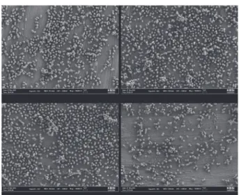
Disks were incubated with bacteria for 5 days and used for surface evaluation under a SEM. SEM im- ages of the specimens are shown in Fig. 2 - 4. Most of bacteria appeared as one, discontinuous layer on the surface of the titanium disks. However, some of bacteria are arranged in multiple layers. The attached bacteria were distributed evenly on the surface of the titanium disks, with no particular orientation with respect to the surface. All bacterial strains examined were attached to the titanium surface by their outer membrane. SEM images of the experimental groups showed no substantial difference compared with that of controls.
Fig. 2. SEM images of P. gingivalis on the titanium surface (Magnification: ×10,000). SEM images showed no substantial difference among the 4 groups.
Table 2. Surface roughness of titanium disk
Group Control APF (pH 3.5) APF (pH 4.0) NaF (pH 7.0)
Group 1 Group 2 Group 3 Group 4 Group 5 Group 6
Ra (μm) 0.19 ± 0.08 0.21 ± 0.08 0.31 ± 0.03* 0.23 ± 0.07 0.22 ± 0.09 0.22 ± 0.08 0.21 ± 0.09
Values are presented as mean ± standard deviation.
* P < 0.05.
APF, acidulated phosphate fluoride; NaF, sodium fluoride.
Fig. 1. Percentage of bacterial protein content on differently treated titanium samples relative to the amount measured on the control samples. The protein content measurements showed no significant differences in the amount of bacteria on the treated samples compared with that of the controls.
150 100 50 0
150 100 50 0
150 100 50 0
% of pr otein content % of pr otein content % of pr otein content
Control Group 2 Group 4 Group 6
Control Group 2 Group 4 Group 6
Control Group 2 Group 4 Group 6
Porphyromonas gingivalis
Fusobacterium nucleatum
Aggregatibacter actinomycetemcomitans Incubation time (days)
Incubation time (days)
Incubation time (days) 1 3 5
1 3 5
1 3 5
Discussion
The recession of soft tissue in the oral cavity due to age, traumatic tooth brushing habits, and peri- odontal disease or treatment results in a greater area of tooth surfaces that are at risk for the development of root caries. According to epidemiologic studies, root caries is prevalent among patients with treated and untreated periodontal disease.
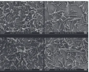
22,23Therefore, professional fluoride application is recommended to prevent root caries. Topical fluoride agents such Fig. 3. SEM images of F. nucleatum on the titanium surface (Magnification: ×10,000). SEM images showed no substantial difference among the 4 groups.
Fig. 4. SEM images of A. actinomycetemcomitans on the titanium surface (Magnification: ×10,000). SEM images showed no substantial difference among the 4 groups.
as stannous fluoride (SnF2), NaF, and APF are used in the professional fluoride application procedure.
APF is produced by acidulation of a NaF solution with phosphoric acid. The acidic pH of APF etches the tooth surface to allow for more uptake of fluo- ride compared with the neutral pH of NaF. In some studies, the topical application of APF agents has been shown to be an effective method to reduce the incidence of dental caries in test populations.
24,25Currently, the use of these agents, particularly as gels, is popular in dental clinics. The pH of the rinses and gels used for caries prevention in dentistry are usually between 3.5 and 7.0, and the fluoride concentration is between 1,000 and 10,000 ppm.
20In this study, titanium disks were treated with fluo- ride gel for 1 or 30 minutes. The application time of the fluoride gel was determined to simulate a clinical situation. In SPT visits, patients were usually treated with fluoride gel for 1 minute. And 30 minutes of application time represent delayed application of fluoride without cleaning. We ordered to patients in SPT without gargling for 30 minutes after application of fluoride.
The influence of fluoride agents on the titanium
surface has been examined previously. High fluoride
concentrations and an acidic pH destroy the corro-
sion resistance of titanium, as crevice and pitting
corrosion can also occur.
26,27Pröbster et al.
21de-
scribed that acidic fluoride agents cause severe sur-
face damage similar to that of hydrofluoric acid. The
authors suggested that acidic fluoride agents should
not be used in patients with titanium implants or
restorations. Stájer et al.
20also suggested that agents
with high fluoride concentrations at an acidic pH
increase the roughness of the titanium surface. On
the other hand, Toniollo et al.
28demonstrated that
the use of a fluoride-containing solution at neutral
pH as a mouthwash does not damage the surface of
cast CP titanium and can be used by patients with
titanium-based restorations. In the present study, the
surface roughness of the disks in the group treated
with APF at pH 3.5 for 30 minutes was significantly
greater than that observed in controls. However, the
disks treated with APF at pH 4.0 and with NaF at
pH 7.0 did not show a significant difference from
controls regardless of the application times.
The increase in surface roughness of the titanium disks is a clinically important finding because it influ- ences the possibility of adhesion of residues and mi- croorganisms to the surface. In many studies, a posi- tive correlation between surface roughness and the rate of plaque accumulation has been reported.
10,11,29Kim et al.
30reported that a 1.23% APF gel caused a significant increase in the adherence of Streptococcus mutans and Streptococcus sobrinus to resin composites.
However, Stájer et al.
20reported no significant dif- ference in the amount of bacteria (P. gingivalis) on fluoride agents-treated titanium disks compared with controls, although the surface roughness of the tita- nium disks was significantly different.
Our results showed no difference in the amount of P. gingivalis, F. nucleatum, and A. actinomycetemcomitans on the treated titanium disks compared with controls, despite the observation that the surface roughness of group 2 titanium disks was significantly greater than the other groups and controls.
While fluoride-containing agents can cause corro- sion and change in the surface structure of titanium, the effect seems to be dependent on pH and the concentration of fluoride ion. The surface roughness of titanium may increase when treated with a high concentration of fluoride at an acidic pH. Although some studies have reported that an increased rough- ness of a titanium surface increases the adherence of bacteria to the surface, our results showed no signifi- cant difference in the amount of bacteria between the treated samples and the controls. Together, the results suggest that, the professional fluoride applica- tion during SPT visits for preventing root caries is reasonable, even in patients with titanium-based res- torations.
Conclusion
The surface roughness of titanium disks is in- creased by treatment with APF at pH 3.5 for 30 min- utes, but not by treatment using other fluoride agents or more basic pH solution. In addition, we found that the amount of P. gingivalis, F. nucleatum, and A.
actinomycetemcomitans was similar among all groups.
ORCID
Sun-Jin Kim http://orcid.org/0000-0002-1251-1567 Jae-Kwan Lee http://orcid.org/0000-0003-1710-1580 Heung-Sik Um http://orcid.org/0000-0002-7986-1019 Beom-Seok Chang http://orcid.org/0000-0002-5280- 3249
Si-Young Lee http://orcid.org/0000-0001-8826-1413
References
1. Adell R, Lekholm U, Rockler B, Brånemark PI. A 15-year study of osseointegrated implants in the treatment of the edentulous jaw. Int J Oral Surg 1981;10:387-416.
2. Buser D, Mericske-Stern R, Bernard JP, Behneke A, Behneke N, Hirt HP, Belser UC, Lang NP. Long- term evaluation of non-submerged ITI implants.
Part 1: 8-year life table analysis of a prospective multi-center study with 2359 implants. Clin Oral Implants Res 1997;8:161-72.
3. Romeo E, Lops D, Margutti E, Ghisolfi M, Chi- apasco M, Vogel G. Long-term survival and success of oral implants in the treatment of full and partial arches: a 7-year prospective study with the ITI den- tal implant system. Int J Oral Maxillofac Implants 2004;19:247-59.
4. Zitzmann NU, Berglundh T. Definition and preva- lence of peri-implant diseases. J Clin Periodontol 2008;35:286-91.
5. Heitz-Mayfield LJ. Peri-implant diseases: diagnosis and risk indicators. J Clin Periodontol 2008;35:292- 304.
6. Quirynen M, Listgarten MA. Distribution of bacte- rial morphotypes around natural teeth and titanium implants ad modum Brånemark. Clin Oral Implants Res 1990;1:8-12.
7. Pontoriero R, Tonelli MP, Carnevale G, Mombelli A, Nyman SR, Lang NP. Experimentally induced peri- implant mucositis. A clinical study in humans. Clin Oral Implants Res 1994;5:254-9.
8. Leonhardt A, Berglundh T, Ericsson I, Dahlén
G. Putative periodontal pathogens on titanium
implants and teeth in experimental gingivitis and
periodontitis in beagle dogs. Clin Oral Implants Res
1992;3:112-9.
9. Leonhardt A, Renvert S, Dahlén G. Microbial find- ings at failing implants. Clin Oral Implants Res 1999;10:339-45.
10. Quirynen M, Marechal M, Busscher HJ, Weerkamp AH, Darius PL, van Steenberghe D. The influence of surface free energy and surface roughness on early plaque formation. An in vivo study in man. J Clin Periodontol 1990;17:138-44.
11. Bollen CM, Papaioanno W, Van Eldere J, Schepers E, Quirynen M, van Steenberghe D. The influence of abutment surface roughness on plaque accumu- lation and peri-implant mucositis. Clin Oral Im- plants Res 1996;7:201-11.
12. Teughels W, Van Assche N, Sliepen I, Quirynen M.
Effect of material characteristics and/or surface topography on biofilm development. Clin Oral Im- plants Res 2006;17 Suppl 2:68-81.
13. Quirynen M, Bollen CM. The influence of surface roughness and surface-free energy on supra- and subgingival plaque formation in man. A review of the literature. J Clin Periodontol 1995;22:1-14.
14. Bollen CM, Lambrechts P, Quirynen M. Compari- son of surface roughness of oral hard materials to the threshold surface roughness for bacterial plaque retention: a review of the literature. Dent Mater 1997;13:258-69.
15. Aykent F, Yondem I, Ozyesil AG, Gunal SK, Avunduk MC, Ozkan S. Effect of different finish- ing techniques for restorative materials on surface roughness and bacterial adhesion. J Prosthet Dent 2010;103:221-7.
16. Dionysopoulos P, Gerasimou P, Tolidis K. The ef- fect of home-use fluoride gels on glass-ionomer, compomer and composite resin restorations. J Oral Rehabil 2003;30:683-9.
17. Benderli Y, Gökçe K, Kazak M. Effect of APF gel on micromorphology of resin modified glass- ionomer cements and flowable compomers. J Oral Rehabil 2005;32:669-75.
18. Walker MP, Ries D, Kula K, Ellis M, Fricke B. Me- chanical properties and surface characterization of beta titanium and stainless steel orthodontic wire following topical fluoride treatment. Angle Orthod 2007;77:342-8.
19. Khoury ES, Abboud M, Bassil-Nassif N, Bouser- hal J. Effect of a two-year fluoride decay protec- tion protocol on titanium brackets. Int Orthod 2011;9:432-51.
20. Stájer A, Urbán E, Pelsõczi IK, Mihalik E, Rakonc- zay Z, Nagy K, Turzó K, Radnai M. Effect of car- ies preventive products on the growth of bacterial biofilm on titanium surface. Acta Microbiol Immu- nol Hung 2012;59:51-61.
21. Pröbster L, Lin W, Hüttemann H. Effect of fluo- ride prophylactic agents on titanium surfaces. Int J Oral Maxillofac Implants 1992;7:390-4.
22. Hix JO, O’Leary TJ. The relationship between ce- mental caries, oral hygiene status and fermentable carbohydrate intake. J Periodontol 1976;47:398-404.
23. Ravald N, Hamp SE. Prediction of root surface caries in patients treated for advanced periodontal disease. J Clin Periodontol 1981;8:400-14.
24. Cobb HB, Rozier RG, Bawden JW. A clinical study of the caries preventive effects of an APF solution and APF thixotropic gel. Pediatr Dent 1980;2:263-6.
25. Horowitz HS, Doyle J. The effect on dental car- ies of topically applied acidulated phosphate- fluoride: results after three years. J Am Dent Assoc 1971;82:359-65.
26. Reclaru L, Meyer JM. Effects of fluorides on tita- nium and other dental alloys in dentistry. Biomate- rials 1998;19:85-92.
27. Schiff N, Grosgogeat B, Lissac M, Dalard F. Influ- ence of fluoride content and pH on the corrosion resistance of titanium and its alloys. Biomaterials 2002;23:1995-2002.
28. Toniollo MB, Tiossi R, Macedo AP, Rodrigues RC, Ribeiro RF, Mattos Mda G. Effect of fluoride- containing solutions on the surface of cast com- mercially pure titanium. Braz Dent J 2009;20:201-4.
29. Amoroso PF, Adams RJ, Waters MG, Williams DW.
Titanium surface modification and its effect on the adherence of Porphyromonas gingivalis: an in vitro study. Clin Oral Implants Res 2006;17:633-7.
30. Kim YJ, Jang KT, Garcia-Godoy F. Effect of acidulated phosphate fluoride (APF) gel on the ad- herence of cariogenic bacteria to resin composites.
Am J Dent 2005;18:91-4.
*교신저자: 엄흥식
(25457)강릉시 죽헌길 7 강릉원주대학교 치과대학 치주과학교실
Tel: 033-640-3184|Fax: 033-642-6410|E-mail: [email protected] 접수일: 2015년 9월 21일|수정일: 2016년 3월 17일|채택일: 2016년 3월 18일
불소함유 겔이 티타늄 표면의 세균성 바이오필름 성장에 미치는 영향