* Corresponding author: Dongsup Lee
Department of Clinical Laboratory Science, Hyejeon College, 25 Daehak-gil, Hongseong 32244, Korea
E-mail: [email protected]
* ORCID: https://orcid.org/0000-0003-4375-2731
ORIGINAL ARTICLE
Evaluation of Commercial Complementary DNA Synthesis Kits for Detecting Human Papillomavirus
Kwangmin Yu 1 , Sunyoung Park 2 , Yunhee Chang 1 , Dasom Hwang 1 , Geehyuk Kim 3 , Jungho Kim 4 , Sunghyun Kim 5 , Eun-Joong Kim 6 , Dongsup Lee 7
1
Department of Biomedical Laboratory Science, College of Health Sciences, Yonsei University, Wonju, Korea
2
Department of Mechanical Engineering, Yonsei University, Seoul, Korea
3
Ministry of Food and Drug Safety Pharmaceutical Safety Bureau, Osong Health Technology Administration Complex, Osong, Korea
4
Clinical Vaccine Research Section, International Tuberculosis Research Center, Seoul, Korea
5
Department of Clinical Laboratory Science, College of Health Sciences, Catholic University of Pusan, Pusan, Korea
6
Department of Clinical Laboratory Science, Chungbuk Health and Science University, Cheongju, Korea
7
Department of Clinical Laboratory Science, Hyejeon College, Hongseong, Korea
인유두종바이러스 검출을 위한 상용화된 cDNA 합성 키트의 평가
유광민 1 , 박선영 2 , 장연희 1 , 황다솜 1 , 김지혁 3 , 김정호 4 , 김성현 5 , 김은중 6 , 이동섭 7
1
연세대학교 임상병리학과,
2연세대학교 기계공학과,
3식품의약품안전처 의약품안전국,
4국제결핵연구소 백신연구부,
5부산카톨릭대학교 임상병리학과,
6
충북보건과학대학교 임상병리과,
7혜전대학교 임상병리과
ARTICLE INFO ABSTRACT
Received July 1, 2019 Revised 1st August 21, 2019 Revised 2nd August 25, 2019 Accepted August 25, 2019
Cervical cancer is the fourth most common malignant neoplasm in women worldwide. Most cases of cervical cancer are caused by an infection by the human papillomavirus. Molecular diagnostic methods have emerged to detect the HPV for sensitivity, specificity, and objectivity. In particular, real-time PCR has been introduced to acquire a more sensitive target DNA or RNA. RNA extraction and complementary DNA synthesis are proceeded before performing real-time PCR targeting RNA.
To identify an adequate and sensitive cDNA synthesis kit, this study evaluated the two commonly used kits for cDNA synthesis. The results show that the R
2and efficiency (%) of the two cDNA synthesis kits were similar in the cervical cancer cell lines. On the other hand, the Takara kit compared to Invitrogen kit showed P <0.001 in the 10
2and 10
3SiHa cell count. The Takara kit compared to the Invitrogen kit showed P <0.001 in the 10
1and 10
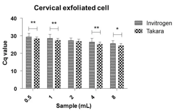
2HeLa cell count. Furthermore, 8, 4, 2, 1, and 0.5 ml of forty exfoliated cell samples were used to compare the cDNA synthesis kits.
The Takara kit compared to the Invitrogen kit showed P <0.01 in 8, 4, and 1 ml and P <0.05 in 0.5 mL. The study was performed to identify the most appropriate cDNA synthesis kit and suggests that a cDNA synthesis kit could affect the real-time PCR results.
Copyright © 2019 The Korean Society for Clinical Laboratory Science. All rights reserved.
Key words
Cervical cancer Complementary DNAComplementary DNA synthesis kit Human papillomavirus
Real-time PCR
INTRODUCTION
Cervical cancer is the fourth common malignant neoplasm in women worldwide [1]. In accordance with
World Health Organization (WHO), cervical cancer occurred with approximately 530,000 new patients and 311,000 deaths per year globally [2]. Annually, approxi- mately 3,500 patients are diagnosed and 960 patients die due to cervical cancer in Korea [3].
Since human papillomavirus (HPV) has been considered as the main cause of cervical cancer [4, 5]. HPV test is used to screen cervical cancer [6, 7]. HPV screening test consists
Korean Society for Clinical Laboratory Science
of cytological diagnostic methods and molecular diagnostic methods. In cytological diagnosis, Pap smear is a procedure to test for HPV screening in women using microscopy [8, 9]. A Pap smear is initiated with collecting cells from the cervix [10]. By screening HPV, a Pap smear allows one to prevent cervical cancer [11]. Detecting these precancerous cells early with a Pap smear is a step in stopping cervical cancer. However, the cytological method is limited by the long-term procedure and the inter- pretation - can be subjective depending on the person [12, 13]. Therefore, molecular diagnostic methods have emerged to detect HPV for sensitivity, specificity, and objectivity [14-16].
HPV screening test is done by molecular diagnostic technology such as PCR, real-time PCR, and NASBA method using HPV DNA or RNA [17-20]. Especially, real-time PCR is more sensitive and specific molecular diagnostic method to detect live HPV in patients compared to other methods [21-24]. Real-time PCR for HPV screening test is performed after RNA extraction, complementary DNA (cDNA) synthesis, and amplification of cDNA [25-27]. Real-time PCR gene expression can depend on efficiency (%) of complementary DNA synthesis even if total RNA is extracted efficiently [28].
In this study, we selected and compared two com- mercially available cDNA synthesis kits for the identi- fication of the most useful kit in HPV screening test by cervical cancer cell lines and exfoliated cell samples.
Reaction efficiency (%), coefficient of variation and cycle threshold (Ct) value analysis was performed.
MATERIALS AND METHODS
1. Cell line culture and clinical samples
Cervical cancer cell lines (HeLa and SiHa) were purchased from American Type Culture Collection ATCC (Manassas, USA) and Korean Cell Line Bank (Seoul, Korea).
SiHa and HeLa cell line were cultured in Dulbecco’s modified Eagle’s Medium (DMEM; Gibco, Carlsbad, USA), with 10% fetal bovine serum (FBS; Gibco, Carlsbad, CA, USA) and 1% streptomycin-penicillin (Gibco, Carlsbad,
CA, USA). All cell lines were incubated at 37°C in humidified 5% CO
2atmosphere. Forty exfoliated cell samples were collected from cervical cancer patients and healthy subjects at Wonju Severance Christian Hospital, Wonju, Korea, from January 2010 to December 2014. All subjects provided clinical information and this study was approved by the Institutional Ethics Committee of Yonsei University Wonju College of Medicine (Approval No.
YWMR-12-4-010).
2. Total RNA extraction
To extract RNA in cervical cancer cell lines, 1 mL of Isol-RNA Lysis Reagent (5Prime, Hamburg, Germany) was added to the cell pellet. Cells were lysed by vortexing or repeated pipetting and left to stand at room temperature for 5 min. Subsequently, 200 L of chloroform was added and the mixture was shaken vigorously and incubated at room temperature for 3 min before centrifugation at 12,000 ×g for 15 min. The resulting aqueous layer was transferred to a new tube and an equal volume of isopropanol was added and mixed by inverting the tube.
After incubation for 10 min at 25°C and centrifugation at 12,000 ×g for 10 min, 1 mL of 75% ethanol was added to the supernatant and mixed by inverting the tube. Finally, the mixture was centrifuged at 7,500 ×g for 5 min and the supernatant was removed. The RNA pellet was dried and eluted in 25 L of diethylpyrocarbonate-treated water (Intron Biotechnology, Seoul, Korea). The purity and concentration of total RNA were determined by measuring the absorbance at 260 nm and 280 nm using an Infinite 200 spectrophotometer (Tecan, Vienna, Austria). All steps in the preparation and handling of total RNA were conducted in a laminar flow hood under RNase-free conditions. The isolated total RNA was stored at −70°C until use.
3. cDNA synthesis
By using MMLV Reverse Transcriptase kits (Invitrogen,
Carlsbad, CA, USA) and random hexamers (Invitrogen,
Carlsbad, CA, USA), complementary DNA (cDNA) was
synthesized according to the manufacturer’s recom-
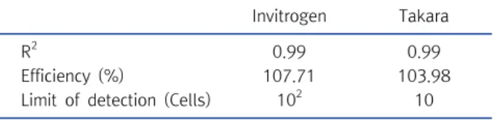
Table 1. R2
, efficiency (%) and limit of detection in SiHa cell line according to cDNA synthesis kits
Invitrogen Takara
R
20.99 0.99
Efficiency (%) 107.71 103.98
Limit of detection (Cells) 10
210
mendation. In short, 5 L of total RNA was added to a mixture containing 1 L of 10 mM dNTP mix at neutral pH, 1 L of 0.25 g/L random hexamers, and 6 L of DEPC-treated water. The PCR mixtures were incubated at 65°C for 5 min and chilled on ice. After adding a mixture of 4 L of First-strand Buffer (5×), 2 L of 0.1 M dithiothreitol (DTT), and 1 L of MMLV reverse transcriptase (at room temperature), cDNA synthesis was performed at 25°C for 10 min, 37°C for 50 min and 70°C for 15 min. The cDNA was stored at −70°C until used. And By using PrimeScript RT Master Mix kits (Takara, Japan), cDNA was synthesized according to the manufacturer’s recommendation. In short, 5 uL of total RNA was added to a mixture containing 5 uL of 5× Master mix and 11 uL of DEPC-treated water.
cDNA synthesis was performed at 37°C for 15 min, and 85°C for 5 s.
4. Reverse transcription quantitative polymerase chain reaction (RT-qPCR) for detecting HPV types
The assay consists of three different sets of HR-HPV and detects 16 HR-HPV genotypes in three tubes (group I: HPV 16 [FAM], 33, 58 [HEX], and 31, 35 [Cy5]; group II: HPV 18 [FAM], 39, 68 [HEX], and 45∼51 [Cy5]; group III: HPV 53, 56, 66 [FAM], 59, 69 [HEX], and 52 [Cy5]), by incorporating specific TaqMan probes labeled with different fluoro- phores. RT-qPCR were performed using 10 L of 2×
Thunderbird probe qPCR mix (Toyobo, Osaka, Japan), 5
L of primer and TaqMan probe mixture, 2 L of template cDNA, and distilled water to a final volume of 20 L per sample. No-template controls as negative controls were included in each run and contained sterile distilled water rather than template DNA. The PCR cycle was run as follows: 95°C for 3 min, followed by 40 cycles of 95°C for 3 s, and 55°C for 30 s. mRNA levels were quantified by determining the cycle threshold (CT), which is defined as the number of PCR cycles required for fluorescence to exceed a value significantly higher than that of the background fluorescence. For internal control, Glyceral- dehyde 3-phosphate dehydrogenase (GAPDH) was used.
5. Data calculation and statistical analysis
The qPCR R
2and efficiency (%) was calculated by qPCR Efficiency Calculator Program (ThermoFisher Scientific, CA, USA). Statistical analysis was conducted using GraphPad Prism software (Version 5.02, La Jolla, CA, USA).
Two way-ANOVA tests were used to determine the statistical significance in cDNA synthesis kits. For all tests, P<0.05 was considered statistically significant. The differences were considered statistically significant when
*P<0.05, **P<0.01, or ***P<0.001.
RESULTS
1. Reaction efficiency (%), R
2and limit of detection in SiHa cell line
To compare the cDNA synthesis efficiency (%) of two commercial kits, SiHa cell line was diluted from 10
6to 10
0and then SiHa cell line cDNA was synthesized by two RT-PCR kits and used for amplification of real-time PCR targeting HPV 16 and GAPDH. R
2of the two cDNA synthesis kits was 0.99 and 0.99, respectively (Table 1).
And efficiency (%) of the two cDNA synthesis kits was 107.71 and 103.98 respectively (Table 1). And limit of detection was 10 and 10
2cells in Takara and Invitrogen kits (Table 1). Takara kit compared to Invitrogen kit showed P<0.001 in 10
2and 10
3cell count (Figure 1). 95%
confidence interval were “−8.004 to 0.004441” “−12.00
to −3.996” “−14.80 to −6.796” “−5.404 to 2.604” “−5.104
to 2.904” “−5.104 to 2.904” from 10
1to 10
6. GAPDH Ct
value range was between 25∼30.
Figure 1. Ct values comparison between Invitrogen and Takara kit
in SiHa cell line. Takara kit compared to Invitrogen kit showed P <0.001 in 10
3and 10
2cell count (bar represents mean and standard deviation).
Figure 2. Ct values comparison between Invitrogen and Takara kit
in HeLa cell line. Takara kit compared to Invitrogen kit showed P <0.001 in 10
2and 10
1cell count (bar represents mean and standard deviation).
Table 2. R2
, efficiency (%) and limit of detection in HeLa cell line according to cDNA synthesis kits
Invitrogen Takara
R
20.95 0.99
Efficiency (%) 96.84 107.23
Limit of detection (Cells) 10
310
Figure 3. Ct values comparison between Invitrogen and Takara kit