ISSN 0378-6471 (Print)⋅ISSN 2092-9374 (Online)
https://doi.org/10.3341/jkos.2018.59.1.44
Original Article
시야결손 전 녹내장군과 정상군에서 표준자동시야검사의 변동성 비교
Comparison of the Variability of Standard Automated Perimetry between Preperimetric Glaucoma Patients and Normal Controls
김성인1⋅박혜영2⋅박찬기2
Sung In Kim, MD1, Hae-Young Lopilly Park, MD, PhD2, Chan Kee Park, MD, PhD2
새빛안과병원1, 가톨릭대학교 의과대학 서울성모병원 안과학교실2 Saevit Eye Hospital1, Goyang, Korea
Department of Ophthalmology, Seoul St. Mary’s Hospital, College of Medicine, The Catholic University of Korea2, Seoul, Korea
Purpose: To compare the variability of standard automated perimetry (SAP) between patients with preperimetric glaucoma (PPG) and normal controls.
Methods: This study included 67 eyes, classified into the following groups: 30 eyes, normal controls; and 37 eyes, PPG. All sub- jects were examined with 24-2 Humphrey static perimetry. The visual field was divided into superonasal, superotemporal, in- feronasal, and inferotemporal sectors. The variability of SAP was obtained using the standard deviation of sensitivity points at each location. We compared the variability of SAP between the normal controls and PPG patients.
Results: The variability of SAP was higher in the PPG group compared with the normal control group (p < 0.001). There was also a significant correlation between the variability of SAP and intraocular pressure fluctuations and retinal nerve fiber layer thick- ness (both p < 0.05). The variability of SAP in the PPG group was higher (p < 0.05) in all areas except the inferotemporal sector compared with the normal control group.
Conclusions: The variability of the SAP increased in PPG patients compared with normal controls, even when the visual field test results were normal.
J Korean Ophthalmol Soc 2018;59(1):44-49
Keywords: Glaucoma, Perimetry, Progression, Variability
■Received: 2017. 9. 28. ■ Revised: 2017. 10. 23.
■Accepted: 2017. 12. 15.
■Address reprint requests to Chan Kee Park, MD, PhD Department of Ophthalmology, The Catholic University of Korea Seoul St. Mary's Hospital, #222 Banpo-daero, Seocho-gu, Seoul 06591, Korea
Tel: 82-2-2258-6199, Fax: 82-2-533-3801 E-mail: [email protected]
*This article was presented at the 31st Korean glaucoma meeting.
*Conflicts of Interest: The authors have no conflicts to disclose.
ⓒ2018 The Korean Ophthalmological Society
This is an Open Access article distributed under the terms of the Creative Commons Attribution Non-Commercial License (http://creativecommons.org/licenses/by-nc/3.0/) which permits unrestricted non-commercial use, distribution, and reproduction in any medium, provided the original work is properly cited.
녹내장은 시신경 유두의 녹내장성 변화와 시신경섬유 층의 결손과 이에 대응하는 시야 결손을 특징으로 하는
진행하는 시신경병증이다.1 녹내장은 비가역적인 신경손 상을 유발하고 전 세계적으로 실명의 주된 원인이기 때문 에 조기 발견이 중요한 질병이다.2,3
시야 검사는 녹내장의 기능적 손상과 진행을 평가하는 데 있어 가장 중요한 진단법으로 현재 표준자동시야검사 계가 가장 흔하게 사용되고 있다.4 그러나 표준시야검사계 는 시신경 섬유의 30-40% 이상 손실이 일어나야 시야결 손이 나타나게 되어 조기 발견에는 적합하지 않으며, 검사 할 때마다 변동성으로 인해 방해를 받을 수 있다.5-7
시야 검사의 변동성은 피검자의 피로, 학습효과, 가양성 률, 가음성률, 주시상실 등과 같은 측정오류에 의해 발생
하지만 녹내장이 진행하면서도 증가하게 된다.7,8 녹내장 이 진행하면서 녹내장 손상에 대응하는 시야검사 구역의 변동성이 증가되었다는 몇몇 보고가 있다.9-12 그러나 보고 들이 대부분 서양인에 대한 보고이며, 국내에서 녹내장 의 증의 시야검사 변동성에 대한 보고는 아직 없는 실정이다.
또한 안압이 정상인 녹내장 의증에 국한하여 연구된 경우 는 많지 않다. 그리하여 본 연구자들은 녹내장 의증, 특히 시야 결손 전 녹내장에서 시야 검사의 변동성에 대해서 알아보기로 하였다. 시야결손 전 녹내장이란 표준자동시 야검사에서는 정상을 보이나 시신경섬유층 결손을 보이 는 녹내장으로 기능 이상을 평가하기가 어렵다. 본 연구에 서는 정상군과 시야결손 전 녹내장의 시야검사의 변동성 을 비교하여 시야결손 전 녹내장에서의 변동성에 대한 임 상적 의미를 알아보고자 하였다.
대상과 방법
2008년 1월부터 2015년 5월까지 가톨릭대학교 서울성 모병원 녹내장 클리닉을 방문한 환자들의 의무기록을 후 향적으로 분석하였다. 이 연구는 헬싱키 선언을 준수하였 고 가톨릭대학교 서울성모병원 임상시험심사위원회의 심 의(IRB)를 통과한 후 수행하였다.
환자들을 대상으로 시력검사, 굴절검사, 세극등현미경 을 통한 전안부검사, 골드만 안압측정, 중심각막두께측정 (Tomey Corporation, Nagoya, Japan), 전방각경검사, 안저 검사, 시신경 유두사진 및 망막신경섬유층사진, 빛간섭단 층촬영 Spectralis OCTTM (Heidelberg Engineering GmbH, Heidelberg, Germany)과 표준자동시야검사(Humphrey Instru- ments, Central 24-2 SITA standard strategy: San Leandro, CA, USA)를 시행하였다. 경과 관찰기간 최소 5회 이상의 시야검사가 시행되었으며, 시야검사 결과 가양성 15%, 가 음성 15%, 주시상실 20%를 초과하는 경우는 대상에서 제 외하였다.
두 명의 녹내장 전문가가 판정한 47명의 시야결손 전 녹 내장 환자 중 5회 이상의 시야 검사가 존재하는 46명 중 약을 쓰고 있는 7명, 고도 근시 2명을 제외하고 37명(37안) 을 포함시켰다. 51명의 정상인 중 시신경유두함몰비 0.4 이하의 35명 중 백내장 1명, 고도근시 4명을 제외하고 30 명(30안)을 정상 대조군으로 비교분석하였다. 시야 결손 전 녹내장은 녹내장성 시야이상을 보이지 않으나 녹내장 성 시신경 변화를 보이고, 망막신경섬유층 사진에서 명백 한 망막신경섬유층의 국소적 결손을 보이는 경우로 정의 하였다. 녹내장성 시신경 변화는 수직 시신경유두함몰비 0.6 이상, 테 얇아짐, 패임 등으로 정의하였다. 녹내장성 시
야 이상은 두 명의 녹내장 전문가가 판정하였으며, pattern deviation plot에서 궁상영역에 있는 3개 이상의 점의 역치 (mean sensitivity)가 정상의 5% 미만으로 나타나고 그중 한 점이 1% 미만일 때이거나, glaucoma hemifield test 상 out- side normal limits가 두 번 연속으로 나타날 때 또는 cor- rected pattern standard deviation가 5% 미만으로 나타날 때 로 정하였다. 시야 결손 전 녹내장은 경과 관찰 기간 동안 의 모든 시야검사에서 녹내장성 시야 이상을 보이지 않는 경우로 하였다. 표준자동시야검사상 정상은 상기 소견에 부합하지 않는 경우로 정의하였다. 안압 변동은 방문 기간 동안의 최고 안압과 최저 안압과의 차이로 구하였다.
최대교정시력이 20/30 미만이거나, 구면렌즈 대응치 값 이 ±5디옵터 이상이거나 난시가 2디옵터 이상의 굴절 이 상을 보이는 경우, 매질 혼탁, 시야검사 결과에 영향을 줄 수 있는 다른 안질환이 있는 경우, 동공의 크기가 3.0 mm 이하인 경우, 안압이 21 mmHg 이상인 경우, 녹내장 약을 사용하고 있는 경우는 대상에서 제외하였다.
시야검사
모든 시야 검사는 표준자동시야검사(Humphrey Instruments, Central 24-2 SITA standard strategy: San Leandro, CA, USA)를 시행하였다. 첫 번째와 두 번째 시야 검사는 학습 효과를 최소화하기 위해 연구에서 제외하였다. 시야 검사 는 수평, 수직 경계선을 기준으로 상비측, 상이측, 하비측, 하이측으로 나누어 분석하였다.
변동성
시야 검사의 변동성은 전체 구역과 위에서 분류한 네 구역에서 구역 내 점들의 역치의 표준 편차를 구한 후, 경 과 관찰 기간 동안의 각 값의 평균값을 사용하였다.
통계분석
Student’s t-test를 이용하여 두 군의 연령, 구면렌즈 대 응치, 안축장, 중심각막두께, 안압, 안압 변동, 신경섬유층 두께를 비교하였다. 시야 검사 지표로는 두 군의 평균편차 (mean deviation, MD), 패턴표준편차(pattern standard de- viation, PSD), 전체 및 구역별 역치 값, 전체 및 구역별 변 동성을 비교하였다. Spearman 상관분석을 이용하여 시야 검사 변동성과 관련된 인자들을 분석하였다. p-value가 0.05 미만인 경우 통계적으로 유의한 것으로 간주하였다.
결 과
연구 대상에는 시야결손 전 녹내장 환자 37명의 37안
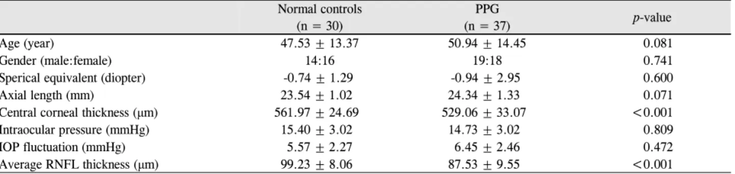
Table 1. Baseline characteristics
Normal controls (n = 30)
PPG
(n = 37) p-value
Age (year) 47.53 ± 13.37 50.94 ± 14.45 0.081
Gender (male:female) 14:16 19:18 0.741
Sperical equivalent (diopter) -0.74 ± 1.29 -0.94 ± 2.95 0.600
Axial length (mm) 23.54 ± 1.02 24.34 ± 1.33 0.071
Central corneal thickness (μm) 561.97 ± 24.69 529.06 ± 33.07 <0.001
Intraocular pressure (mmHg) 15.40 ± 3.02 14.73 ± 3.02 0.809
IOP fluctuation (mmHg) 5.57 ± 2.27 6.45 ± 2.46 0.472
Average RNFL thickness (μm) 99.23 ± 8.06 87.53 ± 9.55 <0.001
Values are presented as mean ± SD unless otherwise indicated.
PPG = preperimetric glaucoma; IOP = intraocular pressure; RNFL = retinal nerve fiber layer.
Table 2. VF characteristics
Normal controls (n = 30)
PPG
(n = 37) p-value
Mean deviation of perimerty (dB) -0.27 ± 0.85 -0.52 ± 1.01 0.090
Pattern standard deviation (dB) 1.14 ± 0.23 1.58 ± 0.35 0.007
Mean VF sensitivity (dB) 30.74 ± 1.48 29.58 ± 1.76 <0.001
VF sensitivity by location (dB)
SN 29.65 ± 1.74 27.64 ± 2.49 <0.001
ST 29.18 ± 1.83 27.59 ± 3.10 <0.001
IN 30.50 ± 1.66 29.01 ± 1.96 <0.001
IT 30.66 ± 1.64 29.53 ± 1.88 <0.001
Mean VF variability (dB) 2.03 ± 0.46 2.83 ± 1.31 <0.001
VF variability by location (dB)
SN 1.85 ± 0.57 2.50 ± 1.52 <0.001
ST 1.76 ± 0.66 2.42 ± 1.58 <0.001
IN 1.70 ± 0.49 1.93 ± 0.78 0.048
IT 1.60 ± 0.62 1.88 ± 1.68 0.063
Correspondence with RNFL defect (dB) 2.64 ± 1.63 <0.001
Values are presented as mean ± SD unless otherwise indicated.
VF = visual field; PPG = preperimetric glaucoma; SN = superonasal; ST = superotemporal; IN = inferonasal; IT = inferotemporal; RNFL
= retinal nerve fiber layer.
(남 19안, 여 18안), 큰 시신경유두함몰이 없는 정상인 30 명의 30안(남 14안, 여 16안, 대조군)이 포함되었다. 시야 결손 전 녹내장 환자의 평균 나이는 50.94 ± 14.45세, 정 상군 47.53 ± 13.37세로 통계학적으로 유의한 차이를 보 이지 않았다. 또한 성별, 구면대응치, 안축장, 중심각막두 께, 안압, 안압 변동성은 시야결손 전 녹내장군과 정상인 두 군 간에 통계적으로 유의한 차이가 없었다. 유두주위 망막신경섬유층 두께는 시야결손 전 녹내장 환자에서 평 균 87.53 ± 9.55 µm, 정상군에서 평균 99.23 ± 8.06 µm 로 시야결손 전 녹내장군에서 통계학적으로 유의하게 감 소되어 있었다(p<0.001) (Table 1).
시야 검사에서 두 군의 MD 값은 차이가 없었으나, 평 균 시야 역치는 시야결손 전 녹내장군에서 정상군에 비해 통계학적으로 유의하게 감소되어 있었다. 시야결손 전 녹 내장군의 PSD 값, 평균 시야 변동성도 정상군에 비해 통 계학적으로 유의하게 증가되어 있었다. 구역별로 비교한
변동성에서는 하이측을 제외한 모든 구역에서 시야결손 전 녹내장군에서 정상군에 비해 통계학적으로 유의하게 증가되어 있었다 (Table 2).
시야 검사 변동성과 관련된 인자들의 상관 관계를 분석 한 결과 안압 변동이 평균 시야 변동성과 유의한 상관 관 계를 보였으며(R=0.266, p=0.038), 시신경 섬유층 두께도 유의한 상관 관계를 보였다(R=-0.244, p=0.042). 시야검사 의 평균 역치 값은 시야검사 변동성과 유의한 상관 관계 를 보였으나(R=-0.787, p<0.001), MD와 PSD 값은 유의한 상관 관계를 보이지 않았다(Table 3).
고 찰
이번 연구는 정상군과 시야결손 전 녹내장의 시야검사 의 변동성을 비교하여 시야결손 전 녹내장의 기능 손상을 기술한 국내 첫 보고라는 데 의의가 있다. 특히 안압이 정
Table 3. Relationship between VF variability and ocular parameters
Normal (n = 30) PPG (n = 37)
R* p-value R* p-value
Axial length
Central corneal thickness IOP
IOP fluctuation RNFL thickness Rim area Vertical CDR Cup volume MD of VF PSD of VF
Total sensitivity of VF Superior sensitivity Inferior sensitivity
0.028 0.004 -0.184 0.232 -0.154 -0.194 0.046 0.126 -0.220 0.127 -0.528 -0.660 -0.360
0.867 0.981 0.269 0.161 0.356 0.244 0.786 0.449 0.185 0.446
<0.001
<0.001 0.026
0.073 0.032 0.346 0.272 -0.271 -0.248 0.237 0.361 -0.136 0.156 -0.874 -0.921 -0.755
0.832 0.307 0.297 0.033 0.037 0.294 0.482 0.275 0.389 0.448
<0.001
<0.001 0.007 VF = visual field; PPG = preperimetric glaucoma; R = correlation coefficient; IOP = intraocular pressure; RNFL = retinal nerve fiber lay- er; CDR = cup to disc ratio; MD = mean deviation; PSD = pattern standard deviation.
*Pearson’s correlation analysis.
A B
Figure 1. Ganglion cells in preperimetric glaucoma. Small circles represent ganglion cells and large circles represent visual field
stimulation. (A) The small circle filled with red color represents sick ganglion cells. (B) The empty space represents dead ganglion cells. The presence of sick or dead ganglion cells leads to a decrease in the action potential for stimulation, which results in a de- crease in mean sensitivity.상인 경우만 포함시키고 약물치료를 받는 군을 배제하여 안압 및 약물에 의한 영항을 배제하려 하였다. 시야검사의 변동성은 정상군에 비해 시야결손 전 녹내장에서 통계적 으로 의미 있게 증가하였다.
시야검사는 피검자가 자극을 인지하여 버튼을 누르는 각 지점별로 단기와 장기 변동성을 가질 수 밖에 없고, 이 러한 변동성은 녹내장에서 더 증가한다고 알려져 있다.13 이전 보고들에서도 녹내장 의증에서 정상군에 비해 더 큰 단기와 장기 변동성을 보이고, 녹내장군에 비해서는 더 작 은 변동성을 보인다고 하였다.14,15 본 연구에서도 시야 결 손 전 녹내장에서 정상군에 비해 더 큰 장기 변동성을 보 였다. 본 연구에서는 시야 검사의 변동성과 관련된 인자들 을 추가적으로 분석하였는데 시야검사의 변동성은 안압 변동이 클수록, 시신경 섬유층의 두께가 얇을수록 증가하 였다. 시야 검사의 변동성은 한 검사 내에서 나타나는 단
기 변동성과 다른 날 시행한 검사에서 나타나는 장기 변 동성이 있다. 단기 변동성은 21° 범위 내에서 지정된 10개 위치에 대해 평가되며, 각 지점의 역치 값 결정과 관련된 측정 오류를 나타내며, 장기 변동성은 2개 이상의 검사 사 이에서 나타나는 각 지점의 역치 값의 변동성을 나타낸 다.16 본 연구에서는 시야 검사 점들의 역치의 표준 편차 를 구한 후, 경과 관찰 기간 동안의 값을 평균하여 변동성 을 도출하였다.
녹내장의 조기 발견을 위한 기능 검사로 주파수배가시 야검사(frequency-doubling technology, FDT), 단파장시야 측정검사(short-wavelength automated perimetry, SWAP) 등이 있다. 주파수배가시야검사는 망막의 M-y 신경세포 절(Y subset of Magnocellular cell)의 기능을 높은 시간 주 파수와 낮은 공간 주파수를 가지는 띠 모양의 자극을 이 용하여 검사하는 방법으로 전체 망막신경절 세포 중 3-5%
밖에 안 되는 M-y 세포가 녹내장에 의해 손상을 입을 경 우 초기에 발견할 수 있다.17-19 단파장시야측정검사는 밝 은 노란색 배경에 파란색 자극을 주어 검사를 함으로써 청색에 민감한 blue cone (S-cone)을 선택적으로 검사할 수 있는 방법으로 이 역시 희박하게 분포하고 있는 S 추 체세포 경로에 손상을 입을 경우 초기에 발견할 수 있
다.20,21 그리고 전기 생리학 검사로 pattern ERG가 녹내장
의 시신경 섬유층 결손이나 시야변화 이전에 이상을 보인 다는 보고도 있다.22-26
시야 검사의 변동성은 정상 대조군에 비해 시야결손 전 녹내장에서 증가하였다. 신경절 세포 활동 전위는 건강 상 태에서는 일정한 진폭을 갖게 되지만, 병이 진행할수록 신 경절 세포의 수가 줄어들거나 기능이 떨어지게 된다(Fig. 1).
그래서 해당 지역의 자극에 대한 활동 전위가 줄어들게 되고, 변동성이 늘어나게 된다.27-30 표준시야검사계는 시 신경 섬유의 30-40% 이상 손실이 일어나야 시야결손이 발생하게 되므로 시야 결손이 발생하기 전에 변동성이 증 가하게 되고, 그 이후에 지속적인 역치의 감소가 되어 시 야 결손이 나타나게 된다.
또한 시야 검사 변동성은 안압 변동과 시신경 섬유층의 두께와 연관성이 있었다. 안압 변동이 클수록, 시신경 섬 유층의 두께가 얇을수록 시야 검사의 변동성이 증가하였 다. 두 가지 인자 모두 여러 연구에서 녹내장 진행에 있어 서 중요한 인자들로31-33 알려져 있으며, 녹내장의 시야 결 손 전 녹내장에서 병이 진행하면서 시야 검사의 변동성이 증가하는 것이라 하겠다. 그러나 시야 검사의 변동성이 직 접적인 녹내장 진행을 예측하는 인자로 사용되기에는 좀 더 많은 연구가 필요할 것으로 사료된다.
본 연구에는 몇 가지 제한점들이 있다. 상대적으로 연 구대상의 수가 적고, 첫 번째, 두 번째 시야검사를 제외하 였지만 시야검사의 학습효과를 완전히 배제하지 못한 점, 또한 변동성에 영향을 줄 수 있는 동공 크기, 피로, 매체 혼탁, 생리적 변동성을 완전히 통제하지 못한 점이 제한 점이라 하겠다.
결론적으로 본 연구에서 시야 검사의 변동성은 정상 대 조군에 비해 시야결손 전 녹내장에서 증가하였다. 따라서 망막신경섬유층 결손이 있는 경우 시야검사상 정상이어 도 시야 검사의 변동성이 정상인보다 크다.
REFERENCES
1) Mavilio A, Scrimieri F, Errico D. Can variability of pattern ERG signal help to detect retinal ganglion cells dysfunction in glaucom- atous eyes? Biomed Res Int 2015;2015:571314.
2) Quigley HA, Broman AT. The number of people with glaucoma
worldwide in 2010 and 2020. Br J Ophthalmol 2006;90:262-7.
3) Tatham AJ, Weinreb RN, Medeiros FA. Strategies for improving early detection of glaucoma: the combined structure-function index. Clin Ophthalmol 2014;8:611-21.
4) Gardiner SK, Johnson CA, Demirel S. The effect of test variability on the structure-function relationship in early glaucoma. Graefes Arch Clin Exp Ophthalmol 2012;250:1851-61.
5) Harwerth RS, Carter-Dawson L, Shen F, et al. Ganglion cell losses underlying visual field defects from experimental glaucoma.
Invest Ophthalmol Vis Sci 1999;40:2242-50.
6) Kerrigan-Baumrind LA, Quigley HA, Pease ME, et al. Number of ganglion cells in glaucoma eyes compared with threshold visual field tests in the same persons. Invest Ophthalmol Vis Sci 2000;41:
741-8.
7) Gillespie BW, Musch DC, Guire KE, et al. The collaborative initial glaucoma treatment study: baseline visual field and test-retest variability. Invest Ophthalmol Vis Sci 2003;44:2613-20.
8) Artes PH, Hutchison DM, Nicolela MT, et al. Threshold and varia- bility properties of matrix frequency-doubling technology and standard automated perimetry in glaucoma. Invest Ophthalmol Vis Sci 2005;46:2451-7.
9) Chauhan BC, Johnson CA. Test-retest variability of frequency- doubling perimetry and conventional perimetry in glaucoma pa- tients and normal subjects. Invest Ophthalmol Vis Sci 1999;40:
648-56.
10) Heijl A, Lindgren A, Lindgren G. Test-retest variability in glau- comatous visual fields. Am J Ophthalmol 1989;108:130-5.
11) Werner EB, Petrig B, Krupin T, Bishop KI. Variability of auto- mated visual fields in clinically stable glaucoma patients.
Investigat Ophthalmol Vis Sci 1989;30:1083-9.
12) Osborne NN, Wood JP, Chidlow G, et al. Ganglion cell death in glaucoma: what do we really know? Br J Ophthalmol 1999;83:
980-6.
13) Spry PG, Johnson CA. Identification of progressive glaucomatous visual field loss. Surv Ophthalmol 2002;47:158-73.
14) Barde MP, Barde PJ. What to use to express the variability of data:
Standard deviation or standard error of mean? Perspect Clin Res 2012;3:113.
15) Flammer J, Drance SM, Fankhauser F, Augustiny L. Differential light threshold in automated static perimetry. Factors influencing short-term fluctuation. Arch Ophthalmol 1984;102:876-9.
16) Hutchings N, Wild JM, Hussey MK, et al. The long-term fluctua- tion of the visual field in stable glaucoma. Invest Ophthalmol Vis Sci 2000;41:3429-36.
17) Johnson CA, Adams AJ, Casson EJ, Brandt JD. Progression of ear- ly glaucomatous visual field loss as detected by blue-on-yellow and standard white-on-white automated perimetry. Arch Ophthalmol 1993;111:651-6.
18) Shabana N, Cornilleau Pérès V, Carkeet A, Chew PT. Motion per- ception in glaucoma patients: a review. Surv Ophthalmol 2003;48:
92-106.
19) Tatham AJ, Medeiros FA, Zangwill LM, Weinreb RN. Strategies to improve early diagnosis in glaucoma. Prog Brain Res 2015;221:
103-33.
20) Dacey DM, Lee BB. The 'blue-on' opponent pathway in primate retina originates from a distinct bistratified ganglion cell type.
Nature 1994;367:731-5.
21) Sample PA, Medeiros FA, Racette L, et al. Identifying glaucoma-
= 국문초록 =
시야결손 전 녹내장군과 정상군에서 표준자동시야검사의 변동성 비교
목적: 시야결손 전 녹내장군과 정상군에서 표준자동시야검사의 변동성을 비교하여 표준자동시야검사 변동성의 임상적 의미를 알아보 고자 하였다.
대상과 방법: 시야결손 전 녹내장 환자 37명의 37안(남 19안, 여 18안), 큰 시신경유두함몰이 없는 정상인 30명의 30안(남 14안, 여 16안, 대조군)을 대상으로 표준자동시야검사와 빛간섭단층촬영(optical coherence tomography, OCT)을 시행하였다. 표준자동시야검 사는 수평, 수직 경계선을 기준으로 상비측, 상이측, 하비측, 하이측으로 나누어 분석하였다. 각 구역별로 변동성을 구하였고, 시야결 손 전 녹내장군을 정상군과 비교 분석하였다.
결과: 시야 검사의 변동성은 정상 대조군에 비해 시야결손 전 녹내장군에서 증가하였다(p<0.001). 또한 시야 검사의 변동성과 안압 변동과 시신경 섬유층 두께와 유의한 상관관계가 있었다(p<0.05). 구역별로 본 분석에서는 하이측을 제외한 모든 구역에서 시야결손 전 녹내장군에서 정상 대조군에 비해 시야 검사의 변동성이 증가하였다(p<0.05).
결론: 시야 검사의 변동성은 정상 대조군에 비해 시야결손 전 녹내장에서 증가하였으며, 망막신경섬유층 결손이 있는 경우 시야검사 상 정상이어도 시야 검사의 변동성이 정상인보다 크다.
<대한안과학회지 2018;59(1):44-49>
2006;47:3381-9.
22) Horn FK, Tornow RP, Jünemann AG, et al. Perimetric measure- ments with flicker-defined form stimulation in comparison with conventional perimetry and retinal nerve fiber measurements.
Investigat Ophthalmol Vis Sci 2014;55:2317-23.
23) Lamparter J, Russell RA, Schulze A, et al. Structure-function rela- tionship between FDF, FDT, SAP, and scanning laser ophthalmo- scopy in glaucoma patients. Invest Ophthalmol Vis Sci 2012;53:
7553-9.
24) Mulak M, Szumny D, Sieja-Bujewska A, Kubrak M. Heidelberg edge perimeter employment in glaucoma diagnosis--preliminary report. Adv Clin Exp Med 2012;21:665-70.
25) Pfeiffer N, Bach M. The pattern-electroretinogram in glaucoma and ocular hypertension. A cross-sectional and longitudinal study.
Ger J Ophthalmol 1992;1:35-40.
26) Bode SF, Jehle T, Bach M. Pattern electroretinogram in glaucoma suspects: new findings from a longitudinal study. Invest Ophthalmol Vis Sci 2011;52:4300-6.
27) Wall M, Woodward KR, Doyle CK, Artes PH. Repeatability of au- tomated perimetry: a comparison between standard automated
28) Anderson JS, Lampl I, Gillespie DC, Ferster D. The contribution of noise to contrast invariance of orientation tuning in cat visual cortex. Science 2000;290:1968-72.
29) Levine MW. Variability of responses to sinusoidal modulation. Vis Neurosci 1994;11:155-63.
30) Redmond T, Garway-Heath DF, Zlatkova MB, Anderson RS.
Sensitivity loss in early glaucoma can be mapped to an enlarge- ment of the area of complete spatial summation. Invest Ophthalmol Vis Sci 2010;51:6540-8.
31) Leske MC, Wu SY, Hennis A, et al. Risk factors for incident open-angle glaucoma: the Barbados Eye Studies. Ophthalmology 2008;115:85-93.
32) Kass MA, Heuer DK, Higginbotham EJ, et al. The Ocular Hypertension Treatment Study: a randomized trial determines that topical ocular hypotensive medication delays or prevents the onset of primary open-angle glaucoma. Arch Ophthalmol 2002;120:
701-13; discussion 829-30.
33) Miglior S, Zeyen T, Pfeiffer N, et al. Results of the European Glaucoma Prevention Study. Ophthalmology 2005;112:366-75.