ISSN 0378-6471 (Print)⋅ISSN 2092-9374 (Online)
http://dx.doi.org/10.3341/jkos.2016.57.3.445
Original Article
특발성 망막전막에서 내경계막제거 시 인도시아닌그린 사용 여부 및 노출시간에 따른 수술 결과 비교
Clinical Outcomes of Indocyanine Green-Assisted Peeling of the Internal Limiting Membrane in Epiretinal Membrane Surgery
최우석⋅정우진⋅권윤형
Woo Seok Choi, MD, Woo Jin Jeong, MD, PhD, Yoon Hyung Kwon, MD
동아대학교 의과대학 안과학교실
Department of Ophthalmology, Dong-A University College of Medicine, Busan, Korea
Purpose: In this study we compared the clinical outcomes of idiopathic epiretinal membrane (ERM) surgery according to the use of indocyanine green (ICG) and ICG exposure time.
Methods: The medical records of 76 patients with an idiopathic ERM that underwent vitrectomy and ERM and internal limiting membrane (ILM) removal were reviewed. We compared the results (best corrected visual acuity [BCVA, log MAR] and central macular thickness [CMT, μm]) of idiopathic ERM surgeries using ILM peeling with (group I, 39 eyes) and without ICG (group II, 37 eyes). Additionally, the correlation of ICG exposure time and clinical outcomes in group I was analyzed.
Results: Gender, age, lens state, preoperative BCVA, and preoperative CMT were not significantly different between the two groups. The postoperative BCVA was significantly improved in both groups but the difference was not statistically significant.
The postoperative CMT was significantly improved in both groups and the change amount of group I was more larger than group II.
Additionally, ICG exposure time was not significantly correlated with changes of BCVA and CMT.
Conclusions: Intravitreal ICG-assisted ILM peeling did not significantly affect the recovery of BCVA, however that impaired the recovery of CMT. ICG exposure time did not affect the postoperative visual outcome.
J Korean Ophthalmol Soc 2016;57(3):445-452
Keywords: Exposure time, Idiopathic epiretinal membrane, Indocyanine green, Internal limiting membrane
■Received: 2015. 6. 5. ■ Revised: 2015. 10. 11.
■Accepted: 2016. 1. 14.
■Address reprint requests to Yoon Hyung Kwon, MD Department of Ophthalmology, Dong-A University Hospital,
#26 Daesingongwon-ro, Seo-gu, Busan 49201, Korea Tel: 82-51-240-2776, Fax: 82-51-254-1987
E-mail: [email protected]
* This study was presented as a narration at the 113th Annual Meeting of the Korean Ophthalmological Society 2015.
* This work was supported by the Dong-A University Research Fund, Busan, Korea.
ⓒ2016 The Korean Ophthalmological Society
This is an Open Access article distributed under the terms of the Creative Commons Attribution Non-Commercial License (http://creativecommons.org/licenses/by-nc/3.0/) which permits unrestricted non-commercial use, distribution, and reproduction in any medium, provided the original work is properly cited.
망막전막은 내경계막 표면에 세포의 증식으로 무혈관성 세포막이 형성되는 질환으로 망막의 주름 및 뒤틀림을 유 발하여 변형시 및 시력저하를 유발하는 질환이다. 전체인 구의 3.5-6.9%에서 발생하며, 발생률은 60세 이전 2%, 70 세 이후 12%로 연령이 증가할수록 증가하는 질환이다.1-3 포도막염, 외상, 망막 혈관 질환, 안내종양, 망막박리 수술 등으로 인해 이차적으로 발생할 수도 있으나, 대부분 그 원 인을 알 수 없는 특발성으로 발생한다. 특발성인 경우 후유 리체박리로 인한 내경계막의 파괴가 망막전막 진행에 중요 한 역할을 하는 것으로 생각되고,4-7 내경계막은 망막과 유 리체 사이의 경계를 이루는 구조로 근섬유세포 증식의 발 판이 되어 망막전막의 재발에 중요한 역할을 하는 것으로
보고되었다.8
1978년 Machemer9에 의해 망막전막의 성공적인 제거가 처 음 보고된 이래로, 여러 연구들에서 망막전막 제거수술의 성 공적인 경과를 보고하였으며,10-15 수술 시 내경계막을 제거해 줌으로써 재발률을 줄이고 황반주름을 호전시킨다는 여러 보 고가 있었다.16-18 하지만 내경계막은 얇고 투명한 조직으로 수 술 시 관찰이 쉽지 않아 수술적 제거의 어려움이 있었으나, 내 경계막만 선택적으로 착색시키는 인도시아닌그린(indocyanine green, ICG)의 사용이 소개된 후19 많은 연구에서 ICG 용액이 내경계막의 시인성과 수술 용이성을 향상시키며 망막손상의 위험성을 줄일 수 있음을 보고하였다.8,19-21
그러나 ICG 용액이 가지는 망막 독성으로 인한 손상이 발생 가능하다는 문제점이 제기되어 왔고, ICG 용액의 농 도와 삼투압, 조직과의 접촉시간, 제거 시 가해진 힘에 의한 견인 등에 의해 영향을 받을 것으로 생각되었다.22,23 ICG의 농도는 높을수록 시인성이 좋으나 광독성이 심하므로, ICG 의 독성을 최소화하며 시인성을 극대화하는 적정 ICG 용액 의 농도에 대해 여러 보고가 있었고, 대체로 0.05-0.5% ICG 용액이 사용되고 있다.24-27 또한 용매로는 Balanced salt sol- ution (BSS)과 5% Dextrose가 주로 사용되고, 최근 연구에 서 삼투압이 더 큰 5% Dextrose가 BSS에 비해 ICG의 독성 이 적으며, 내경계막의 시인성을 높인다고 보고되었다.28-30 이에 저자들은 기존의 보고를 토대로 망막 독성 가능성 을 최소화할 수 있을 것으로 생각되는 5% Dextrose를 용매 로 한 0.05% 농도의 ICG 용액을 이용한 수술과 ICG 없이 Triamcinolone만을 이용한 수술 간의 결과를 후향적으로 비교해 보고자 하였으며, ICG 용액의 술 중 노출시간과의 연관성도 알아보고자 하였다.
대상과 방법
본원에서 2010년 3월부터 2015년 3월까지 특발성 망막 전막으로 진단 후 3개월 이상 관찰이 가능했던 76명 76안 을 대상으로 의무기록을 후향적으로 조사하였으며 수술 전 망막박리, 망막혈관질환, 안내염증질환, 당뇨망막병증, 그 외 황반부 질환이 동반된 경우는 제외하였다. 유리체 절제술 시 망막전막 및 내경계막 염색을 위해 트리암시놀 론(Triamcinolone, TA)과 ICG를 함께 사용한 39안을 I군, 트리암시놀론(TA)만을 사용한 37안을 II군으로 나누어 분 석하였다. 대상군은 수술 시기별로 구분하여, 2012년 6월 이전에 수술을 시행한 환자들에서는 트리암시놀론만을 이 용하여 수술을 시행하였고(II군), 그 이후의 환자들은 모두 인도시아닌그린과 트리암시놀론을 함께 사용하여 수술을 시행하였다(I군). 수술 전 특발성 망막전막의 증등도와는
무관하게 일괄적으로 적용하였다.
두 군 모두 Triamcinolone (Triamcinolone acetonide, Dong Kwang Pharm Co., Seoul, Korea)을 사용하였고, ICG 용액은 ICG (DID-Indocyanine green® injection: Dongindang Phar- maceutical, Seoul, Korea) 25 mg을 5% Dextrose 10 mL로 완전히 용해시킨 후 이 중 1 mL를 5% Dextrose 4 mL와 혼합하여 최종적으로 0.5 mg/mL (0.05%) 농도가 되도록 하여 사용하였다.
수술 전 최대교정시력, 전안부 및 수정체 검사, 안저검사 를 시행하였으며, 스펙트럼영역 빛간섭단층촬영(spectral do- main optical coherence tomography [SD-OCT], Cirrus HD OCT® Model 4000, Carl Zeiss Meditec Inc., Dublin, CA, USA)을 시행하여 중심황반두께(central macular thick- ness, CMT)를 측정하였다.
수술은 숙련된 2명의 술자에 의해 시행되었으며, 수술방 법의 차이는 없었다. 3개의 모양체 평면부 공막창을 통한 표준 3-port 유리체절제술(Accurus®, Alcon, Fort Worth, TX, USA)을 시행하였고, 중심부 유리체 절제술 시행 후 Triamcinolonea acetonide (TA)를 주입하여 후유리체 박리 를 확인하고 박리가 없으면 박리를 일으킨 후 유리체를 제 거하였다. I군에서는 0.05% ICG 용액을 황반부에 0.1 mL 주입하였다. 약 10-15초 후 재관류하여 ICG를 제거하고, 염색된 내경계막을 확인한 후 안내겸자를 사용하여 중심와 로부터 약 3-4배 유두직경 크기의 내경계막을 원형으로 제 거하였다. ICG 1회의 주입으로 내경계막 염색이 충분치 않 을 경우 위 과정을 반복하여 염색하였다. II군에서는 관류 액의 주입을 멈추고 트리암시놀론 용액을 황반부에 0.1 mL 주입하였다. 약 10-15초 후 재관류하여 유리체내 트리암시 놀론을 제거하였고, 그 후 내경계막 표면의 콜라겐섬유와 부착된 트리암시놀론을 통해 망막전막 및 내경계막을 확인 하고 안내겸자를 사용하여 중심와로부터 약 3-4배 유두직 경 크기의 내경계막을 원형으로 제거하였다. 내경계막이 불완전하게 제거될 가능성이 있어 1-2회 추가적으로 트리 암시놀론을 주입함으로서 충분한 내경계막의 제거를 확인 하였다. 수정체안은 모든 경우에서 백내장 수술을 시행하 였으며, 백내장 수술이 필요한 경우에서는 유리체 절제술 전 12시 방향에 각막절개를 통하여 초음파유화술을 먼저 시행하고 유리체절제술과 망막전막 제거술이 모두 끝난 다 음 마지막에 접형 인공수정체를 삽입하였다.
수술 후 1주일, 1개월, 3개월째 경과관찰을 시행하였으 며, 각 시기의 최대교정시력과 빛간섭단층촬영을 통한 중 심황반두께를 측정하였다. 최대교정시력은 술 전과 비교하 기 위해 logMAR 스케일(logMAR)로 환산하였다. ICG 총 노출 시간은 ICG 주입 시마다 유리체내에 머무른 시간을
Table 1. Comparison of patient’s demographics between the two groups
Group I* (39 eyes) Group II† (37 eyes) p-value
Age (years) 66.23 ± 9.48 (44-92) 68.18 ± 8.79 (47-83) 0.692‡
Male:female 18:21 16:21 0.721§
Phakia:pseudophakia 31:8 29:8 0.953§
Preop BCVA (log MAR) 0.55 ± 0.22 0.61 ± 0.22 0.223‡
Preop CMT (μm) 419.69 ± 79.66 392.71 ± 109.25 0.219‡
Total ICG exposure time (seconds) 37.74 ± 15.09 (14-74) (-) (-)
Numbers of ICG injection 2.33 ± 0.83 (1-4) (-) (-)
Values are presented as mean ± SD unless otherwise indicated.
Preop = preoperative; BCVA = best corrected visual acuity; CMT = central macular thickness; ICG = indocyanine green.
*Using triamcinolone with indocyanine green to stain the internal limiting membrane; †Using only triamcinolone; ‡Student t-test; §Chi-square test.
Table 2. Comparison of the BCVA (log MAR) changes after surgery between the two groups
BCVA (log MAR) Baseline Postoperative
1 week
Postoperative 1 month
Postoperative
3 months p-value‡
Group I* 0.003
BCVA 0.55 ± 0.22 0.66 ± 0.37 0.51 ± 0.34 0.36 ± 0.36
△ - -0.08 ± 0.30 0.03 ± 0.28 0.20 ± 0.31
Group II† <0.001
BCVA 0.61 ± 0.22 0.79 ± 0.32 0.56 ± 0.24 0.48 ± 0.31
△ - -0.16 ± 0.33 0.04 ± 0.25 0.13 ± 0.25
p-value§ - 0.254 0.898 0.318
Values are presented as mean ± SD unless otherwise indicated.
BCVA = best corrected visual acuity; △ = baseline value-postoperative value.
*Using triamcinolone with indocyanine green to stain the internal limiting membrane; †Using only triamcinolone; ‡Compared to baseline with the value of postoperative 3 months, paired t-test; §Comparison of the BCVA change amount between the two groups at each point, Student t-test.
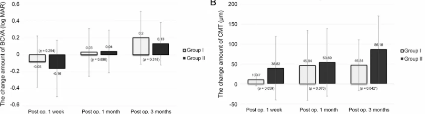
Figure 1. Comparison of the change amount of BCVA (log MAR) and the CMT (μm) in the two groups. (A) The change amount
of BCVA (log MAR) did not differ significantly between the two groups at any point. (B) The change amount of CMT (μm) in the‘ICG dye with triamcinolone’ group was significantly smaller than that in the ‘triamcinolone only’ group at 3 months after surgery (p = 0.042, *Student t-test). BCVA = best corrected visual acuity; CMT = central macular thickness; Post op. = postoperative;
ICG = indocyanine green.
측정하였으며, 수회 주입 시 각 노출시간을 합산하여 총 노 출시간을 계산하였다.
통계학적 분석은 SPSS statistics v. 12.0 (SPSS Inc., Chicago, IL, USA)을 사용하여 두 군 간의 비교는 Student t-test, Chi-squre test를, 수술 전후 결과 비교는 paired t-test 를 이용하였으며 ICG 총 노출시간과 시력 및 중심황반두께
변화량의 상관관계는 Pearson 상관계수를 이용하였다. p값 이 0.05 미만인 경우에 통계적으로 유의하다고 판단하였다.
결 과
전체 76명의 대상안 중 I군이 39안, II군이 37안이었으며,
A B
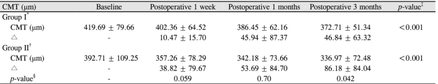
Table 3. Comparison of the CMT (μm) changes after surgery between the two groups
CMT (μm) Baseline Postoperative 1 week Postoperative 1 months Postoperative 3 months p-value‡ Group I*
CMT (μm) 419.69 ± 79.66 402.36 ± 64.52 386.45 ± 62.16 372.71 ± 51.34 <0.001
△ - 10.47 ± 15.70 45.94 ± 87.37 46.84 ± 63.32
Group II†
CMT (μm) 392.71 ± 109.25 357.26 ± 78.29 342.18 ± 73.66 336.97 ± 72.48 <0.001
△ - 38.82 ± 79.67 53.69 ± 84.70 86.18 ± 84.04
p-value§ - 0.059 0.70 0.042
Values are presented as mean ± SD unless otherwise indicated.
CMT = central macular thickness; △ = baseline value-postoperative value.
*Using triamcinolone with indocyanine green to stain the internal limiting membrane; †Using only triamcinolone; ‡Compared to baseline with the value of postoperative 3 months, paired t-test; §Comparison of the CMT change amount between the two groups at each point, Student t-test.
Table 4. Correlation of ICG exposure time with best corrected visual acuity and central macular thickness changes
ICG time △BCVA △CMT
1 week 1 month 3 months 1 week 1 month 3 months
Pearson coefficients (r)* -0.312 -0.3 -0.55 -0.245 -0.062 -0.312
p-value* 0.053 0.86 0.74 0.139 0.341 0.71
ICG = indocyanine green; BCVA = best corrected visual acuity; CMT = central macular thickness; △ = baseline value-postoperative value.
*Pearson's correlation test.
Figure 2. Scatter plots of ICG exposure time versus BCVA change and CMT change of 3 months value after operation. (A) ICG ex-
posure time is not significantly correlated with BCVA change (r* = ‑0.55, p = 0.74). (B) ICG exposure time is not significantly correlated with CMT change (r* = ‑0.312, p = 0.71). ICG = indocyanine green; BCVA = best corrected visual acuity; CMT = central macular thickness. *Pearson's correlation test.남자와 여자의 비율은 I군이 18:21, II군이 16:21이었고, 평 균연령은 I군이 66.23 ± 9.48세(44-92세), II군이 68.18 ± 8.79세(47-83세)로 두 군 간 통계학적으로 유의한 차이는 없 었다. 수정체안과 인공수정체안의 비율은 I군이 31:8, II군 은 29:8로 두 군 간 유의한 차이는 없었으며, 초진 시 최대 교정시력은 I군이 0.55 ± 0.22 logMAR, II군이 0.61 ± 0.22 logMAR로 I군이 더 좋았으나 유의한 차이는 없었다. 수술 전 중심황반두께 또한 I군은 419.69 ± 79.66 μm, II군은 392.71 ± 109.25 μm로 유의한 차이가 없었으며, ICG를 사
용한 I군에서 평균 ICG 주사횟수는 2.33 ± 0.83회(1-4회), 평균 총 망막과의 노출시간은 37.74 ± 15.09초(14-74초)였 다(Table 1).
수술 전후 최대교정시력의 변화는 수술 후 1주일째 I군 은 0.55에서 0.66 logMAR로, II군에서는 0.61에서 0.79 logMAR로 두 군 모두 통계학적으로 유의하게 악화되었으 며(paired t-test, p=0.024, 0.002), 술 후 3개월째 I군은 0.36 logMAR, II군은 0.48 logMAR로 술 전과 비교하여 유의하 게 호전되었다(p=0.003, 0.001). 그러나 술 후 경과관찰 기
A B
간 동안의 최대교정시력 변화량은 두 군 간 유의한 차이가 없었다(Student t-test, p>0.05) (Table 2, Fig. 1A).
술 전과 수술 3개월 후 중심황반두께의 변화는 I군에서 는 419.69 μm에서 372.71 μm로, II군에서는 392.71 μm에서 336.97 μm로 두 군 모두 통계적으로 유의하게 호전되었다 (Paired t-test, p=0.001, 0.001). 술 전과 비교해 중심황반두 께는 수술 후 3개월째 I군은 46.84 μm, II군은 86.18 μm의 감소를 보여 I군이 II군에 비해 중심황반두께의 호전량이 유의하게 적었다(Student t-test, p=0.042) (Table 3, Fig. 1B).
I군을 대상으로 ICG 총 노출시간과 각 시기별 최대교정 시력(logMAR) 변화량, 중심황반두께(μm) 변화량의 상관관 계(Pearson's correlation test)를 분석하였다. 모든 시기에서 총 노출시간이 길어질수록 최대교정시력 변화량이 작아지 는 음의 상관관계를 보였으나 통계학적으로 유의하지 않았 으며(r=-0.55, p=0.74), 중심황반두께의 변화량도 음의 상관관 계를 보였지만, 유의하지 않았다(r=-0.312, p=0.71)(Table 4, Fig. 2).
고 찰
현재 망막전막 수술에서 술 후 재발률을 줄일 수 있는 내 경계막의 제거가 흔히 시행되고 있으며, 수술 중 내경계막 의 시인성을 높이기 위한 염색제로 Indocyanine green, Trephan blue, Brilliant blue G 등이 사용되고 있다. 가장 흔 히 사용하는 ICG는 망막에 대한 독성을 가지고 있어 수술 후 결과를 저해한다는 여러 보고가 있었고,22,27,31-33 망막손 상을 일으키는 기전으로 ICG 용액의 농도와 삼투압, 조직 과의 접촉시간, 제거 시 가해진 힘에 의한 견인 등과 관계 된다고 보고하였다.22
Yam et al34은 0.25% (2.5 mg/mL) ICG 용액에 망막이 노출된 후, 망막색소상피세포의 세포사멸유전자 발현을 관 찰하였고, 그 발현은 농도가 높을수록 증가한다고 보고하 였다. 또한 Engelbrecht et al35은 ICG 사용 후 망막색소상 피 세포의 위축을 관찰하였다. 이에 ICG의 독성을 최소화 하며 시인성을 극대화하는 적정 ICG 용액의 농도에 대한 연구가 진행되었고, Kwok et al25은 내경계막 염색 시 ICG 용액 농도에 따른 염색 정도 및 수술 결과를 비교하였고, 0.025%, 0.05%, 0.125%의 농도에서 수술 후 시력호전에는 유의한 차이가 없는 반면, 0.125%의 농도가 내경계막 염색 의 시인성이 가장 좋다고 보고하였다.
또한 ICG 용액에 사용한 용매의 종류, 용매의 삼투압에 따라서도 망막 독성의 차이가 보고되었는데, Stalmans et al36은 ICG 용액은 삼투압이 낮을수록 망막색소상피세포에 더욱 독성을 띄어, 낮은 삼투압의 BSS보다 높은 삼투압을
가지는 5% Dextrose가 망막색소상피세포에 독성이 적음을 보고하였고, Ho et al29은 Sodium이 없는 용매를 사용하면 ICG로 인한 광독성을 줄일 수 있다고 보고하였다. 또한 Haritoglou et al28은 다양한 농도로 서로 다른 종류의 용매 를 사용한 ICG 용액의 흡수스펙트럼을 측정하였고, ICG 용 액의 농도가 높을수록 광민감성이 증가하여 망막색소상피세 포의 광독성이 증가한다고 하였으며, BSS보다 5% Dextrose 를 사용하는 것이 광독성을 줄일 수 있다고 보고하였다. 저 자들은 이러한 연구들을 토대로 ICG 독성을 최소화하는 5% Dextrose를 용매로 한 0.05% ICG 용액을 수술에 사용 한 경우와 ICG 없이 Triamcinolone만 사용한 경우의 수술 전후 최대교정시력 및 중심황반두께의 변화를 후향적으로 비교분석하였다.
Oh et al37은 특발성 망막 전막 수술을 시행한 43안을 대 상으로 본 연구보다 더 높은 농도인 0.5% ICG 사용 여부에 따라 최대교정시력, 중심황반두께, 다초점 망막전위도검사 를 비교분석하였으나 의미 있는 차이를 보이지 않았다. 또 한 Choi et al38과 Hahm et al39은 황반 원공에서 ICG 사용 여부에 따른 원공폐쇄율 및 최대교정시력을 비교하였고, 두 연구 모두 해부학적 성공 및 기능적인 시력회복에 유의 한 차이를 보이지 않았다. 이와는 달리 Shiono et al40은 황 반 원공 환자에서 ICG 사용 여부에 따른 술 전후 수술결과 를 비교하였고, ICG를 사용한 군에서 술 후 초기 수개월간 최대교정시력 및 광수용체 내절과 외절 손상의 회복이 지연 됨을 보고하였다. 또한 Choi et al41 의 연구에서도 ICG 염색 이 기능적인 시력회복을 방해한다고 보고하였다. Haritoglou et al42은 전자현미경을 이용하여 ICG 사용 여부에 따른 내 경계막의 경직도를 측정하였고, ICG의 사용이 내경계막의 경직도를 높이며 이로 인해 내경계막 제거 시 접해 있는 내 측 망막의 손상을 발생시킬 수 있다고 보고하였다. 또한 Gandorfer et al22은 ICG 사용에 따른 내경계막 시인성의 향상으로 인해 술자가 내경계막 제거 시 그 아래 주변망막 층까지 제거할 수도 있다고 보고하였다.
본 연구에서 최대교정시력은 두 군 모두 술 후 1주째 유 의하게 악화되었고, 이는 유리체 절제술 후 생긴 전방 내 염증 및 일시적 각막 부종 등으로 인해 시력저하가 나타난 것으로 생각된다. 술 후 3개월째 두 군 모두 술 전에 비해 통계적으로 유의하게 호전을 보였지만, 두 군 간 유의한 차 이를 보이진 않았다. 중심황반두께 역시 술 전에 비해 두 군 모두 유의한 호전을 보였지만, 술 후 3개월째에 측정한 중심황반두께의 변화량은 I군이 II군에 비해 중심황반두께 의 호전량이 유의하게 적었다. ICG의 사용은 수술 전후 최 대교정시력의 회복에는 유의한 영향을 끼치지 않았지만 수 술 후 중심황반두께의 호전량을 유의하게 감소시켰다. 이
러한 결과는 ICG의 망막에 대한 독성, ICG에 의한 내경계 막 경직도의 상승과 그로 인한 내경계막 제거 시 내측 망막 세포의 손상, 그리고 트리암시놀론만 사용 시 상대적으로 불완전한 내경계막의 제거 등과 같은 차이로 인해 나타난 것으로 생각된다.
기존의 연구8,25,31들에서 내경계막 제거를 위한 ICG 용액 사용 시 유리체내 용액이 머무르는 시간은 약 30-60초였다.
본 연구에서도 ICG 용액 총 노출시간은 평균 37.74 ± 15.09초(14-74초)로 기존의 연구들과 유사하였다. Enaida et al33은 Rat의 망막을 대상으로 상대적으로 낮은 농도인 0.025% ICG 용액을 망막에 노출시켰으며, 노출 후 10일이 경과하여 망막의 형태학적 변화가 나타남을 보고하였다.
낮은 농도에서도 노출 시간이 길어지면 망막의 손상을 나 타낼 수 있음을 뜻하고, 이에 본 연구에서는 노출 시간과 술 후 결과의 상관관계를 분석하고자 하였다. 모든 시기에 서 총 노출시간이 길어질수록 최대교정시력, 중심황반두께 의 변화량이 적어지는 음의 상관관계를 보였지만 통계학적 으로 유의하지 않았다. 망막전막 수술 시에는 망막색소상 피층이 직접적으로 ICG에 접촉하지 않기 때문에 약 10-60 초가량 적은 시간의 ICG 노출은 수술 후 결과와 상관성을 띄지 않을 수 있을 것으로 생각된다. 그러나 황반원공 수술 에서는 ICG에 망막색소상피층이 직접 노출될 가능성이 높 으므로 기존의 보고들과 같이 상대적으로 수술 후 시력예 후에 더욱 영향을 끼칠 수 있을 것으로 생각된다.
저자들은 특발성 망막전막에서 내경계막 제거 시 내경계 막 염색을 위한 인도시아닌그린의 사용이 수술결과에 미치 는 영향을 분석하고자 하였다. 그러나 본 연구는 후향적 연 구였고, 상대적으로 짧은 3개월의 경과관찰 기간으로 중심 황반두께 호전량의 장기적인 경과를 확인할 수 없는 제한 점이 있었다. 또한 망막전막이 심할수록 시세포 내절/외절 접합부 파열의 가능성이 높아지고, 불량한 시력예후를 보
이므로43-45 술 전 망막전막의 심한 정도를 고려한 추가연구
가 필요할 것으로 생각된다. 하지만 한국인에서 ICG 노출 시간에 따른 술 후 결과를 비교한 첫 결과물이며, 현재 보 편적으로 사용되는 ICG의 효과와 그 위험성을 알아보고자 하는 데 의의가 있겠다.
인도시아닌그린의 사용은 수술 후 시력의 예후에는 의미 있는 영향을 끼치지 않았으나, 수술 후 초기 중심황반두께 의 호전을 방해하는 것으로 생각된다. 그러나 인도시아닌 그린의 노출시간에 따른 차이는 보이지 않았다. 수술 후 초 기 중심황반두께 호전은 ICG의 망막에 대한 독성, ICG에 의한 내경계막 경직도의 상승, 그로 인한 내경계막 제거 시 내측 망막세포 손상 정도의 차이 등에 의해 저해되는 것으 로 생각된다. 추후 내경계막의 완전한 제거 및 내측 망막손
상 여부를 수술 중 확인할 수 있는 실시간 빛간섭단층촬영 등을 이용한 장기간의 전향적인 추가 연구가 필요할 것으 로 생각된다.
REFERENCES
1) Kawasaki R, Wang JJ, Sato H, et al. Prevalence and associations of epiretinal membranes in an adult Japanese population: the Funagata study. Eye (Lond) 2009;23:1045-51.
2) Klein R, Klein BE, Wang Q, Moss SE. The epidemiology of epi- retinal membranes. Trans Am Ophthalmol Soc 1994;92:403-25;
discussion 425-30.
3) Mitchell P, Smith W, Chey T, et al. Prevalence and associations of epiretinal membranes. The Blue Mountains Eye Study, Australia.
Ophthalmology 1997;104:1033-40.
4) Poliner LS, Olk RJ, Grand MG, et al. Surgical management of pre- macular fibroplasia. Arch Ophthalmol 1988;106:761-4.
5) de Bustros S, Thompson JT, Michels RG, et al. Vitrectomy for idio- pathic epiretinal membranes causing macular pucker. Br J Ophthalmol 1988;72:692-5.
6) de Bustros S, Rice TA, Michels RG, et al. Vitrectomy for macular pucker. Use after treatment of retinal tears or retinal detachment.
Arch Ophthalmol 1998;106:758-60.
7) Pesin SR, Olk RJ, Grand MG, et al. Vitrectomy for premacular fibroplasia. Prognostic factors, long-term follow-up, and time course of visual improvement. Ophthalmology 1991;98:1109-14.
8) Kwok AK, Lai TY, Li WW, et al. Indocyanine green-assisted in- ternal limiting membrane removal in epiretinal membrane surgery:
a clinical and histologic study. Am J Ophthalmol 2004;138:194-9.
9) Machemer R. The surgical removal of epiretinal macular mem- branes (macular puckers) (author's transl). Klin Monbl Augenheilkd 1978;173:36-42.
10) Michels RG. Vitrectomy for macular pucker. Ophthalmology 1984;91:1384-8.
11) Margherio RR, Cox MS Jr, Trese MT, et al. Removal of epimacular membranes. Ophthalmology 1985;92:1075-83.
12) McDonald HR, Verre WP, Aaberg TM. Surgical management of idiopathic epiretinal membranes. Ophthalmology 1986;93:978-83.
13) Donati G, Kapetanios AD, Pournaras CJ. Complications of surgery for epiretinal membranes. Graefes Arch Clin Exp Ophthalmol 1998;236:739-46.
14) Benhamou N, Massin P, Spolaore R, et al. Surgical management of epiretinal membrane in young patients. Am J Ophthalmol 2002;
133:358-64.
15) Massin P, Paques M, Masri H, et al. Visual outcome of surgery for epiretinal membranes with macular pseudoholes. Ophthalmology 1999;106:580-5.
16) Park DW, Dugel PU, Garda J, et al. Macular pucker removal with and without internal limiting membrane peeling: pilot study.
Ophthalmology 2003;110:62-4.
17) Kwok AKh, Lai TY, Yuen KS. Epiretinal membrane surgery with or without internal limiting membrane peeling. Clin Experiment Ophthalmol 2005;33:379-85.
18) Bovey EH, Uffer S, Achache F. Surgery for epimacular membrane:
impact of retinal internal limiting membrane removal on functional outcome. Retina 2004;24:728-35.
19) Burk SE, Da Mata AP, Snyder ME, et al. Indocyanine green-assisted
peeling of the retinal internal limiting membrane. Ophthalmology 2000;107:2010-4.
20) Gandorfer A, Messmer EM, Ulbig MW, Kampik A. Indocyanine green selectively stains the internal limiting membrane. Am J Ophthalmol 2001;131:387-8.
21) Da Mata AP, Burk SE, Riemann CD, et al. Indocyanine green-as- sisted peeling of the retinal internal limiting membrane during vi- trectomy surgery for macular hole repair. Ophthalmology 2001;
108:1187-92.
22) Gandorfer A, Haritoglou C, Gass CA, et al. Indocyanine green-as- sisted peeling of the internal limiting membrane may cause retinal damage. Am J Ophthalmol 2001;132:431-3.
23) Lee JE, Yoon TJ, Oum BS, et al. Toxicity of indocyanine green in- jected into the subretinal space: subretinal toxicity of indocyanine green. Retina 2003;23:675-81.
24) Lai MM, Williams GA. Anatomical and visual outcomes of idio- pathic macular hole surgery with internal limiting membrane re- moval using low-concentration indocyanine green. Retina 2007;
27:477-82.
25) Kwok AK, Lai TY, Yew DT, Li WW. Internal limiting membrane staining with various concentrations of indocyanine green dye un- der air in macular surgeries. Am J Ophthalmol 2003;136:223-30.
26) Haritoglou C, Gandorfer A, Gass CA, Kampik A. Histology of the vitreoretinal interface after staining of the internal limiting mem- brane using glucose 5% diluted indocyanine and infracyanine green. Am J Ophthalmol 2004;137:345-8.
27) Gandorfer A, Haritoglou C, Gandorfer A, Kampik A. Retinal dam- age from indocyanine green in experimental macular surgery.
Invest Ophthalmol Vis Sci 2003;44:316-23.
28) Haritoglou C, Gandorfer A, Schaumberger M, et al. Light-absorb- ing properties and osmolarity of indocyanine-green depending on concentration and solvent medium. Invest Ophthalmol Vis Sci 2003;44:2722-9.
29) Ho JD, Chen HC, Chen SN, Tsai RJ. Reduction of indocyanine green-associated photosensitizing toxicity in retinal pigment epi- thelium by sodium elimination. Arch Ophthalmol 2004;122:871-8.
30) Kim MR, Park JH, Sagong M, Chang WH. Effect of solvent in in- docyanine green-assisted internal limiting membrane peeling dur- ing idiopathic epiretinal membrane surgery. J Korean Ophthalmol Soc 2014;55:847-53.
31) Haritoglou C, Gandorfer A, Gass CA, et al. The effect of in- docyanine-green on functional outcome of macular pucker surgery.
Am J Ophthalmol 2003;135:328-37.
32) Sippy BD, Engelbrecht NE, Hubbard GB, et al. Indocyanine green effect on cultured human retinal pigment epithelial cells: implication
for macular hole surgery. Am J Ophthalmol 2001;132:433-5.
33) Enaida H, Sakamoto T, Hisatomi T, et al. Morphological and func- tional damage of the retina caused by intravitreous indocyanine green in rat eyes. Graefes Arch Clin Exp Ophthalmol 2002;240:
209-13.
34) Yam HF, Kwok AK, Chan KP, et al. Effect of indocyanine green and illumination on gene expression in human retinal pigment epi- thelial cells. Invest Ophthalmol Vis Sci 2003;44:370-7.
35) Engelbrecht NE, Freeman J, Sternberg P Jr, et al. Retinal pigment epithelial changes after macular hole surgery with indocyanine green-assisted internal limiting membrane peeling. Am J Ophthalmol 2002;133:89-94.
36) Stalmans P, Van Aken EH, Veckeneer M, et al. Toxic effect of in- docyanine green on retinal pigment epithelium related to osmotic effects of the solvent. Am J Ophthalmol 2002;134:282-5.
37) Oh HN, Lee JE, Kim HW, Yun IH. Clinical outcomes of double staining and additional ILM peeling during ERM surgery. Korean J Ophthalmol 2013;27:256-60.
38) Choi ES, Choi YR, Yoon HS. Comparison of outcomes of ILM peeling using triamcinolone and indocyanine green during idio- pathic macular hole surgery. J Korean Ophthalmol Soc 2006;47:
1589-96.
39) Hahm IR, Tae KS, Cho SW, et al. The outcomes after indocyanine green-assisted peeling of the internal limiting membrane in mac- ular hole surgery. J Korean Ophthalmol Soc 2005;46:1361-7.
40) Shiono A, Kogo J, Klose G, et al. Effects of indocyanine green staining on the recovery of visual acuity and macular morphology after macular hole surgery. Ophthalmologica 2013;230:138-43.
41) Choi YH, Park JW, Cho YW. Internal limiting membrane peeling with or without indocyanine green in macular hole surgery. J Korean Ophthalmol Soc 2005;46:1342-50.
42) Haritoglou C, Mauell S, Benoit M, et al. Vital dyes increase the ri- gidity of the internal limiting membrane. Eye (Lond) 2013;27:
1308-15.
43) Cobos E, Arias L, Ruiz-Moreno J, et al. Preoperative study of the inner segment/outer segment junction of photoreceptors by spec- tral-domain optical coherence tomography as a prognostic factor in patients with epiretinal membranes. Clin Ophthalmol 2013;7:1467-70.
44) Inoue M, Morita S, Watanabe Y, et al. Inner segment/outer segment junction assessed by spectral-domain optical coherence tomog- raphy in patients with idiopathic epiretinal membrane. Am J Ophthalmol 2010;150:834-9.
45) Kim JH, Kim YM, Chung EJ, et al. Structural and functional pre- dictors of visual outcome of epiretinal membrane surgery. Am J Ophthalmol 2012;153:103-10.e1.
= 국문초록 =
특발성 망막전막에서 내경계막제거 시 인도시아닌그린 사용 여부 및 노출시간에 따른 수술 결과 비교
목적: 특발성망막전막에서 내경계막 제거 시 0.05% indocyanine green (ICG) 사용 여부 및 사용 시 노출시간에 따른 수술 결과를 비교해 보고자 하였다.
대상과 방법: 특발성 망막전막으로 유리체 절제술을 시행한 환자 76명 76안을 대상으로 후향적 비교연구를 시행하였다. 내경계막 제 거 시 ICG 용액을 사용한 군(I군 39안)과 사용하지 않은 군(II군 37안)으로 나누어 술 전후의 최대교정시력 및 중심황반두께의 변화를 비교하였다. 또한 ICG 용액을 사용한 군(I군 39안)에서 ICG 노출 시간에 따른 수술 전후 최대교정시력 및 중심황반두께의 변화를 분석 하였다.
결과: 술 전에 비해 술 후 3개월째 시력(logMAR)은 I군에서 0.55에서 0.36으로, II군은 0.61에서 0.48로 두 군 모두 유의하게 증가하였 으나 두 군 간 유의한 차이는 없었다. 중심황반두께는 술 전에 비해 술 후 3개월째 I군은 419.36 μm에서 372.71 μm로, II군은 392.71 μm에서 336.97 μm로 두 군 모두 유의하게 감소하였으나 중심황반두께변화량 비교 시 I군이 II군에 비해 중심황반두께의 호전량이 유의하게 적었다. I군을 대상으로 ICG 총 노출시간과 각 시기별 최대교정시력, 중심황반두께변화량을 분석하였으나 유의한 상관 관계 를 보이지 않았다.
결론: 특발성 망막전막 환자에서 내경계막 제거를 위한 ICG의 사용이 최대교정시력의 호전에는 차이를 보이지 않았으나, 중심 황반 두께의 호전을 방해하는 것으로 생각된다. 그러나 ICG 노출시간은 술 전후 최대 교정 시력 및 황반 중심 두께의 변화에 의미 있는 영향을 끼치지 않는 것으로 생각된다.
<대한안과학회지 2016;57(3):445-452>