www.ophthalmology.org 712
대한안과학회지 2012년 제 53 권 제 5 호 J Korean Ophthalmol Soc 2012;53(5):712-715 pISSN: 0378-6471
eISSN: 2092-9374
http://dx.doi.org/10.3341/jkos.2012.53.5.712
= 증례보고 =
안검에 발생한 호산구 증가증을 동반한 혈관림프증식증 1예
윤제환⋅지미정 가천대학교 길병원 안과학교실
목적: 안검에 발생한 호산구 증가증을 동반한 혈관림프증식증(angiolymphoid hyperplasia with eosinophilia, ALHE) 1예를 경험하여 이를 보고하고자 한다.
증례요약: 63세 남자환자가 1개월 전 우연히 발견된 우상안검의 결절로 내원하였다. 환자는 우상안검 비측부 피부에 약 1.0×0.5 cm 크기의 연갈색의 단독성 결절이 있었다. 전신검사상 림프절비대 소견은 없었으며 혈액검사상 호산구 증가증을 보였다. 이에 저자들은 정확한 진단 및 치료를 위하여 상안검 결절에 대한 절제 생검을 시행하였다. 수술 후 얻은 조직은 크기 1.0×0.6×0.5 cm의 표면이 매끄러운 황적색 결절이었다. 현미경 검사상 크기가 증가된 혈관 내피세포로 이루어진 다수의 모세혈관과 이를 둘러싸고 침윤되어 있는 림프구 및 호산구가 관찰되었다. 위의 소견을 종합하여 최종적으로 호산구 증가증을 동반한 혈관림프증식증으로 진단되었다.
결론: 상안검의 결절을 주소로 내원한 환자에서 호산구 증가증을 동반한 혈관림프증식증을 감별 진단으로 고려하여야 할 것으로 생각 되며 안검을 침범한 드문 증례를 보고하는 바이다.
<대한안과학회지 2012;53(5):712-715>
■ 접 수 일: 2011년 4월 25일 ■ 심사통과일: 2011년 9월 2일
■ 게재허가일: 2012년 3월 24일
■ 책 임 저 자: 지 미 정
인천광역시 남동구 남동대로 774번길 21 가천대길병원 안과
Tel: 032-460-3364, Fax: 032-460-3358 E-mail: [email protected]
* 이 논문의 요지는 2010년 대한안과학회 제104회 학술대회에서 포스터로 발표되었음.
호산구 증가증을 동반한 혈관림프증식증(Angiolymphoid hyperplasia with eosinophilia, ALHE)은 주로 두경부에 발 생하는 혈관 증식성 종양으로 가슴, 사지, 서혜부 등에도 발 생한 보고가 있으나 안검에서의 발생은 드물다. ALHE 병 변은 적색이나 갈색의 구진 혹은 결절로 나타나며 소양증 및 통증을 흔히 동반하고 조직학적으로 혈관의 증식과 그 주변으로 침윤된 호산구와 림프구를 관찰할 수 있다. 1969 년 Well and Whimster1에 의해 처음 보고되었으나 발병원 인에 대해서는 명확히 밝혀져 있지 않으며 남자보다 여자 에서 더 흔한 것으로 알려져 있다.
저자들은 상암검의 종물을 주소로 내원한 환자에서 조직 생 검을 시행하여 ALHE를 확인하였기에 이를 보고하고자 한다.
증례보고
63세된 남자 환자가 1개월 전 우연히 발견된 우상안검의
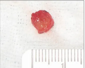
결절을 주소로 본원 안과 외래에 내원하였다(Fig. 1). 환자 는 감염이나 외상의 과거력은 없었으나 약 1년 전 혈전성 혈소판 감소 자반증 및 전신 림프절 비대로 내과에 입원하 여 치료 받았으며 당시 서혜부의 결절로 조직 생검을 시행 하여 조직학적으로 ALHE 의증을 진단받았고 이후 피부과 에 정기적으로 경과 관찰하고 있었다. 결절은 우상안검의 내측 피하에서 약 1.0×0.5 cm 크기로 촉진이 가능하였으 며 연부조직 경도의 유동성이 있는 형태로 통증이나 소양 증은 동반하지 않았다. 안과적 검사에서 특이 소견은 없었 으며 가족력에도 특이 사항은 없었다. 진단 및 치료를 위해 절제 생검을 시행하였다. 수술 후 얻은 조직은 크기 1.0×0.6×0.5 cm의 표면이 매끄러운 황적색 결절이었으며 (Fig. 2) 조직 병리학적 검사에서 일정한 타원형 핵 및 세 포질 내 소공포를 지닌 크기가 증가된 염주알 모양의 혈관 내피세포로 이루어진 다수의 모세혈관이 관찰되었고 이를 둘러싸고 림프구 및 호산구가 대량으로 침윤되어 있었다 (Fig. 3, 4). 임상 소견과 조직 병리학적 소견에 근거하여 ALHE로 확진되었다.
전신적인 검사를 위해 내과에 의뢰하여 신체 촉진, 혈액 검사를 시행하였으며 말초 혈액의 호산구가 31.3%로 호산 구 혈증을 보이는 것 이외에는 특이 사항은 없었다. 수술 후 다른 치료 없이 정기적 추적 관찰 중에 있으며 절제 후 13개월 현재까지 재발 소견은 보이지 않고 있다.
www.ophthalmology.org 713 - 윤제환⋅지미정 : 호산구 증가증을 동반한 혈관림프증식증 -
Figure 1. A relaltively well-demarcated, erythematous, firm,
subcutaneous nodule in the upper eyelid.Figure 2. A yellowish red color mass (size 1.0 × 0.6 × 0.5
cm).Figure 3. There is marked proliferation of irregular vascular
channels surrounded by perivascular lymphocytic infiltration with eosinophils (H&E, ×200).Figure 4. The vascular channels are lined by round and cuboi-
dal endothelial cells, which have protruded into the lumens of the vessels (H&E, ×200).고 찰
ALHE는 20-50대에서 주로 발생하는 질환으로 남자보 다 여자에서 호발한다고 하나 성별에 따른 차이가 없거나 남자에서 호발한다는 보고도 있다.2,3호발 부위로는 두경부 가 약 85%로 가장 흔하고 그중 대부분이 귀 주변이나 이 마, 두피에 발생하며 그 외 구강, 흉벽, 사지, 서혜부 등에도 나타날 수 있다.1국내에선 구강, 이마, 서혜부, 손등에 발생 한 보고가 있으나 안검에 발생한 경우는 해외에서 3예가 보 고 되었고 국내 보고는 없었다.4-11
임상증상으로는 유동성이 있는 연부조직 경도의 무통성 의 종물이 결절이나 구진의 형태로 피하조직 내에 단일 하 거나 혹은 다발성으로 나타나는 것이 특징적이며 병변은 보 통 갈색이나 적색으로 0.5-2.0 cm의 크기를 가진다.3전신 적인 증상은 신증후군이 드물게 보고된 경우가 있으나 연관 성이 명확하지 않으며 약 20%에서 호산구 증가증이 동반된 다.12본 증례에서와 같은 혈소판 감소 자반증후 발생은 1예 있으나 연관성은 알려진 바 없다.13ALHE는 생검을 통한 조
직학적 소견으로 확진할 수 있으며 소혈관의 증식 및 증식 된 혈관을 구성하는 내피세포의 변화가 관찰된다. 내피 세 포의 변화는 거품핵, 한 개 이상의 핵소체, 풍부한 세포질, 조직구모양 또는 상피세포모양의 변화와 같은 특징적인 소 견을 가지고 있으며 그 외 부어있거나, 불룩한, 말뚝 울타리 모양(picket-fenced), 혹은 징 모양(hobnailed)의 양상으로 나타나기도 하고 혈관 내로 자라 들어오거나 혈관벽 및 혈 관주위 간질부에 침윤되기도 한다.14-17특히 큰 혈관주위의 내피세포 송이의 존재는 ALHE의 특징적인 소견 중 하나이
다.17-19또한 증식된 혈관 주변으로 림프구, 중성구, 호산구,
비만세포 등의 침착이 관찰되며 호산구의 침착 정도는 비만 세포의 화학주성인자의 분비 정도에 따라 달라진다.3
ALHE은 이러한 조직학적 특징상 혈관내피세포의 양성 신생물의 일종일 가능성이 높을 것으로 생각되나 ALHE에
www.ophthalmology.org 714
- 대한안과학회지 2012년 제 53 권 제 5 호 -
서 병변 내에 면역글로불린의 침착, 혈청 내 한랭 글로불린의 증가, 칸디다 속에 대한 피부 반응 검사 시 혈청 IgE의 상승과 아토피 환자에 호발한다는 점 등이 ALHE가 면역학적 기전에 의한 반응성 과정에 의한 질환이라는 견해도 있다.8,14,15,20
가장 중요하게 감별 진단해야 할 질환으로 기무라병은 1948년 Kimura et al21에 의해 처음 발표되었으며 초기에 는 ALHE가 기무라병의 일종으로 생각되었으나 1979년 Rosai et al14이 두 질환이 조직학적 특성상 각기 다른 질환 이라는 주장을 편 이후 구분하여 보고 있다. 기무라병은 임 상적으로 10-20대의 젊은 남성의 두경부에 평균 3 cm의 피하결절 양상으로 나타나며 말초 혈액내 호산구 및 혈청 IgE의 증식 소견이나 국소적 림프절 비대가 ALHE에 비해 흔하다. 또한 두 질환 모두 비슷한 양상의 조직학적 소견을 보이기는 하나 기무라병은 ALHE에 비해 내피세포의 변화 는 없으나 배중심을 동반한 림프양의 소절이 다발성으로 나타나며 호산구성 농양으로 불리는 괴사조직 내 호산구의 침착이 부분적 혹은 전반적으로 나타나기도 하고 두드러진 조직의 경화가 관찰된다.22 이와 같은 소견들로 미루어 보 아 기무라병은 ALHE와는 달리 병리학적 기전상 림프유사 조직의 양성 신생물 과정이나 혈관종의 변종 혹은 알러지 반응의 일종인 것으로 생각한다.23 본 증례에서는 종물의 크기가 1.0 cm 이하인 점과 전신 림프선 비대가 없는 점에 서 ALHE에 더 가까운 양상을 보였으며 조직학적 소견상 혈관내피세포의 크기 증가로 ALHE로 확진할 수 있었다.
ALHE의 치료로 일부에서 자연관해된 보고도 있으나 대부 분 수술적 절제술이 가장 흔히 사용된다.24-26이외에도 부 신 피질 호르몬제의 도포나 병변 내 주입 및 경구 복용, 방 사선 요법, 화학 요법, 이산화탄소 레이저, 아르곤 레이저, 전기 건조법(electrodesiccation), 세포 독성제, 냉동요법 등이 있으나 그 치료 효과는 수술적 절제술에 비해 떨어지 며 치료 후 재발 빈도도 높은 편이다.27,28 수술적 치료 시 초기에 불완전하게 절제되는 경우 재발 가능성이 높으며 2 차 수술이 필요하다. 본 증례의 경우 종괴를 완전히 제거하 였고 이후 약 13개월 경과한 현재까지 재발 소견은 관찰되 지 않아 성공적으로 치료된 것으로 판단된다.
본 증례는 드물게 발생한 상안검의 ALHE로 수술적 절제 술에 의해 수술 후 13개월까지 재발 없이 유지되었으며, 앞 으로도 지속적인 추적 관찰을 통해 장기적 재발 유무에 대 한 연구가 필요하고 안검 종양의 감별 진단 시 ALHE를 고 려하는 것이 필요하다고 생각한다.
참고문헌
1) Wells GC, Whimster IW. Subcutaneous angiolymphoid hyper-
plasia with eosinophilia. Br J Dermatol 1969;81:1-14.
2) Kim SM, Yoon J, Yoon TJ. Angiolymphoid hyperplasia with eosi- nophilia on the palm. Ann Dermatol 2010;22:358-61.
3) Olsen TG, Helwig EB. Angiolymphoid hyperplasia with eosinophilia. A clinicopathologic study of 116 patients. J Am Acad Dermatol 1985;12(5 Pt 1):781-96.
4) Park Y, Chung J, Cho CG. Angiolymphoid hyperplasia with eosi- nophilia of the tongue: report of a case and review of the literature.
Oral Oncol 2002;38:103-6.
5) Park JS, Lee MJ. A case of angiolymphoid hyperplasia with eosi- nophilia (ALHE) in the genital area accompanied by varicocele.
Int JDermatol 2009;48:1264-6.
6) Lee WJ, Kim MS, Lee MW, et al. Angiolymphoid hyperplasia with eosinophilia associated with arteriovenous malformation. Clin Exp Dermatol 2009;34:e272-3.
7) Kim SM, Yoon J, Yoon TJ. Angiolymphoid hyperplasia with eosi- nophilia on the Palm. Ann Dermatol 2010;22:358-61.
8) Jang KA, Lee JY, Kim CH, et al. Angiolymphoid hyperplasia with eosinophilia and Kimura’s disease: a clinico-pathologic study in Korea. Korean J Dermatol 2001;39:309-17.
9) Thompson MJ, Whitehead J, Gunkel JL, Kulkarni AD.
Angiolymphoid hyperplasia with eosinophilia affecting the eyelids. Arch Ophthalmol 2007;125:987.
10) Lin B, Tan SH, Looi A. Angiolymphoid hyperplasia with eosino- philia of eyelid with spontaneous regression. Ophthal Plast Reconstr Surg 2008;24:308-10.
11) Mariatos G, Gorgoulis VG, Laskaris G, Kittas C. Epithelioid hae- mangioma (angiolymphoid hyperplasia with eosinophilia) in the inner canthus. J Eur Acad Dermatol Venereol 2001;15:90-1.
12) Jang KA, Lee JY, Kim CH, et al. Angiolymphoid hyperplasia with eosinophilia and Kimura's disease: a clinico-pathologic study in Korea. Korean J Dermatol 2001;39:309-17.
13) Kitamura H, Ito S, Kuwana N, Yutani C. Epithelioid hemangioma of the temporal artery clinically mimicking temporal arteritis.
Pathol Int 1999;49:831-5.
14) Rosai J, Gold J, Landy R. The histiocytoid hemangiomas. A unify- ing concept embracing several previously described entities of skin, soft tissue, large vessels, bone, and heart. Hum Pathol 1979;10:707-30.
15) Rosai J. Angiolymphoid hyperplasia with eosinophilia of the skin.
Its nosological position in the spectrum of histiocytoid hemangioma. Am J Dermatopathol 1982;4:175-84.
16) Weiss SW, Enzinger FM. Epithelioid hemangioendothelioma: a vascular tumor often mistaken for a carcinoma. Cancer 1982;50:
970-81.
17) Chung DH, Kim BJ, Kim YD. Kimura’s disease involving the eye- lid and orbit. J Korean Ophthalmol Soc 2002;43:1789-96.
18) Urabe A, Tsuneyoshi M, Enjoji M. Epithelioid hemangioma versus Kimura’s disease. A comparative clinicopathologic study. Am J Surg Pathol 1987;11:758-66.
19) Googe PB, Harris NL, Mihm MC Jr. Kimura’s disease and angio- lymphoid hyperplasia with eosinophilia: two distinct histopatho- logical entities. J Cutan Pathol 1987;14:263-71.
20) Googe PB, Harris NL, Mihm MC Jr. Kimura’s disease and angio- lymphoid hyperplasia with eosinophilia: two distinct histopatho- logical entities. J Cutan Pathol 1987;14:263-71.
21) Kimura T, Yoshimura S, Ishikawa E. Unusual granulation com- bined with hyperplastic change of lymphoid tissues. Trans Soc
www.ophthalmology.org 715
=ABSTRACT=
A Case of Angiolymphoid Hyperplasia with Eosinophilia (ALHE) of the Eyelid
Je Hwan Yoon, MD, Mijung Chi, MD, PhD
Department of Ophthalmology, Gachon University Gil Hospital, Incheon, Korea
Purpose: The authors of the present study describe a rare case of angiolymphoid hyperplasia with eosinophilia (ALHE) of the eyelid.
Case summary: A 63-year-old male who was diagnosed with ALHE based on biopsy of an inguinal mass presented with an eyelid mass of 1 month duration. A light brown, solitary, 1.0 × 0.5 cm-sized mass involved the right upper eyelid. There was no lymphadenopathy, but eosinophilia was present. An excisional biopsy of the mass was performed for diagnosis and management. Macroscopic examination of the excised mass revealed a well-defined, smooth, firm, yellowish-red colored lesion measuring 1.0 × 0.6 × 0.5 cm. Histopathology showed the proliferation of small blood vessels, many of which were lined by enlarged endothelial cells with uniform ovoid nuclei and intracytoplasmic vacuoles. The distinctive endothelial cells were described as having a cobblestone appearance. In addition, a perivascular and interstitial infiltrate composed primarily of lymphocytes and eosinophils was present. ALHE was finally confirmed with clinical and microscopic examination.
Conclusions: The authors of the present study report a rare case of ALHE of the eyelid and suggest that a differential diag- nosis should be considered.
J Korean Ophthalmol Soc 2012;53(5):712-715
Key Words: Angiolymphoid hyperplasia with eosinophilia, Cobblestone appearance, Enlarged endothelial cell, Eyelid mass, Lymphadenopathy
Address reprint requests to Mijung Chi, MD, PhD
Department of Ophthalmology, Gachon University Gil Hospital
#21 Namdong-daero 774beon-gil, Namdong-gu, Incheon 405-760, Korea Tel: 82-32-460-3364, Fax: 82-32-460-3358, E-mail: [email protected]
- 윤제환⋅지미정 : 호산구 증가증을 동반한 혈관림프증식증 -
Pathol Jpn 1948;37:179-80.
22) Kung IT, Gibson JB, Bannatyne PM. Kimura’s disease: a clin- ico-pathological study of 21 cases and its distinction from angio- lymphoid hyperplasia with eosinophilia. Pathology 1984;16:39-44.
23) Takenaka T, Okuda M, Usami A, et al. Histological and immuno- logical studies on eosinophilic granuloma of soft tissue, so-called Kimura’s disease. Clin Allergy 1976;6:27-39.
24) Baghestani S, Firooz A, Ghazisaidi MR. A refractory case of an- giolymphoid hyperplasia with eosinophilia successfully treated by surgery. J Dermatolog Treat 2011;22:49-51.
25) Satpathy A, Moss C, Raafat F, Slator R. Spontaneous regression of a rare tumour in a child: angiolymphoid hyperplasia with eosino-
philia of the hand: case report and review of the literature. Br J Plast Surg 2005;58:865-8.
26)Lin B, Tan SH, Looi A. Angiolymphoid hyperplasia with eosino- philia of the eyelid with spontaneous regression. Ophthal Plast Reconstr Surg 2008;24:308-10.
27) Archer KF, Hurwitz JJ, Heathcote G. Orbital angiolymphoid hy- perplasia with eosinophilia. Presentation as chalazion. Ophthalmic Plast Reconstr Surg 1991;7:208-21.
28) Ruckenstein MJ, Birt BD, Gruss JS. Angiolymphoid hyperplasia with eosinophilia: a case report and literature review. J Otolaryngol 1989;18:236-40.