www.e-arms.org 89 The past several decades have seen a marked improvement
in the management and reconstruction of complex chest wall defects, with long-term success rates improved from 50%
to 90% to 99% and hospital stays reduced from an average of 84 days to less than 13 days.1-3 Widespread acceptance of muscle and musculocutaneous flaps such as those of the rectus abdominis, latissimus dorsi, pectoralis major, and serratus anterior has led to a sharp decrease in infections and mortality. The case reported here involved the use of sternal reconstruction with a pedicled transverse rectus abdominis myocutaneous (TRAM) flap on titanium plates to maintain sternal closure and stability in a pediatric heart transplantation
patient of high donor-recipient ratio.
CASE REPORT
A female patient was diagnosed with Russell-Silver syndrome at the age of 6 years in Asan Medical Center. At the age of 8 years, the patient was admitted to our hospital with dilated cardiomyopathy. The patient’s height and weight were 112 cm and 15 kg with a body surface area of 0.68 m2. The donor was a 46-year-old woman. Her height and weight were 150 cm and 50.6 kg with a body surface area of 1.5 m2. The ratio of donor to recipient weight was 3.37:1. The transplantation was performed
Chest Wall Reconstruction with a Transverse Rectus Abdominis Musculocutaneous Flap in an Extremely Oversized Heart Transplantation
Ji Hong Yim, Jin Sup Eom*, Deok Yeol Kim1
Department of Plastic and Reconstructive Surgery, Asan Medical Center, University of Ulsan College of Medicine, Seoul,
1Department of Plastic and Reconstructive Surgery, Korea University Guro Hospital, Korea University College of Medicine, Seoul, Korea
CC This is an open-access article distributed under the terms of the Creative Commons Attribution Non-Commercial License (http://creativecommons.org/licenses/by-nc/3.0) which permits unrestricted noncommercial use, distribution, and reproduction in any medium, provided the original work is properly cited.
Copyright © 2014 by the Korean Society for Microsurgery. All Rights Reserved.
Received October 1, 2014 Revised November 4, 2014 Accepted November 4, 2014
*Correspondence to: Jin Sup Eom Department of Plastic and Reconstructive Surgery, Asan Medical Center, University of Ulsan College of Medicine, 88 Olympic- ro 43-gil, Songpa-gu, Seoul 138-736, Korea
Tel: +82-2-3010-3600 Fax: +82-2-476-7471 E-mail: [email protected]
Financial support: None.
Conflict of interest: None.
An 8-year-old girl diagnosed with dilated cardiomyopathy and Russell-Silver syndrome was admitted to our pediatric intensive care unit due to low cardiac output and multiple-organ dysfunction. The patient was placed on the heart transplant waiting list and extracorporeal membrane oxygenation was performed as a bridge to transplantation. After 17 days, heart transplantation was performed. The donor was a 46-year-old female (weight, 50 kg;
height, 150 cm). The donor:recipient weight ratio was 3.37:1. Because the dimension and volume of the recipient’s thoracic cage were insufficient, the sternum could not be closed.
Nine days after transplantation, the patient underwent delayed sternal closure. To obtain adequate space, we left the sternum 4.5 cm apart from each margin using four transverse titanium plates. A transverse rectus abdominis musculocutaneous flap was chosen to cover the wound. Due to the shortage of donors, a size-mismatched pediatric heart transplantation is sometimes unavoidable. Closure of the opened sternum of a transplant recipient can be challenging. Sternal reconstruction after an extremely oversized heart transplantation with transverse titanium plate fixation and a musculocutaneous flap can effectively achieve sternal closure and stability.
Key Words: Chest wall, Transverse rectus abdominis musculocutaneous flap
ARMS
Archives of Reconstructive MicrosurgeryCase Report
pISSN 2383-5257 eISSN 2288-6184 Arch Reconstr Microsurg 2014;23(2):89-92 http://dx.doi.org/10.15596/ARMS.2014.23.2.89
Arch Reconstr Microsurg Vol. 23. No. 2. November 2014
90
using the orthotopic transplantation method on March 11, 2012. The first operation was completed with the sternum left open because it was pressing against the transplanted heart.
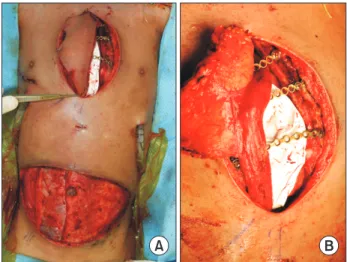
Nine days after the first procedure, sternal reconstruction was performed. Four titanium plates were placed across the sternum and ribs using screws so that the lower sternal body could be fixed with enough space.
For soft tissue coverage of the sternal defect, a TRAM flap was planned. Because the defect size was determined to be 11×7 cm (Fig. 1), and considering that the donor site needed to be enough to obtain primary closure, we designed the
TRAM flap to be 13.0×6.5 cm (Fig. 2). The myocutaneous flap was separated from the surrounding structures across the whole width of the abdomen (as designed). These tissues were, however, left attached to the right rectus abdominis muscles underneath, through which the tissues received their blood supply via superior epigastric vessels. The muscle was then cut at the bottom of the abdomen and the deep inferior epigastric artery was ligated. The pedicled TRAM flap was next transferred to the sternal area through the xiphoid tunnel and zone 2 and 70% of zone 4 of the TRAM flap were cut (Fig.
3). The pedicled TRAM flap was inserted into the pre-cardiac open space with a 90o counter-clockwise rotation (Fig. 4). The
Fig. 2. The preoperative design of the transverse rectus abdominis myocutaneous flap.
Fig. 3. Transfer of the transverse rectus abdominis myocutaneous flap through the xiphoid tunnel.
A B
Fig. 4. An immediate postoperative view.
Fig. 1. The defect in the presternal area after the heart transplantation.
Ji Hong Yim, et al. Chest wall Reconstruction in Heart Transplantation
www.e-arms.org 91
donor site fascial repair was performed with a double horizontal mattress suture and fascial plication.
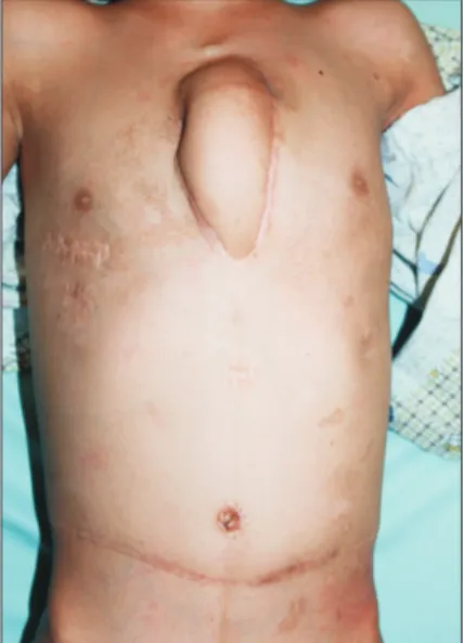
After the chest wall reconstruction, the large donor heart compressed the left bronchial tube, complicating patient extubation. Eleven days after the second surgery, the patient could be extubated and, after 27 days, the patient was transferred to a general ward. After 43 days, the patient was discharged without any complications. The patient currently receives treatment as an outpatient. The postoperative clinical photo was taken after 14 weeks (Fig. 5).
DISCUSSION
Russell-Silver syndrome is a pattern of malformation first described by Silver et al. in 1953 and then by Russell in 1954.4 Russell-Silver syndrome is a clinically and genetically heterogenous condition characterized by severe intrauterine and postnatal growth retardation, craniofacial disproportion and normal intelligence downward curvature of the corner of the mouth, syndactyly and webbed fingers, cardiac defects including congenital heart disease, and pericarditis, etc. In this case, the patient was diagnosed with Russell-Silver syndrome at the age of 6 years and admitted to our hospital with dilated cardiomyopathy. The patient was required to receive immediate heart transplantation and chest wall reconstruction.
Chest wall stabilization begins with a fundamental
understanding of the defect id est, what is missing, what remains, what needs to be replaced, and what resources are available for the reconstruction. In addition to reestablishing the normal functions of the chest wall (respiration, protection, support, and aesthetics), evidence suggests that bony or plate fixation results in reduced ventilator dependence, shorter overall hospital stay, and improved upper extremity function.5 Small to moderate defects can be reconstructed with autologous tissue alone.
Except in the case of previously irradiated defects, larger defects are reconstructed with methyl methacrylate composite meshes and covered with vascularized tissue. Contaminated wounds are generally not reconstructed with synthetic mesh techniques but rather with bioprosthetic mesh.
Using these principles, reconstructive plastic surgeons can devise comprehensive and safe repair plans for tremendous defects of the chest wall. In our case, we planned to use autologous tissue with titanium plates for reconstruction of the chest wall defect. Since the introduction of rectus abdominis flaps in 1977, these flaps have been heavily used in reconstructive surgery due to their ease of elevation, their reliability, and the large amount of tissue that may be harvested while still permitting primary closure of the donor site.6-8 The rectus abdominis muscle is a type III flap according to the Mathes and Nahai classification, with the broad vertical muscle of the abdominal wall originating from the pubic symphysis and inserting on the xiphoid process and fifth to seventh costal cartilages. Rectus abdominis myocutaneous flaps are supplied primarily by the deep inferior and superior epigastric arteries, which communicate at the peri-umbilical watershed area, permitting a variety of potential flap configurations. In addition, the rectus abdominis muscle may be pedicled superiorly in the absence of the internal mammary system by additional blood supply from the eighth subcostal artery9 and can easily cover the lower third of the sternum, but can also reach as high as the sternal notch.10
The superiorly based single-pedicled TRAM flap is most commonly used for autologous tissue for breast reconstruction following mastectomy due to the amount of tissue that may be harvested and the relatively aesthetic donor site scar. The TRAM flap, however, is generally a less well-vascularized flap than the vertical rectus abdominis myocutaneous flap because a large part of the skin paddle does not lie over the muscle and is dependent on the integrity of communicating perforating
Fig. 5. A postoperative view of the patient, 14 weeks after the reconstruction.
Arch Reconstr Microsurg Vol. 23. No. 2. November 2014
92
vessels. Our experience with pediatric patients under the age of 10 years indicates that the TRAM flap is sufficiently vascularized and has enough capacity and adaptability to limit any potential vascular compromise. In addition to the relatively aesthetic donor site scar of the TRAM flap and the abundant tissue and stable blood supply, the chest wall flexibility is likely to be maintained. However, with the growth of the patient, the flap size should also increase and close follow-up of the bony defect of the sternum is required to ensure that the physiological function of the heart is maintained and that the chest cavity remains completely covered. In our present patient, a stable flap statuswas seen after 2-years follow-up in outpatient clinic.
Due to the shortage of donors, size-mismatched pediatric heart transplantations are inevitable. Closing the recipient’s open sternum can pose a considerable challenge. A pedicled TRAM flap allowed us to cover the entire protuberant heart without placing tension on the heart. Thus, chest wall reconstruction after an extremely oversized heart transplantation using a TRAM flap is an effective technique for achieving sternal closure and stability.
REFERENCES
1. Breyer RH, Mills SA, Hudspeth AS, Johnston FR, Cordell AR. A prospective study of sternal wound complications. Ann Thorac
Surg 1984;37:412-6.
2. Yuen JC, Zhou AT, Serafin D, Georgiade GS. Long-term sequelae following median sternotomy wound infection and flap reconstruction. Ann Plast Surg 1995;35:585-9.
3. Schulman NH, Subramanian V. Sternal wound reconstruction:
252 consecutive cases. The Lenox Hill experience. Plast Reconstr Surg 2004;114:44-8.
4. Russell A. A syndrome of intra-uterine dwarfism recognizable at birth with cranio-facial dysostosis, disproportionately short arms, and other anomalies (5 examples). Proc R Soc Med 1954;
47:1040-4.
5. Cicilioni OJ Jr, Stieg FH 3rd, Papanicolaou G. Sternal wound reconstruction with transverse plate fixation. Plast Reconstr Surg 2005;115:1297-303.
6. Schwabegger A, Ninković M, Brenner E, Anderl H. Seroma as a common donor site morbidity after harvesting the latissimus dorsi flap: observations on cause and prevention. Ann Plast Surg 1997;38:594-7.
7. Delay E, Gounot N, Bouillot A, Zlatoff P, Rivoire M. Autologous latissimus breast reconstruction: a 3-year clinical experience with 100 patients. Plast Reconstr Surg 1998;102:1461-78.
8. Titley OG, Spyrou GE, Fatah MF. Preventing seroma in the latissimus dorsi flap donor site. Br J Plast Surg 1997;50:106-8.
9. Fernando B, Muszynski C, Mustoe T. Closure of a sternal defect with the rectus abdominis muscle after sacrifice of both internal mammary arteries. Ann Plast Surg 1988;21:468-71.
10. Iacobucci JJ, Stevenson TR, Hall JD, Deeb GM. Sternal osteomyelitis: treatment with rectus abdominis muscle. Br J Plast Surg 1989;42:452-9.