Biomedical Science Letters 2018, 24(3): 168~174 https://doi.org/10.15616/BSL.2018.24.3.168 eISSN : 2288-7415
The Improving Effect of Gastrodia elata Blume on DSS-induced Colitis in Mice
Eun-Mi Ahn
*and Su-Jin Kim
†,*Department of Biotechnology and Convergence, DaeguHanny University, Gyeongsan 38453, Korea
Ulcerative colitis (UC) is a chronic inflammatory bowel disease characterized by abdominal pain, rectal bleeding and diarrhea. Gastrodia elata Blume (GE) has been used for the treatment of various diseases including neurodegenerative diseases and inflammatory disease. However, there has been no information on whether GE regulates intestinal inflammation.
The aim of this study is to elucidate whether GE can protect against dextran sulfate sodium (DSS)-induced colitis in a mouse model. The colitis mice were induced by drinking water containing 5% DSS for 7 days. Body weight, colon length and clinical score were assessed to determine the effects on colitis. The levels of inflammatory cytokines, tumor necrosis factor (TNF)-α and interleukin (IL)-6 in colitis tissue were also measured. The results showed that mice administrated with DSS showed clinical signs including weight loss and reduced colon length. GE inhibited the DSS-induced loss of body weight and shortening of colon and increased Disease activity index score. Additionally, we observed that GE suppressed the levels of TNF-α and IL-6 in DSS-treated colon tissues. Collectively, these findings provide experimental evidence that GE might be a useful therapeutic agent for patients with UC.
Key Words: Gastrodia elata Blume, Ulcerative colitis, Dextran sulfate sodium, Tumor necrosis factor-α, Interleukin-6
INTRODUCTION
Ulcerative colitis (UC) is a typical inflammatory intestinal disease belonging to the inflammatory bowel diseases (IBD).
It is characterized by bloody diarrhea, colonic mucosal ul- ceration and, in severe cases, systemic symptoms (Hyams, 2000; Danese et al., 2004). The incidence and prevalence rates of IBD in Korea are still low compared with those of the Western countries, but have been increasing rapidly during the past decades (Yang, 2002). Recently, in spite of extensive research implicating genetic susceptibility and environmental factors associated with development of the
disease, its pathogenesis is still obscured. Nowadays, most therapies for UC include glucocorticosteroids, sulfasalazine, and other such drugs (Domènech, 2006). However, these treatments cause serious side effects such as easy to relapse, long-term medication side effects, refractory characteristics, and there is a pressing need for developing effective drugs for the treatment of UC.
Recent studies have reported that inflammatory cytokines are involved in the initiation of the inflammatory response in colitis (Li et al., 2010). Other studies have reported that interleukin (IL)-6 level is elevated in inflammatory bowel conditions, and it suggests that IL-6 plays an integral role in the pathogenesis of these conditions (Zheng et al., 2005). It
Original Article
Received: April 16, 2018 / Revised: September 1, 2018 / Accepted: September 13, 2018
*Professor.
†Corresponding author: Su-Jin Kim. Department of Biotechnology and Convergence, DaeguHanny University, Gyeongsan 38453, Korea Tel: +82-53-819-1389, Fax: +82-53-819-1389, e-mail: [email protected]
○CThe Korean Society for Biomedical Laboratory Sciences. All rights reserved.
○CCThis is an Open Access article distributed under the terms of the Creative Commons Attribution Non-Commercial License (http://creativecommons.org/licenses/by-nc/3.0/) which permits unrestricted non-commercial use, distribution, and reproduction in any medium, provided the original work is properly cited.
was reported that tumor necrosis factor (TNF)-α was expres- sed at high levels in UC patients and TNF-α can stimulate the ROS production in intestinal epithelial cell (Rokutan et al., 2008). Hence, there have been attempts to develop the agents that can suppress the generation or action of inflam- matory cytokines.
Traditional herbal medicine has been the subject of in- creased interest for its potential in the treatment of inflam- mation (Talhouk et al., 2007; Lin et al., 2009). Gastrodia elata (GE) Blume is a belong to the to the Orchidaceae family and used for treating nervous diseases such as headaches, migraine, dizziness, and epilepsy (Matias et al., 2016; Zhan et al., 2016). A number of studies have shown that GE pos- sess a variety of pharmacological activities including anti- inflammatory, analgesic, anti-oxidative, memory-improving and anti-aging, anti-virus and anti-tumor activities (Zhan et al., 2016, Kim et al., 2017). However, there has been no precise information on whether GE regulates intestinal in- flammation. In this study, we were interested in determining whether GE has anti-colitis activity and chose the dextran sulfate sodium (DSS)-induced mouse colitis model as the subject for this study. This model resembles human IBD and is used for pharmacological analysis of potentially effective anti-inflammatory agents (Camuesco et al., 2005; Ramakers et al., 2007). In present study, to provide experimental evi- dence that GE may be a useful therapeutic drug, we examined the effects of GE on clinical signs and inflammatory cyto- kines level in DSS-induced colitis in mice.
MATERIALS AND METHODS Reagents
DSS (mol wt; 36,000~50,000) was purchased from MP Biomedicals (Solon, OH, USA). Purified anti-mouse IL-6 and TNF-α, recombinant mouse (rm) IL-6 and TNF-α and biotinylated anti-mouse IL-6 and TNF-α were obtained from BD-Pharmingen (San Diego, CA). Sulfasalazine, avidin- peroxidase (AP) and other chemical reagents were obtained from Sigma-Aldrich Co. (St. Louis, MO, USA).
Animals
Male BALB/c mice (six weeks old) were obtained from
the Daehan biolink Co., Ltd. (Chungbuk, Korea). Animals were housed 6 heads per cage, allowed spontaneous take in food and water. Animals were kept under a 12-h light/dark cycle (light on 08:00~20:00) at room temperature (23 ± 2℃) and humidity (55 ± 10%). All animal procedures and experiments were approved by the Daegu Haany university guidelines (DHU2013-086).
Preparation of GE
The dried of GE were purchased from the Human herb (Gyeongbuk, Korea). The GE (100 g) was chopped using a blender with 1 L of 70% aqueous ethanol solution under room temperature for 24 h and then concentrated under a vacuum. Then the extract solution obtained was filtered, concentrated on a water bath under vacuo, frozen and lyophi- lized (yield: 15.3%). The samples were dissolved in distilled water and then filtered through 0.22 μm syringe filter.
Induction of colitis by DSS and experimental procedures A widely used experimental model of colitis involves oral consumption of DSS dissolved in drinking water. Acute colitis in mice was induced by administration drinking water containing 5% (w/v) DSS for 7 days. Mice were randomized into groups (n=7) receiving GE (50 mg/kg), sulfasalazine (150 mg/kg) as a positive control, or water as a negative control. GE and sulfasalazine were orally administrated once a day for 7 days prior to DSS treatment. Mice finally were sacrificed after DSS treatment for 7 days.
Disease activity index (DAI)
The activity of intestinal disease was assessed through manifestations, comprising loss of weight, diarrhea accom- panied with blood and mucus, and shortening of colon (Hendrickson et al., 2002). As described by Murthy et al.
(1993), DAI was obtained from score of three major clinical
signs (weight loss, diarrhea, and rectal bleeding). DAI was
calculated using the following formula: DAI = (weight loss
score) + (diarrhea score) + (rectal bleeding score). The clinical
parameters used here are comprehensive functional measures
that are analogous to the subjective clinical symptoms ob-
served in human ulcerative colitis (Cooper et al., 1993). This
method of scoring has been validated by repeated studies.
Cytokine analysis
Distal colon specimens were chopped with scissor and homogenized in ice-cold lysis buffer (20 mM HEPES, 1.5 mM MgCl
2, 0.2 mM EDTA, 0.1 M NaCl, 0.2 mM DTT, 0.5 mM Na
3VO
4, 1% protease inhibitor cocktail) and centri- fuged for 30 min. The levels of IL-6 and TNF-α were meas- ured in the colon protein extracts using a modification of the enzyme-linked immunosorbent assay (ELISA), as pre- viously described (Kim et al., 2010). Briefly, 96-well plates were coated with anti-mouse monoclonal antibodies and incubated overnight at 4℃. After additional washes, sample or an IL-6 and TNF-α standard were added and incubated at room temperature for 2 h. After washing the wells, bio- tinylated anti-mouse IL-6 and TNF-α was added and incu- bated for 2 h. After washing, AP was added and plates were incubated for 30 min at 37℃. Wells were again washed and 2,2'-azino-bis-(3-ethylbenzthiazoline-6-sulfonic acid) sub- strate was added. The optical density was measured by using a microplate ELISA reader.
Statistical analysis
The experiments were shown a summary of the data from at least-three experiments and presented as the mean ± S.D.
Statistical evaluation of the results was performed by inde- pendent t-test. A value of P < 0.05 was considered statisti- cally significant.
RESULTS
Effects of GE on DSS-induced the weight loss and DAI increase in mice
DSS-induced colitis in mice has a phenotype similar to that of human acute and chronic UC. In this study, the clinical effects of GE on DSS-induced experimental colitis were investigated. Firstly, the effects of GE on DSS-induced the weight loss for 7 days were evaluated. The result showed that mice treated with DSS showed a significant weight loss compared to the control. However, GE treatment showed a significant attenuation of body weight loss caused by DSS (Fig. 1A). As sulfasalazine has been used as a treatment for colitis, it was used as a positive control. Any other side effects except colitic symptoms did not detected in GE treated group.
The common feature of the DSS-induced colitis model is an increase in DAI (Ardizzone and Bianchi, 2005). The physiological signs (weight loss, colon shortening, diarrhea, and occult/gross bleeding) induced by 5% DSS treatment for 7 days were observed, and their DAIs were evaluated.
We analyzed the clinical effect of oral administration GE in DSS-induced DAI. Increased DAI score in DSS group was remarkably inhibited in the group administered with GE (Fig.
1B). The inhibitory effect of GE on colon DAI was similar to the sulfasalazine group. These results suggest that GE
Fig. 1. Effect of GE on DSS-induced the body weight loss and DAI increase in mice. Experimental colitis in mice (n = 7/ group) was induced by a 5% DSS dissolved in the drinking water for 7 days. GE was administered orally at doses of 50 mg/kg once a day for 7 days prior 5% DSS supplement. (A) Body weight of mice was measured. (B) DAI was calculated as described in Materials and Methods. Data were represented in the mean ± SD. (n = 7) from triplicate experiments (#P < 0.05 vs. control, *P < 0.05 vs. DSS alone).A B
effectively inhibits the symptoms of colitis caused by DSS.
Effects of GE on DSS-induced the colon length in mice The DSS-induced model of colitis is associated with a sig- nificant decrease in colon length (Fiocchi, 1998; Hendrickson et al., 2002). Thus, the measurement of colon length has been used as a parameter of intestinal inflammation in DSS- induced colitis. As shown in Fig. 2A, the colon length in the DSS-treatment mice was significantly shorter than that of control and this phenomenon was significantly alleviated by GE treatment. Relative colon lengths are shown in Fig. 2B.
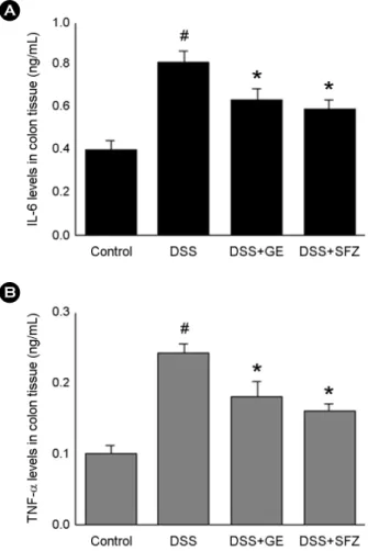
Effect of GE on IL-6 and TNF-α level in DSS-induced colitis
Inflammatory cytokine is considered important inflam-
matory mediators that play a key role in the pathogenesis of UC (Hibi et al., 2003; Neurath et al., 2014). Therefore, re- search on new biological therapies for UC has focused on blocking components of the inflammatory cascade such as cytokines. To determine the effect of GE on inflammatory cytokine in colitis tissues, ELISA was performed. At the end of the experiment, colon tissues were excised and homogen- ized. As shown in Fig. 3, the levels of IL-6 and TNF-α were significantly increased in the colon tissues of DSS-treated mice compared to that of control. However, administration of GE reduced these increased levels induced by DSS. The inhibition rates of IL-6 and TNF-α level by GE were approxi-
Fig. 2. Effect of GE on DSS-induced the colon length shorteningin mice. (A) Experimental colitis in mice (n = 7/ group) was induced by 5% DSS dissolved in the drinking water for 7 days.
GE was administered orally at doses of 50 mg/kg once a day for 7 days prior to 5% DSS supplement. The colons were removed at day 7 after DSS treatment, and the colon lengths were measured.
(B) Relative colon lengths were represented. SFZ (150 mg/kg) was used as a positive control Data were represented in the mean ± SD. (n = 7) from triplicate experiments (#P < 0.05 vs. control, *P
< 0.05 vs. DSS alone).
A
B
Fig. 3. Effect of GE on the levels of IL-6 and TNF-α in DSS- induced colitis. Experimental colitis was induced by 5% DSS drinking water for seven days in mice. At the end of the experiment, the colon tissues were excised and homogenized. The levels of IL-6 and TNF-α in colitis tissue were quantified by ELISA. All data are expressed as the means mean ± SD. of three independent experi- ments (#P < 0.05 vs. control,*P < 0.05 vs. DSS alone).
A
B
mately 47.14 % and 45.31%, respectively.
DISCUSSION
Although traditional herbal medicines have long been used effectively based on traditional knowledge, the pharma- cologic actions of most herbal have not been discovered. In this study, we attempted to provide experimental evidence that GE might be a useful therapeutic drug for patients with UC. The finding of this study demonstrated that GE inhibits inflammatory response and colon injury provoked by DSS treatment, and suggested an important effect by which GE attenuated intestinal inflammation.
UC is an idiopathic disease characterized by the develop- ment of intestinal inflammation (Bouma and Strober, 2003).
UC is an idiopathic disease characterized by the development of intestinal inflammation (Bouma and Strober, 2003). Even if the exact pathogenesis of UC is barely understood, recent evidence suggests that heredity, infection, environmental factors and immune dysfunction have been proposed as pos- sible causes. Generally, UC symptoms can include the bloody diarrhea, colonic mucosal ulceration and weight loss. Ther- apies for UC include glucocorticosteroids, sulfasalazine, and other such drugs. However, it cannot be administered over the long-term, owing to its deleterious side-effects (Sandborn and Targan, 2002; Ishiguro et al., 2006). Consequently, there is a need for anti-colitis agents that cause fewer side effects.
Recently, traditional herbal medicine has been increased interest for the treatment of these disorders. DSS-induced colitis has a phenotype similar to that of human UC char- acterized by bloody stools, ulcerations and infiltration of in- flammatory cells. The concentration of DSS between 1.5 and 5% is accepted for the Balb/c strain in acute colitis (Eichele et al., 2017; Wirtz et al., 2017). Therefore, we tried to in- vestigate the effect of GE on DSS-induced colitis (5% DSS administration for 7 days in Balb/c mice) in present study.
Treatment with GE reduced the weight loss and colon shor- tening caused by DSS. In addition, the DAI, scored using three major clinical signs (weight loss, diarrhea and rectal bleeding), was remarkably inhibited in the group given GE.
The ameliorative effect of GE on colon shortening and DAI was similar to the sulfasalazine group. These results suggest
that GE effectively inhibits the symptoms of colitis caused by DSS.
Inflammatory cytokines are associated in the early stage of the inflammatory response in colitis. Accumulated ex- perimental evidence shows that increase levels of IL-6 and TNF-α destroy the mucous layer and progress the intestinal inflammation. It was also reported that the level of IL-6 and TNF-α in serum is remarkably elevated in UC patients (Li et al., 2010) and that it plays an integral role in their patho- genesis (Zheng et al., 2005). Therefore, development of new biological therapies for UC has focused on inhibition com- ponents of the inflammatory cytokines. In present study, we found that the levels of IL-6 and TNF-α increased in DSS treated-colon tissues compared with those of the control and that treatment with GE reduced these increased levels. These results demonstrated that the anti-inflammatory effect of GE via the suppression of inflammatory cytokines in DSS- induced colitis.
In conclusion, we demonstrated that a treatment with GE reduced the weight loss, colon shortening and DAI, scored using three major clinical signs (weight loss, diarrhea, and rectal bleeding) caused by DSS. These results suggest that GE effectively attenuated the symptoms of colitis. Add- itionally, we showed that the levels of IL-6 and TNF-α were increased in DSS treated-colon tissues compared to those of a normal group; however, these levels were inhibited in the colon tissues by treatment with GE. These results in- dicated that the anti-inflammatory effect of GE is due to the regulation of inflammatory mediators in colitis tissue. Taken together, these suggested that GE may be a useful thera- peutic candidate for colitis. However, the further studies must be performed to elucidate the precise mechanism of GE for the treatment of intestinal inflammatory disorders.
ACKNOWLEDGEMENTS None.
CONFLICT OF INTEREST
No conflict of interests exists for any of the authors.
REFERENCES
Ardizzone S, Bianchi PG. Biologic therapy for inflammatory bowel disease. Drugs. 2005. 65: 2253-2286.
Bouma G, Strober W. The immunological and genetic basis of in- flammatory bowel disease. Nature Reviews Immunology. 2003.
3: 521-533.
Camuesco D, Gálvez J, Nieto A, Comalada M, Rodríguez-Cabezas ME, Concha A, Xaus J, Zarzuelo A. Dietary olive oil supple- mented with fish oil, rich in EPA and DHA (n-3) polyun- saturated fatty acids, attenuates colonic inflammation in rats with DSS-induced colitis. The Journal of Nutrition. 2005. 135:
687-694.
Cooper HS, Murthy SN, Shah RS, Sedergran DJ. Clinicopathologic study of dextran sulfate sodium experimental murine colitis.
Laboratory Investigation. 1993. 69: 238-249.
Danese S, Sans M, Fiocchi C. Inflammatory bowel disease: the role of environmental factors. Autoimmunity Reviews. 2004.
3: 394-400.
Domènech E. Inflammatory bowel disease: current therapeutic options. Digestion. 2006. 73: 67-76.
Eichele DD, Kharbanda KK. Dextran sodium sulfate colitis murine model: an indispensable tool for advancing our understanding of inflammatory bowel diseases pathogenesis. World Journal of Gastroenterology. 2017. 23: 6016-6029.
Fiocchi C. Inflammatory bowel disease: etiology and pathogenesis.
Gastroenterology. 1998. 115: 182-205.
Hendrickson BA, Gokhale R, Cho JH. Clinical aspects and Patho- physiology of inflammatory bowel disease. Clinical Micro- biology Reviews. 2002. 15: 79-94.
Hibi T, Inoue N, Ogata H, Naganuma M. Introduction and review:
recent advances in the immunotherapy of inflammation bowel disease. Journal of Gastroenterology. 2003. 38: 36-42.
Hyams JS. Inflammatory bowel disease. Pediatrics in Review. 2000.
21: 291-295.
Ishiguro K, Ando T, Maeda O, Hasegawa M, Kadomatsu K, Ohmiya N, Niwa Y, Xavier R, Goto H. Paeonolattenuates TNBS- induced colitis by inhibiting NF-κB and STAT1 transactivation.
Toxicology and Applied Pharmacology. 2006. 217: 35-42.
Kim SJ, Kim MC, Um JY, Hong SH. The beneficial effect of vanillic acid on ulcerative colitis. Molecules. 2010. 15: 7208 -7217.
Kim NH, Xin MJ, Cha JY, Ji SJ, Kwon SU, Jee HK, Park MR, Park YS, Kim CT, Kim DK, Lee YM. Antitumor and Immuno-
modulatory Effect of Gastrodia elata on Colon Cancer In vitro and In vivo. American Journal of Chinese Medicine. 2017. 45:
319-335.
Li Y, de Haar C, Chen M, Deuring J, Gerrits MM, Smits R, Xia B, Kuipers EJ, van der Woude J. Disease-related expression of the IL-6/STAT3/SOCS3 signaling pathway in ulcerative colitis and ulcerative colitis-related carcinogenesis. Gut. 2010. 59:
227-235.
Lin TY, Liu YC, Jheng JR, Tsai HP, Jan JT, Wong WR, Horng JT.
Anti-enterovirus 71 activity screening of Chinese herbs with anti-infection and inflammation activities. The American Journal of Chinese Medicine. 2009. 37: 143-158.
Matias M, Silvestre S, Falcao A, Alves G. Gastrodia elata and epilepsy: rationale and therapeutic potential. Phytomedicine.
2016. 23: 1511-1526.
Murthy SN, Cooper HS, Shim H, Shah RS, Ibarahim SA, Sedergran DJ. Treatment of dextran sulfate sodium-induced murine colitis by intracolonic cyclosporin. Digestive Diseases and Sciences.
1993. 38: 1722-1734.
Neurath MF. Cytokines in inflammatory bowel disease. Nature.
2014. 14: 329-342.
Ramakers JD, Verstege MI, Thuijls G, Te Velde AA, Mensink RP, Plat J. The PPARgamma agonist rosiglitazone impairs colonic inflammation in mice with experimental colitis. Journal of Clinical Immunology. 2007. 27: 275-283.
Rokutan K, Kawahara T, Kuwano Y, Tominaga K, Nishida K, Teshima-Kondo S. Nox enzymes and oxidative stress in the immunopathology of the gastrointestinal tract. Seminars in Immunopathology. 2008. 30: 315-327.
Sandborn, WJ, Targan SR. Biologic therapy of inflammatory bowel disease. Gastroenterology. 2002. 122: 1592-1608.
Talhouk RS, Karam C, Fostok S, El-Jouni W, Barbour EK.
Anti-inflammatory bioactivities in plant extracts. Journal of Medicinal Food. 2007. 10: 1-10.
Wirtz S, Popp V, Kindermann M, Gerlach K, Weigmann B, Fichtner- Feigl S, Neurath MF. Chemically induced mouse models of acute and chronic intestinal inflammation. Natrue Protocols.
2017. 12: 1295-1309.
Yang SK. Current status and clinical characteristics of inflammatory bowel disease in Korea. The Korean Journal of Gastroentero- logy. 2002. 40: 1-14.
Zhan HD, Zhou HY, Sui YP, Du XL, Wang WH, Dai L, Sui F, Huo HR, Jiang TL. The rhizome of Gastrodia elata Blume - an ethnopharmacological review. Journal of Ethnopharmacology.
2016. 189: 361-385.
Zheng P, Niu F, Liu W, Shi Y, Lu L. Anti-inflammatory mechanism of oxymatrine in dextran sulfate sodium-induced colitisofrats.
World Journal of Gastroenterology. 2005. 11: 4912-4915.