JCS
Journal of Chest SurgeryClinical Research Implementation of Venoarterial Extracorporeal Membrane
Oxygenation in Nonintubated Patients
Hyeon A Kim, M.D., Young Su Kim, M.D., Yang Hyun Cho, M.D., Ph.D., Wook Sung Kim, M.D., Ph.D., Kiick Sung, M.D., Ph.D., Dong Seop Jeong, M.D., Ph.D.
Department of Thoracic and Cardiovascular Surgery, Samsung Medical Center, Sungkyunkwan University School of Medicine, Seoul, Korea
ARTICLE INFO Received June 9, 2020 Revised September 30, 2020 Accepted October 5, 2020 Corresponding author Yang Hyun Cho Tel 82-2-3410-2213 Fax 82-2-3410-0089 E-mail [email protected] ORCID
https://orcid.org/0000-0003-1685-3641
Background: Although extracorporeal membrane oxygenation (ECMO) is generally performed percutaneously, the technology is deployed under sedation and necessitates endotracheal intubation. However, in some patients, the use of venoarterial (VA) ECMO without intubation may be beneficial. Herein, we describe our experiences with VA ECMO performed without prior endotracheal intubation.
Methods: A total of 783 patients treated with VA ECMO at a single center between Jan- uary 2013 and July 2018 were reviewed retrospectively. We included patients who un- derwent successful VA ECMO implementation without prior endotracheal intubation, and excluded those who were younger than 18 years, had ongoing cardiopulmonary resusci- tation status, and had poor quality of the vessels needed for percutaneous cannulation.
The primary study outcome was in-hospital survival.
Results: In total, 50 patients were included in this study, 94% of whom showed cardio- genic shock. The mean age of the study participants was 56.3±14.5 years. The median VA ECMO support time was 7 days (range, 2–13 days). Twenty-one patients (42%) did not receive ventilator care during the VA ECMO support period, while 29 patients (58%) pro- gressed to intubation after VA ECMO implementation. The rates of survival at discharge and weaning success were 82% (n=41) and 92% (n=46), respectively, and 80% (n=40) of patients presented good Glasgow–Pittsburgh Cerebral Performance Categories scores at discharge.
Conclusion: Even in patients with cardiogenic shock, percutaneous VA ECMO can be introduced safely without prior endotracheal intubation by an experienced care team. The application of nonintubated VA ECMO might be a feasible strategy in selected cases.
Keywords: Extracorporeal membrane oxygenation, Intubation, Endotracheal
Copyright © The Korean Society for Thoracic and Cardiovascular Surgery. 2021. All right reserved.
This is an Open Access article distributed under the terms of the Creative Commons Attribution Non-Commercial License (http://creativecommons.org/licenses/
Introduction
Extracorporeal membrane oxygenation (ECMO) is wide- ly used for pulmonary or cardiopulmonary support of un- stable patients. Recently, a dramatic increase in the fre- quency of ECMO use has occurred; catheterization can now be performed at the bedside or in a catheterization laboratory, and medical personnel have more experience with the technology. Usually, ECMO deployment is com- pleted percutaneously using Seldinger’s technique under local anesthesia [1].
Given the increasing number of ECMO cases and grow- ing concerns about the outcomes of long-term mechanical
ventilator care [2,3], several studies have attempted to as- certain whether prior endotracheal intubation or mechani- cal ventilator support is necessary for the success of ECMO treatment. Investigations of ECMO in awake patients have mainly involved venovenous (VV) ECMO during sponta- neous breathing. Notably, most of these studies were con- ducted in patients with respiratory failure from acute re- spiratory distress syndrome or patients waiting for lung transplantation [4-6]. Separately, although few cases were included, some studies have also explored awake venoarte- rial (VA) ECMO in patients in cardiogenic shock [7-10].
There are definitive benefits associated with prior endo- tracheal intubation before VA ECMO implementation, in-
https://doi.org/10.5090/kjtcs.20.070
pISSN: 2765-1606 eISSN: 2765-1614
J Chest Surg. 2021;54(1):17-24
https://doi.org/10.5090/kjtcs.20.070
JCS
cluding the establishment of optimal conditions for the de- livery of therapy with relatively well-controlled vital signs, the absence of concerns regarding respiratory failure, and avoidance of patient agitation or movement. However, in some patients experiencing cardiogenic shock, the precon- ditioning stage required for endotracheal intubation might deteriorate the vital signs by reducing native sympathetic tone and may be a time-consuming aspect of the procedure [4]. If the etiology of shock is obviously cardiogenic and quick decision-making by an experienced care team is fea- sible, the straightforward implementation of VA ECMO without prior intubation might be of greater benefit to the patient. Based on this hypothesis, herein, we describe our experiences with VA ECMO implementation preceding en- dotracheal intubation. Notably, we seek to distinguish this protocol from previous “awake ECMO” protocols as specif- ically referring to nonintubated VA ECMO implementa- tion.
Methods
Study population and procedures
We retrospectively reviewed the records of 783 patients who were treated with ECMO at Samsung Medical Center, Seoul, Republic of Korea between January 2013 and July 2018. We included patients who underwent successful per- cutaneous VA ECMO introduction without prior endotra- cheal intubation. Nonintubated VA ECMO deployment was performed in selected patients with spontaneous breathing and peripheral vessels deemed suitable for the necessary percutaneous access. A multidisciplinary team composed of cardiac surgeons, cardiologists, and critical care specialists made the decision to pursue nonintubated VA ECMO implementation, weighing the risks and bene-
fits of nonintubated VA ECMO use in each case, albeit rap- idly given the urgent context. The contraindications for nonintubated VA ECMO implementation included ongo- ing cardiopulmonary resuscitation (CPR) status and poor quality of the vessels needed for percutaneous cannulation.
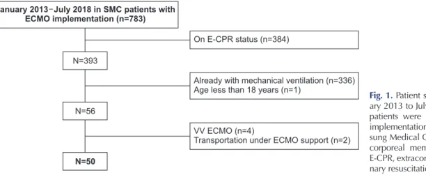
Patients who were already intubated, who required VV ECMO implementation, who were younger than 18 years of age, and/or who were transported from another hospital under ECMO support were excluded from our study anal- ysis (Fig. 1). The implementation of VA ECMO was per- formed in the catheterization laboratory, intensive care unit (ICU), or operating room (OR). Candidates were placed under local anesthesia (2% lidocaine, subcutaneous injection) together with intravenous pain-control medica- tions (fentanyl or pethidine). Spontaneous breathing was facilitated with oxygen administered by a mask. All proce- dures were performed percutaneously using Seldinger’s technique; ultrasound-guided cannulation and fluoroscopy were used in the catheterization laboratory and blind or ultrasound-guided procedures were performed in the ICU or OR. Immediately after VA ECMO introduction, a Dop- pler sensor was applied to the distal part of the arterial cannulation site and a distal perfusion catheter was added if needed.
This study was approved by the Institutional Review Board of Samsung Medical Center (IRB approval no., 2018-09-099-002). The requirement for informed consent was waived because this was a retrospective study. All per- sonal patient data, along with clinical, laboratory, and out- comes data, were collected from patients’ medical records.
Definitions and outcomes
Nonintubated VA ECMO is defined as the introduction of VA ECMO without prior endotracheal intubation at the
January 2013 July 2018 in SMC patients with ECMO implementation (n=783)
Already with mechanical ventilation (n=336) Age less than 18 years (n=1)
On E-CPR status (n=384)
VV ECMO (n=4)
Transportation under ECMO support (n=2) N=393
N=56
N=50
Fig. 1. Patient selection. From Janu- ary 2013 to July 2018; in total, 783 patients were treated with ECMO implementation at SMC. SMC, Sam- sung Medical Center; ECMO, extra- corporeal membrane oxygenation;
E-CPR, extracorporeal cardiopulmo-
nary resuscitation; VV, venovenous.
Hyeon A Kim, et al. VA ECMO in Nonintubated Patients JCS
time of VA ECMO implementation. “Awake ECMO” is a relatively broad concept that defines a status in which me- chanical ventilation support is removed during the mainte- nance period regardless of whether it was present at the time of VA ECMO or VV ECMO implementation. There- fore, we distinguished our patient pool from those receiv- ing awake ECMO by using the term “nonintubated VA ECMO.”
The primary study outcome was survival to hospital dis- charge and the secondary outcomes were successful VA
ECMO weaning and neurological status at the time of dis- charge. The Glasgow–Pittsburgh Cerebral Performance Categories (CPC) scale was used to measure patients’ neu- rological status at discharge. CPC scores of 1 or 2 points were regarded as suggesting good neurological outcomes.
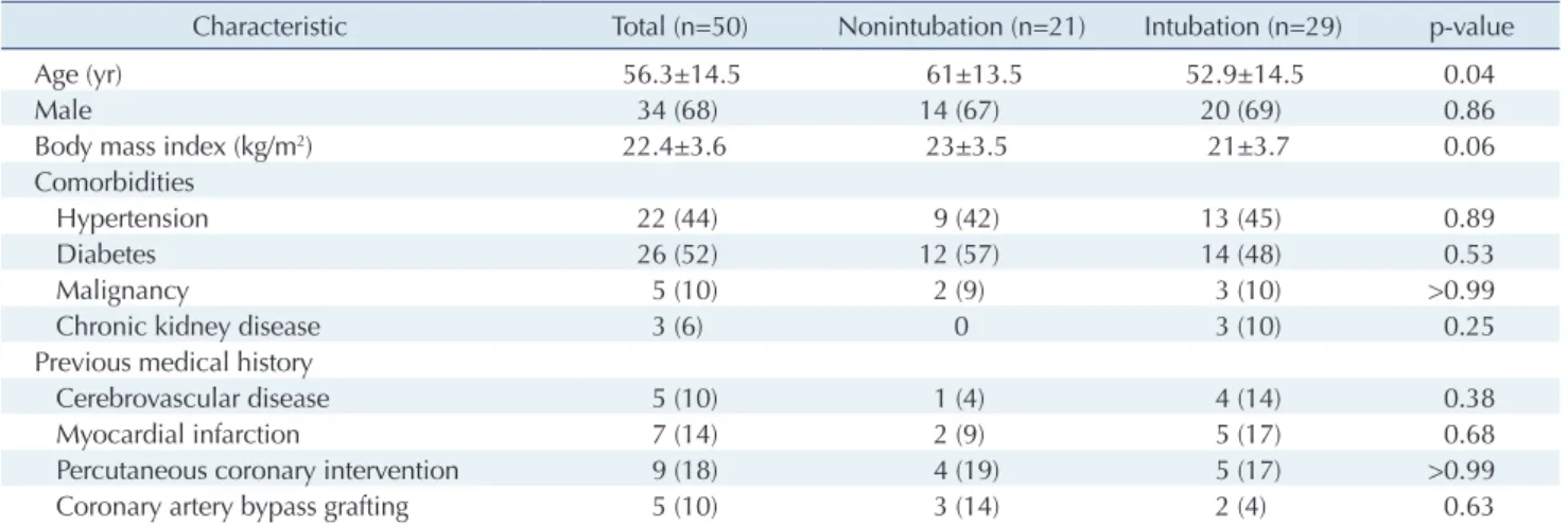
All included patients who underwent nonintubated VA ECMO implementation were divided into 2 groups: those who eventually required intubation during the VA ECMO maintenance period (intubation group) and those who did not (nonintubation group). In addition, patients were strat- Table 1. Demographics of patients
Characteristic Total (n=50) Nonintubation (n=21) Intubation (n=29) p-value
Age (yr) 56.3±14.5 61±13.5 52.9±14.5 0.04
Male 34 (68) 14 (67) 20 (69) 0.86
Body mass index (kg/m
2) 22.4±3.6 23±3.5 21±3.7 0.06
Comorbidities
Hypertension 22 (44) 9 (42) 13 (45) 0.89
Diabetes 26 (52) 12 (57) 14 (48) 0.53
Malignancy 5 (10) 2 (9) 3 (10) >0.99
Chronic kidney disease 3 (6) 0 3 (10) 0.25
Previous medical history
Cerebrovascular disease 5 (10) 1 (4) 4 (14) 0.38
Myocardial infarction 7 (14) 2 (9) 5 (17) 0.68
Percutaneous coronary intervention 9 (18) 4 (19) 5 (17) >0.99
Coronary artery bypass grafting 5 (10) 3 (14) 2 (4) 0.63
Values are presented as mean±standard deviation or number (%).
Table 2. The location of VA ECMO implementation and other variables
Variable Total (n=50) Nonintubation (n=21) Intubation (n=29) p-value
VA ECMO implementation
Location of implementation 0.68
Intensive care unit 4 (8) 3 (14) 1 (3)
Catheterization lab 44 (88) 18 (86) 26 (90)
Operating room 2 (4) 0 2 (7)
Reasons for implementation >0.99
Cardiogenic shock 47 (94) 19 (90) 28 (97)
Obstructive shock 2 (4) 2 (9) 0
For the operation 1 (2) 0 1 (3)
VA ECMO duration (day) 7 (2–13) 5.2 (1.4–13.9) 8.1 (2.7–17.8) 0.40
Events during VA ECMO implementation
Stroke events 4 (8) 0 4 (14) 0.1
Limb ischemia 2 (4) 0 2 (7) 0.3
Bleeding events
VA ECMO site 7 (14) 3 (14) 4 (14) 0.6
Gastrointestinal bleeding 2 (4) 0 2 (7) 0.3
Intubation duration
<24 hr 17 (58.6)
1≤ day ≤7 6 (20.7)
>7 day 6 (20.7)
Values are presented as number (%) or median (range).
VA ECMO, venoarterial extracorporeal membrane oxygenation.
https://doi.org/10.5090/kjtcs.20.070
JCS
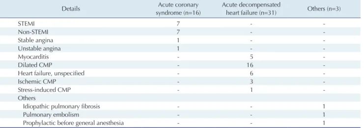
ified into 3 groups—acute coronary syndrome (ACS), acute decompensated heart failure (HF), and other—according to the etiology underlying VA ECMO implementation. The primary and secondary outcomes were also compared ac- cording to these etiologies.
Statistical analysis
Mean values and standard deviations were used to eval- uate patients’ basic characteristics and outcomes. The Fish- er exact test and the chi-square test were used to compare primary and secondary endpoints between the groups. The values for survival at discharge and CPC scores at dis- charge were calculated for all patients. Kaplan-Meier curves were constructed to determine the estimated sur- vival and was drawn based on the last follow-up date rath- er than the discharge date. The log-rank test was per- formed to compare the results of each group. Statistical analyses were executed using SAS ver. 9.4 (SAS Institute Inc., Cary, NC, USA). A p-value of less than 0.05 was con- sidered to indicate statistical significance.
Results
Patient characteristics
A total of 50 patients were enrolled in this study. The mean age of the study participants was 56.3±14.5 years and 34 (68%) were men. The mean body mass index of patients was 22.4±3.6 kg/m
2and a majority of the patients were ei- ther normal body weight or overweight (Table 1). Forty-four
patients (88%) had VA ECMO initiated in the catheteriza- tion laboratory. The cause of VA ECMO implementation was mainly cardiogenic shock (n=47), followed by obstruc- tive shock (n=2), and as part of a preoperative preparation protocol (n=1) (Table 2). Among patients with cardiogenic shock, 16 patients had ACS and 31 patients had acute de- compensated HF. The specific origins of cardiogenic shock are described in detail in Table 3. The median duration of VA ECMO maintenance was 7 days (range, 2–13 days).
During the VA ECMO maintenance period, 29 patients (58%; intubation group) required endotracheal intubation
Intubation causes after VA ECMO implementation
Respiratory failure For operation Sequential procedure Uncontrolled cardiac events Others
Respiratory failure
14%
For operation 41%
Sequential procedure
10%
Uncontrolled cardiac events
21%
Others 14%
Fig. 2. Reasons for endotracheal intubation after VA ECMO im- plementation. In patients with nonintubated VA ECMO imple- mentation, 29 patients underwent endotracheal intubation for the reasons shown above. Twelve patients underwent intubation for an operation (9 for heart transplantation, left ventricular assisted device implantation, or coronary artery bypass grafting and 3 for other operations), 6 for uncontrolled cardiac events, 4 due to re- spiratory failure, and 3 for sequential procedures (septal puncture procedures in the catheterization laboratory). VA ECMO, venoar- terial extracorporeal membrane oxygenation.
Table 3. Details of various origins of cardiogenic shock
Details Acute coronary
syndrome (n=16)
Acute decompensated
heart failure (n=31) Others (n=3)
STEMI 7 - -
Non-STEMI 7 - -
Stable angina 1 - -
Unstable angina 1 - -
Myocarditis - 5 -
Dilated CMP - 16 -
Heart failure, unspecified - 6 -
Ischemic CMP - 3 -
Stress-induced CMP - 1 -
Others
Idiopathic pulmonary fibrosis - - 1
Pulmonary embolism - - 1
Prophylactic before general anesthesia - - 1
Values are presented as number of patients.
STEMI, ST-elevation myocardial infarction; CMP, cardiomyopathy.
Hyeon A Kim, et al. VA ECMO in Nonintubated Patients JCS
with mechanical ventilator support. The causes of intuba- tion were for operation in 12 patients, respiratory failure in 4 patients, persistent cardiac events after VA ECMO imple- mentation in 6 patients, and as sequential treatment imme- diately after VA ECMO implementation in 3 patients (Fig. 2).
Outcomes
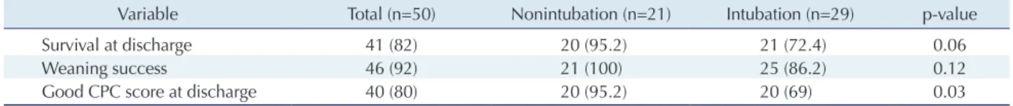
The rate of survival at discharge was 82%, the weaning success rate among all patients was 92% (n=46), and 80%
(n=40) of patients presented good CPC scores at the time of discharge. Notably, the rates of survival at discharge (95.2% versus 72.4%, p=0.06) and the VA ECMO weaning success rate (100% versus 86.2%, p=0.12) were not signifi- cantly different between the nonintubation and intubation groups. However, the proportion of patients with good
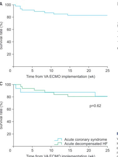
CPC scores at discharge (95.2% versus 69%, p=0.03) was higher in the nonintubation group (Table 4). The primary and secondary outcomes were also compared according to the etiology underlying VA ECMO implementation; among the 47 patients with cardiogenic shock, 16 patients had ACS and 31 patients had acute decompensated HF. The rate of survival at discharge was not significantly different between patients with ACS and with acute decompensated HF (87.5% versus 77.4%, p=0.69). The rates of weaning suc- cess (100% versus 87.1%, p=0.28) and good CPC score at discharge (81.3% versus 77.4%, p>0.99) were also not sig- nificantly different between the 2 groups (Table 5). Fig. 3 shows the de-cannulation methods for VA ECMO. Of the 50 patients, 46 achieved weaning success from VA ECMO.
De-cannulation of VA ECMO was performed by surgical removal in 20 patients (44%), device closure in 20 patients
De-cannulation methods of VA ECMO
Surgical removal Device closure Manual compression
Surgicalremoval, Device 44%
closure, 44%
Manual compression, 12%
A De-cannulation methods of VA ECMO in the
intubation group
Surgical removal Device closure
Surgicalremoval, 24%
Device closure, 76%