Nosocomial Infection in Adult Patients Undergoing Veno-Arterial Extracorporeal Membrane Oxygenation
Data on the frequency of nosocomial infections during extracorporeal membrane oxygenation (ECMO) in adult populations remain scarce. We investigated the risk factors for nosocomial infections in adult patients undergoing venoarterial ECMO (VA-ECMO) support. From January 2011 to December 2015, a total of 259 patients underwent ECMO.
Of these, patients aged 17 years or less and patients undergoing ECMO for less than 48 hours were excluded. Of these, 61 patients diagnosed with cardiogenic shock were evaluated. Mean patient age was 60.6 ± 14.3 years and 21 (34.4%) patients were female.
The mean preoperative Sequential Organ Failure Assessment (SOFA) score was 8.6 ± 2.2.
The mean duration of ECMO support was 6.8 ± 7.4 days. The rates of successful ECMO weaning and survival to discharge were 44.3% and 31.1%, respectively. There were 18 nosocomial infections in 14 (23.0%) patients. These included respiratory tract infections in 9 cases and bloodstream infections in a further 9. In multivariate analysis, independent predictors of infection during ECMO were the preoperative creatinine level (hazard ratio [HR], 2.176; 95% confidence interval [CI], 1.065–4.447; P = 0.033) and the duration of ECMO support (HR, 1.400; 95% CI, 1.081–1.815; P = 0.011). A higher preoperative creatinine level and an extended duration of ECMO support are risk factors for infection.
Therefore, to avoid the development of nosocomial infections, strategies to shorten the length of ECMO support should be applied whenever possible.
Keywords: Extracorporeal Membrane Oxygenation Support; Infection Gwan Sic Kim,1* Kyo Seon Lee,1*
Choung Kyu Park,2 Seung Ku Kang,1 Do Wan Kim,1 Sang Gi Oh,1
Bong-Suk Oh,1 Yochun Jung,1 Seok Kim,1 Ju Sik Yun,1 Sang Yun Song,1
Kook Joo Na,1 In Seok Jeong,1 and Byoung Hee Ahn1
1Department of Thoracic and Cardiovascular Surgery, Chonnam National University Hospital, Chonnam National University Medical School, Gwangju, Korea; 2Department of Thoracic and Cardiovascular Surgery, University of Ulsan College of Medicine, Asan Medical Center, Seoul, Korea
* Gwan Sic Kim and Kyo Seon Lee contributed equally to this work.
Received: 18 August 2016 Accepted: 20 January 2017 Address for Correspondence:
Byoung Hee Ahn, MD
Department of Thoracic and Cardiovascular Surgery, Chonnam National University Hospital, Chonnam National University Medical School, 42 Jebong-ro, Dong-gu, Gwangju 61469, Republic of Korea
E-mail: [email protected]
https://doi.org/10.3346/jkms.2017.32.4.593 • J Korean Med Sci 2017; 32: 593-598
INTRODUCTION
In the time since the first successful application of extracorpo- real membrane oxygenation (ECMO) support in 1972 (1), ECMO has become one of the most important therapeutic modalities for severe cardiopulmonary failure (2). The use of ECMO to treat adults has increased greatly since the H1N1 influenza pandem- ic of 2009 and publication of the findings of a multicenter ran- domized trial of treatments for severe adult respiratory failure (the CESAR trial) (3,4).
Although the experience of clinicians with ECMO therapy in adult populations is growing, ECMO is still associated with high- level mortality and many potential complications. Of these, nos- ocomial infection (NI) is associated with high-level mortality (5,6). Adult patients undergoing ECMO are at higher risk for nos- ocomial infection than neonate and pediatric patients (6.1% of neonates and pediatric patients vs. 20.5% of adults develop cul- ture-proven infections during ECMO) (5).
However, data on NIs acquired during ECMO in the adult pop- ulations remain scarce. Although a few studies have explored this topic (7,8), the works have been limited in terms of patient
numbers considerable heterogeneity in patient baseline char- acteristics including the ECMO mode employed (venovenous vs. venoarterial), the indications for ECMO, and the ECMO can- nulation site (percutaneous vs. intrathoracic). Such heteroge- neity may affect the incidence of NI development during ECMO.
Thus, further work with a homogeneous patient group is required to precisely identify risk factors for NI in patients on ECMO sup- port.
Therefore, we investigated the risk factors for NIs in patients with cardiogenic shock who underwent percutaneous venoar- terial ECMO (VA-ECMO) support.
MATERIALS AND METHODS Patients
From January 2011 to December 2015, 259 consecutive patients underwent ECMO at Chonnam National University Hospital.
Patients aged 17 years or less were excluded. Patients who un- derwent ECMO for < 48 hours were not included in this study since they were not exposed to ECMO long enough to identify ECMO-related infection events. A total of 95 adult patients un- Infectious Diseases, Microbiology & Parasitology
derwent VA-ECMO for cardiogenic shock. Of these, 34 adult patients underwent ECMO support for < 48 hours. Among the 34 patients, 2 patients survived and 32 patients were non-survi- vors. The cause of death in the 32 non-survivor patients includ- ed hypoxic brain death in 8 patients and multi-organ failure in 24. However, there was no sepsis-related death in these patients.
Finally, cardiogenic shock patients (n = 61) undergoing ECMO for ≥ 48 hours were evaluated. Weaning success rate, survival to discharge, and infection incidence on ECMO were assessed through hospital records. Baseline characteristics and operative information including age, gender, underlying conditions, pre- ECMO laboratory findings, Sequential Organ Failure Assess- ment (SOFA) score, chest X-ray state (1 point per quadrant in- filtrated) before ECMO, transfusion history before ECMO, hos- pital and intensive care unit (ICU) stay before ECMO, type and mode of ECMO, type of anticoagulation, and duration of ECMO support were collected and evaluated. Each patient’s survival at the time of ECMO de-cannulation and at the time of discharge was also evaluated in terms of weaning success rate and surviv- al to discharge.
ECMO procedures
Cannulation of ECMO was performed in a sterile fashion with chlorhexidine painting. In ECMO-eligible patients, an emer- gency bypass system (n = 38, 62.3%; Terumo Inc., Tokyo, Japan) or a permanent life support system (n = 23, 37.7%; Maquet Inc., Hirrlingen, Germany) was applied. The oxygenator of the per- manent life support system is a polymethylpentene (PMP) type.
The emergency bypass system is a non-PMP type oxygenator.
Recently, the Maquet system has been more commonly used than the Terumo system, which is known to be the more dura- ble and thrombo-resistant device (9). Heparin or nafamostat mesilate was used as an anticoagulant. Cannulation mode, size, and approach site were determined by the surgeon considering the patient’s body weight, height, and vessel size. A peripheral approach using femoral artery and vein was the predominant approach in the study. The management protocol for ECMO follows the Extracorporeal Life Support Organization (ELSO) guidelines (10).
Definition of nosocomial infection during ECMO support The definition of nosocomial infections during ECMO support was based on the Centers for Disease Control and Prevention definitions for nosocomial infections (11). Nosocomial infec- tion during ECMO support was defined as a case with confirmed organisms from one or more blood, respiratory, or urinary cul- tures during the period 48 hours after the initiation of ECMO to 24 hours after ECMO weaning. Microbiological isolations were correlated with clinical symptoms and typical inflammatory characteristics in blood samples and radiographic findings.
Antibiotics strategy
Prophylactic antibiotics with broad spectrum drug was prescrib- ed to patients at the initiation of ECMO support. In patients who had already been taking antibiotics, antibiotic agents were main- tained. If infection developed during ECMO support, antibiot- ics were changed according to the culture results. With regard to dosing of antibiotics, conventional dose was started and ad- justed according to the drug concentration monitoring. Every antibiotic therapy was discussed with the department of infec- tion and adjusted according to the patient’s clinical situations.
Statistical analysis
Categorical variables are presented as frequencies and percent- ages and continuous variables are expressed as means ± standard deviations. Student’s t-tests and χ2 tests were used to compare factors related to nosocomial infection. We performed the lo- gistic regression using nosocomial infection as the dependent variable. P values < 0.05 were considered statistically signifi- cant. SPSS version 24.0 (SPSS Inc., Chicago, IL, USA) was used for all statistical analyses.
Ethics statement
This study was approved by Institutional Review Board of Chon- nam National University Hospital, which waived the require- ment for informed patient consent based on the retrospective nature of the work (IRB No: CNUH-2016-228).
RESULTS
Baseline characteristics
The 61 patients with cardiogenic shock underwent ECMO for a total of 416.0 days. The mean age of patients at the time of ECMO support was 60.6 ± 14.3 years, and 34.4% (n = 21) were female.
A total of 42.6% (n = 26) of patients had hypertension and 41.0%
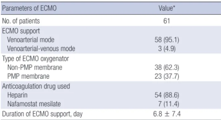
(n = 25) diabetes mellitus. The infiltration status of pre-ECMO chest X-rays was 1.6 ± 1.4. Eighteen patients (29.5%) had a trans- fusion history before ECMO. Pre-ECMO hospital and ICU stays before ECMO were 4.4 ± 6.5 and 1.8 ± 2.5 days, respectively (Ta- ble 1). Venoarterial type was the predominant ECMO type in 95.1% (n = 58) of the patients. Venoarterial-venous type of ECMO was applied in 3 patients (4.9%). According to the ECMO oxy- genator type, the non-PMP oxygenator was used in 38 patients (62.3%) and the PMP type in 23 patients (37.7%). Anticoagula- tion was performed using heparin in 88.6% (n = 54) of patients.
The duration of ECMO support was 6.8 ± 7.4 days (Table 2).
Early outcomes and infection incidence
The rates of weaning success and survival to discharge were 44.3% (27/61) and 31.1% (19/61), respectively. There were 18 nosocomial infection events of 14 (23.0%) patients. The incidence of ECMO-related nosocomial infection was 43.3 cases per 1,000
ECMO-days. These included bloodstream infection (BSI) in 9 cases, and respiratory tract infection (RTI) in 9 cases (Table 3).
However, there was no urinary tract infection (UTI) and surgi- cal site wound infection.
Causative microorganisms of nosocomial infection The offending organisms associated with nosocomial infection
are shown in Table 3. Gram-negative pathogens were predomi- nant in both RTI and BSI. Acinetobacter baumannii (n = 8, 88.9%) was the most common respiratory tract pathogens. In terms of BSI, A. baumannii (n = 2), and Pseudomonas aeruginosa (n = 2) were the most common pathogens.
Table 1. Baseline characteristics before ECMO support
Characteristics Value*
No. of patients 61
Age, yr 60.6 ± 14.3
Female gender 21 (34.4)
Body weight, kg 62.1 ± 11.8
Underlying condition
Hypertension 26 (42.6)
Diabetes mellitus 25 (41.0)
Coronary artery disease 10 (16.4)
Chronic renal disease 5 (8.2)
Acute kidney injury 3 (4.9)
Dialysis 3 (4.9)
COPD 1 (1.6)
Cerebrovascular accident 2 (3.3)
Laboratory findings
White blood cell (103/mm3) 12.8 ± 5.9
Hemoglobin, g/dL 11.9 ± 2.8
Platelets (103/mm3) 204.7 ± 77.8
CRP, mg/dL 4.6 ± 6.2
Lactate, mmol/L 8.9 ± 4.7
Total bilirubin, mg/dL 1.1 ± 0.9
Creatinine, mg/dL 1.5 ± 1.2
SOFA score 8.6 ± 2.2
Infiltration status on CXR† 1.6 ± 1.4
Transfusion history 18 (29.5)
Pre-ECMO ICU stay, day 1.8 ± 2.5
Pre-ECMO hospital stay, day 4.4 ± 6.5
ECMO = extracorporeal membrane oxygenation, COPD = chronic obstructive lung disease, CXR = chest X-ray, CRP = C-reactive protein, ICU = intensive care unit, SOFA
= Sequential Organ Failure Assessment.
*Data are presented as numbers of cases (%) for categorical variables or as means ± standard deviations for continuous variables; †One point for each quadrant infiltrated (0–4).
Table 2. ECMO data of patients
Parameters of ECMO Value*
No. of patients 61
ECMO support Venoarterial mode
Venoarterial-venous mode 58 (95.1)
3 (4.9) Type of ECMO oxygenator
Non-PMP membrane
PMP membrane 38 (62.3)
23 (37.7) Anticoagulation drug used
Heparin
Nafamostat mesilate 54 (88.6)
7 (11.4)
Duration of ECMO support, day 6.8 ± 7.4
ECMO = extracorporeal membrane oxygenation, PMP = polymethylpentene.
*Data are presented as numbers of cases (%) for categorical variables or as means ± standard deviations for continuous variables.
Table 3. Microorganisms causing infections during ECMO support
Microorganism species RTI (n = 9) BSI (n = 9)
Gram-negative pathogens A. baumannii P. aeruginosa Enterobacter cloacae Serratia marcescens Klebsiella pneumonia
8 0 0 0 0
2 2 0 1 1 Gram-positive pathogens
S. aureus
Staphylococcus epidermidis Corynebacterium striatum
0 0 0
1 1 1 Fungi
Candida albicans
Candida tropicalis 1
0 0
0 ECMO = extracorporeal membrane oxygenation, RTI = respiratory tract infection, BSI = bloodstream infection.
Table 4. The characteristics of patients with and without nosocomial infections dur- ing ECMO
Risk factors Without infection*
(n = 47) With infection*
(n = 14) P value
Age, yr 61.3 ± 14.9 58.5 ± 12.5 0.529
Female gender 14 (29.8) 7 (50.0) 0.206
Body weight, kg 62.9 ± 12.1 59.6 ± 10.5 0.369
Underlying condition Hypertension Diabetes mellitus Coronary artery disease Chronic renal disease Acute kidney injury Dialysis COPD
Cerebrovascular accident
19 (40.4) 18 (38.3) 7 (14.9) 3 (6.4) 3 (6.4) 2 (4.3) 0 2 (4.3)
7 (50.0) 7 (50.0) 3 (21.4) 2 (14.3) 0 1 (7.1) 1 (7.1) 0
0.553 0.540 0.683 0.322
> 0.999 0.549 0.230
> 0.999 Laboratory findings
White blood cell (103/mm3) Hemoglobin, g/dL Platelets (103/mm3) CRP, mg/dL Lactate, mmol/L Total bilirubin, mg/dL Creatinine, mg/dL
12.5 ± 6.1 12.1 ± 2.8 204.8 ± 82.2
4.8 ± 6.6 8.7 ± 4.4 1.1 ± 0.9 1.4 ± 0.8
13.7 ± 5.2 11.2 ± 3.0 204.4 ± 63.5
3.7 ± 5.1 9.7 ± 5.7 1.1 ± 1.1 2.1 ± 2.1
0.490 0.310 0.989 0.563 0.499 0.819 0.227
SOFA score 8.5 ± 2.4 9.0 ± 1.7 0.473
Infiltration status on CXR 1.6 ± 1.4 1.6 ± 1.6 0.874 Peak body temperature, °C 37.3 ± 1.3 37.2 ± 1.0 0.667 Transfusion history 17 (36.2) 1 (7.1) 0.047
Pre-ECMO ICU stay, day 1.7 ± 2.0 2.1 ± 3.7 0.599
Pre-ECMO hospital stay, day 4.5 ± 7.0 3.9 ± 4.3 0.747 ECMO = extracorporeal membrane oxygenation, COPD = chronic obstructive lung disease, CRP = C-reactive protein, CXR = chest X-ray, ICU = intensive care unit, SOFA
= Sequential Organ Failure Assessment.
*Data are presented as numbers of cases (%) for categorical variables or as mean ± standard deviation for continuous variables.
The characteristics and ECMO outcomes of patients with and without nosocomial infections
The baseline characteristic data for 47 patients without nosoco- mial infection and 14 patients with nosocomial infection are shown in Tables 4 and 5. There were no significant differences between the 2 groups in terms of age, sex, body weight, under- lying condition, laboratory findings, infiltration state in chest X- ray, ICU and hospital stay before ECMO, ECMO oxygenator type and anticoagulation drug type. However, patients with nosoco- mial infection underwent ECMO for a longer period than pa- tients without nosocomial infection (14.5 ± 12.4 days vs. 4.5 ± 2.4 days, P = 0.010). The rates of weaning success and survival to discharge were higher in patients without nosocomial infection than in patients with nosocomial infection, (48.9% vs. 28.6% and 36.2% vs. 14.3%, respectively); however, the differences were not significant (P = 0.228 and P = 0.190, respectively).
Multivariate analysis of risk factors for nosocomial infection
In the logistic regression analysis, pre-specified covariates were included in this analysis. Independent predictors of infection during ECMO were a higher preoperative creatinine level (ad- justed odds ratio [OR], 2.176; 95% confidence interval [CI], 1.065–
4.447; P = 0.033) and the duration of ECMO support (adjusted
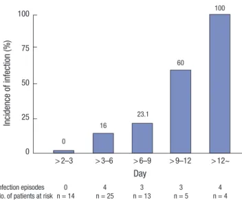
OR, 1.400; 95% CI, 1.081–1.815; P = 0.011) (Table 6). Fig. 1 shows the incidence of infection according to the duration of ECMO.
DISCUSSION
We found that the incidence of nosocomial infection in an adult population undergoing ECMO was 43.3 cases per 1,000 ECMO- days. In multivariate analyses, both a higher preoperative creat- inine level and the duration of ECMO support were indepen- dently associated with nosocomial infection. ECMO patients are at an increased risk for infections as a consequence of being in an ICU. Mechanical ventilators, central venous catheters, in- dwelling urinary catheters, surgical incisions, and health care providers are all sources of infection for ECMO patients. In gen- eral, the rate of nosocomial infection in the ICU has ranged from 13.9% to more than 44.0% (12,13).
In terms of the site of nosocomial infection, Vincent et al. (12) analyzed data from 1,417 ICUs in Europe to determine the inci- dence of pathogens and infections; pneumonia and other low- er RTIs (64.7%) were the most common diseases, followed by UTI (17.6%) and BSI (12.0%). However, among infections dur- ing ECMO support, BSI was the most common, occurring in 32.6% to 89.4%, and BSIs were predominant in most studies, fol- lowed by RTIs, UTIs, and surgical site infections (7,14,15). In our study, the infection site differed from that of the general ECMO cohort. We found that the incidence of RTI was similar to that of BSI.
We found that the hazard ratio for nosocomial infection in patients with higher levels of preoperative creatinine was 2.176 (P = 0.033). Although the precise mechanism by which increas- ed preoperative creatinine increases the risk of nosocomial in- fection remains unknown, some data suggest that increased serum levels of creatinine correlate with nosocomial infection.
Table 5. Outcomes of ECMO support in patients with and without nosocomial infec- tions
Outcomes Without infection*
(n = 47)
With infection*
(n = 14) P value ECMO oxygenator type
Non-PMP membrane
PMP membrane 30 (63.8)
17 (36.2) 8 (57.1) 6 (42.9)
0.756
Anticoagulation drug given Heparin
Nafamostat mesilate 42 (89.3)
5 (10.7) 12 (85.7) 2 (14.3)
0.710
ECMO support duration, day 4.5 ± 2.4 14.5 ± 12.4 0.010 ECMO weaning
Success Failure
23 (48.9) 24 (51.1)
4 (28.6) 10 (71.4)
0.228
Survival to discharge Yes
No
17 (36.2) 30 (63.8)
2 (14.3) 12 (85.7)
0.190
ECMO = extracorporeal membrane oxygenation, PMP = polymethylpentene.
*Data are presented as numbers of cases (%) for categorical variables or as means ± standard deviations for continuous variables.
Table 6. Multivariate analysis of risk factors for nosocomial infection
Variables Adjusted OR (95% CI) P value
Pre-ECMO creatinine level, mg/dL 2.176 (1.065–4.447) 0.033 Duration of ECMO support, day 1.400 (1.081–1.815) 0.011 OR = odds ratio, CI = confidence interval, ECMO = extracorporeal membrane oxy- genation.
*Variables with a probability valve < 0.20 in univariate analyses were candidates for multivariate analysis. The candidate variables were gender, pre-ECMO creatinine lev- el, transfusion history, and duration of ECMO support.
Fig. 1. The incidence of infection according to the duration of ECMO.
ECMO = extracorporeal membrane oxygenation.
Incidence of infection (%)
Day
Infection episodes 0 4 3 3 4
No. of patients at risk n = 14 n = 25 n = 13 n = 5 n = 4 > 2–3 > 3–6 > 6–9 > 9–12 > 12~
100
75
50
25
0
0
16
23.1
60
100
An increase in creatinine levels reflects the presence of kidney disease; an injured kidney may compromise the immune re- sponse via systemic release from leukocytes in the kidneys and renal tubular cells (16-20). Such changes in the host immune response may be associated with nosocomial infection.
We also found that the duration of ECMO was a risk factor for infection. Many previous studies showed that the duration of ECMO support was the most important risk factor of acquired infection during ECMO and was associated with a significantly increased rate of death (7,14,15). Burket et al. (21) reported that the rate of BSI increased with the duration of ECMO support.
Among patients who underwent ECMO for 3–10 days, the rate of BSI was 9.5 cases per 1,000 ECMO-days, and among those who underwent ECMO for 11–20 days and 21–30 days, the rates of BSI increased to 27.2 cases per 1,000 ECMO-days and 64.5 cases per 1,000 ECMO-days, respectively. We found that a lon- ger duration of ECMO was associated with nosocomial infec- tion. The receiver operation characteristic (ROC) curve showed that a cut-off point of 3.7 days was significant. The incidence of nosocomial infection was higher in patients who underwent ECMO for > 3.7 days than for shorter times. However, nosoco- mial infection did not affect survival to discharge. This may be because of our small patient numbers. Further studies on larger cohorts are required.
In terms of the causative microorganisms, large-scale inter- national data show that Staphylococcus aureus and other Staphylococcus species (38%) are the most common pathogens causing BSIs, followed by Escherichia coli (24%), in critically ill patients without ECMO support (12,22). In addition, Candida species are often isolated in the ICU and can cause pneumonia and BSIs with a mortality rate of up to 47% (12,22). On the other hand, microbes are commonly associated with medical device- related infections in patients with ECMO support, such as co- agulase-negative Staphylococcus, Pseudomonas, and Candida species (7). However, the balance is shifting from multi-resis- tant staphylococci to highly resistant classes of A. baumannii and P. aeruginosa (23). In our study, A. baumannii was the most common pathogen in both blood and the respiratory tract, which would be a common pathogen in the ICU environment. We en- countered no case of fungal infection (for example, Candida) in the present study. This may have been due to the short duration of ECMO compared with those in other ECMO cohort studies.
This study had the limitations inherent in retrospective work using observational data from a single center. Although our pa- tient numbers were too low to allow us to draw strong conclu- sions, we sought to identify precise risk factors in a homogeneous group.
A higher preoperative creatinine level and an extended dura- tion of ECMO support were risk factors for nosocomial infec- tion. Therefore, to avoid the development of nosocomial infec- tions, strategies shortening the length of ECMO support should
be applied whenever possible.
DISCLOSURE
The authors have no potential conflicts of interest to disclose.
AUTHOR CONTRIBUTION
Conceptualization: Kim GS, Lee KS, Jeong IS, Ahn BH. Data cu- ration: Kim GS, Kang SK, Kim S. Investigation: Kim GS, Lee KS, Park CK, Kang SK, Kim DW. Supervision: Kim GS, Lee KS, Park CK, Kang SK, Kim DW, Oh SG, Oh BS, Jung Y, Kim S, Yun JS, Song SY, Na KJ, Jeong IS, Ahn BH. Writing - original draft: Kim GS, Lee KS, Oh SG, Oh BS, Kim S, Yun JS, Song SY, Na KJ, Jeong IS, Ahn BH. Writing - review & editing: Kim GS, Lee KS, Oh SG, Oh BS, Kim S, Yun JS, Song SY, Na KJ, Jeong IS, Ahn BH.
ORCID
Gwan Sic Kim http://orcid.org/0000-0002-0648-0178 Kyo Seon Lee http://orcid.org/0000-0001-7397-4680 Choung Kyu Park http://orcid.org/0000-0002-9898-2545 Seung Ku Kang http://orcid.org/0000-0001-7558-9102 Do Wan Kim http://orcid.org/0000-0003-2262-2882 Sang Gi Oh http://orcid.org/0000-0001-9394-4980 Bong-Suk Oh http://orcid.org/0000-0001-8389-190X Yochun Jung http://orcid.org/0000-0001-7165-3007 Seok Kim http://orcid.org/0000-0002-0850-169X Ju Sik Yun http://orcid.org/0000-0002-5167-3454 Sang Yun Song http://orcid.org/0000-0002-2084-8143 Kook Joo Na http://orcid.org/0000-0003-0923-1414 In Seok Jeong http://orcid.org/0000-0002-2249-0667 Byoung Hee Ahn http://orcid.org/0000-0002-7579-1359 REFERENCES
1. Hill JD, O’Brien TG, Murray JJ, Dontigny L, Bramson ML, Osborn JJ, Ger- bode F. Prolonged extracorporeal oxygenation for acute post-traumatic respiratory failure (shock-lung syndrome). Use of the Bramson membrane lung. N Engl J Med 1972; 286: 629-34.
2. Bartlett RH. Extracorporeal life support: history and new directions. ASAIO J 2005; 51: 487-9.
3. Paden ML, Conrad SA, Rycus PT, Thiagarajan RR; ELSO Registry. Extra- corporeal life support organization registry report 2012. ASAIO J 2013;
59: 202-10.
4. Peek GJ, Mugford M, Tiruvoipati R, Wilson A, Allen E, Thalanany MM, Hibbert CL, Truesdale A, Clemens F, Cooper N, et al. Efficacy and eco- nomic assessment of conventional ventilatory support versus extracor- poreal membrane oxygenation for severe adult respiratory failure (CE- SAR): a multicentre randomised controlled trial. Lancet 2009; 374: 1351- 63.
5. Aubron C, Cheng AC, Pilcher D, Leong T, Magrin G, Cooper DJ, Scheink-
estel C, Pellegrino V. Infections acquired by adults who receive extracor- poreal membrane oxygenation: risk factors and outcome. Infect Control Hosp Epidemiol 2013; 34: 24-30.
6. Haneke F, Schildhauer TA, Schlebes AD, Strauch JT, Swol J. Infections and extracorporeal membrane oxygenation: incidence, therapy, and outcome.
ASAIO J 2016; 62: 80-6.
7. Bizzarro MJ, Conrad SA, Kaufman DA, Rycus P; Extracorporeal Life Sup- port Organization Task Force on Infections, Extracorporeal Membrane Oxygenation. Infections acquired during extracorporeal membrane oxy- genation in neonates, children, and adults. Pediatr Crit Care Med 2011;
12: 277-81.
8. Schmidt M, Bréchot N, Hariri S, Guiguet M, Luyt CE, Makri R, Leprince P, Trouillet JL, Pavie A, Chastre J, et al. Nosocomial infections in adult car- diogenic shock patients supported by venoarterial extracorporeal mem- brane oxygenation. Clin Infect Dis 2012; 55: 1633-41.
9. Undar A, Wang S, Palanzo DA. Impact of polymethylpentene oxygenators on outcomes of all extracorporeal life support patients in the United States.
Artif Organs 2013; 37: 1080-1.
10. Annich G, Lynch W, MacLaren G, Wilson J, Bartlett R. ECMO: Extracor- poreal Cardiopulmonary Support in Critical Care. 4th ed. Ann Arbor, MI, Extracorporeal Life Support Organization, 2012.
11. Garner JS, Jarvis WR, Emori TG, Horan TC, Hughes JM. CDC definitions for nosocomial infections, 1988. Am J Infect Control 1988; 16: 128-40.
12. Vincent JL, Bihari DJ, Suter PM, Bruining HA, White J, Nicolas-Chanoin MH, Wolff M, Spencer RC, Hemmer M. The prevalence of nosocomial infection in intensive care units in Europe. Results of the European Prev- alence of Infection in Intensive Care (EPIC) Study. EPIC International Advisory Committee. JAMA 1995; 274: 639-44.
13. Maillet JM, Guérot E, Novara A, Le Guen J, Lahjibi-Paulet H, Kac G, Diehl JL, Fagon JY. Comparison of intensive-care-unit-acquired infections and their outcomes among patients over and under 80 years of age. J Hosp Infect 2014; 87: 152-8.
14. Schutze GE, Heulitt MJ. Infections during extracorporeal life support. J Pediatr Surg 1995; 30: 809-12.
15. Brown KL, Ridout DA, Shaw M, Dodkins I, Smith LC, O’Callaghan MA, Goldman AP, Macqueen S, Hartley JC. Healthcare-associated infection in pediatric patients on extracorporeal life support: the role of multidisciplinary surveillance. Pediatr Crit Care Med 2006; 7: 546-50.
16. Bihorac A, Efron PA, Ang D, Maier RV, Moldawer LL. Acute kidney injury is associated with nosocomial infections and surgical site infections after trauma. Surg Infect (Larchmt) 2011; 12: S017.
17. Bihorac A, Baslanti TO, Cuenca AG, Hobson CE, Ang D, Efron PA, Maier RV, Moore FA, Moldawer LL. Acute kidney injury is associated with early cytokine changes after trauma. J Trauma Acute Care Surg 2013; 74: 1005- 13.
18. Hoste EA, De Corte W. Clinical consequences of acute kidney injury. Con- trib Nephrol 2011; 174: 56-64.
19. Grigoryev DN, Liu M, Hassoun HT, Cheadle C, Barnes KC, Rabb H. The local and systemic inflammatory transcriptome after acute kidney injury.
J Am Soc Nephrol 2008; 19: 547-58.
20. Lee DW, Faubel S, Edelstein CL. Cytokines in acute kidney injury (AKI).
Clin Nephrol 2011; 76: 165-73.
21. Burket JS, Bartlett RH, Vander Hyde K, Chenoweth CE. Nosocomial infec- tions in adult patients undergoing extracorporeal membrane oxygenation.
Clin Infect Dis 1999; 28: 828-33.
22. Pappas PG, Kauffman CA, Andes D, Benjamin DK Jr, Calandra TF, Edwards JE Jr, Filler SG, Fisher JF, Kullberg BJ, Ostrosky-Zeichner L, et al. Clinical practice guidelines for the management of candidiasis: 2009 update by the Infectious Diseases Society of America. Clin Infect Dis 2009; 48: 503- 35.
23. Doyle JS, Buising KL, Thursky KA, Worth LJ, Richards MJ. Epidemiology of infections acquired in intensive care units. Semin Respir Crit Care Med 2011; 32: 115-38.