책임저자:우고운, 서울시 도봉구 쌍문동 388-1
132-073, 한일병원 외과 Tel: 02-901-3068, Fax: 02-901-3063 E-mail: cadebo@nate.com
재조합 표피성장인자
우 고 운 한일병원 외과
Recombinant Human Epidermal Growth Factor
Go Woon Woo, M.D.
Department of Surgery, Hanil Hospital, Seoul, Korea
Wound healing has three physiological steps; inflammation, proliferation and remodeling. It is a very complicated and sys- tematic process involving inflammation, synthesis and matu- ration of collagen, fibroblast activity, neovascularization and factor epithelial migration, and there are many unknown informations. The growth factors such as Epidermal growth factor (EGF), platelet-derived growth factor (PDGF), fibro- blast growth factor, keratinocyte growth factor, transforming growth factor-b, vascular endothelial growth factor and gran- ulocyte colony-stimulating take an important role in this process. Epidermal growth factor (EGF) derived from plate- lets, macrophages and monocytes stimulates epithelial cells, fibroblasts and smooth muscle cells to make a wound healing. EGF had found out in submandibular gland of mouse in 1962, by Cohen and many studies have inves- tigated the role of EGF as a strong mitogen related to re- ceptors of epidermal cells and fibroblasts to make an epitheli- alization and as a chemoattractant to make a neova- scularization. EGF also helps a wound healing in a chronic radiation ulcer, diabetic foot ulcer and Op wound like a gstrojejunostomy. Therefore, now it is time to make results from the role of EGF in burn. (Journal of Korean Burn Society 2008;11:79-84)
Key Words: Wound healing, Epidermal growth factor, Epithelialization
서 론
상처가 생기면, 우리 몸은 복합적이고 총체적인 여러 과 정을 거쳐서 복구하려고 하는데, 이는 상처의 깊이나 정도
에 따라 수개월이나 수년이 걸릴 수도 있다. 상처 치유 과정 은 급성 염증기(inflammatory phase), 증식기(proliferative phase), 개형기(remodelling phase)의 3단계로 구분되어서 설명되어지고 있다. 급성 염증기는 상처, 조직손상이라는 외부의 충격이 생기면서 방출되는 여러 염증성 매개체에 대응하는 시기이고, 증식기는 염증성 매개체의 작용을 극 복하여 항상성(homeostasis)을 유지하기위해 상처 치유에 관여하는 여러 인자를 동원하여 손상된 조직을 재생하는 시기이다. 개형기는 일단 손상된 조직을 재생하여 형태를 유지한 것을 기능을 향상하기위해 조직을 다시 정리하는 시기라고 할 수 있겠다.
빠른 조직 재생을 위하여 많은 연구와 방법이 진행되었 다. 상처가 치유되는 과정에서 EGF의 역할은 절대적이다.
현재까지 Epithelial growth factor (EGF)에 대한 분자구조 및 생물학적 작용기전에 대하여 많은 연구가 이루어지고 있다. 최근에는 당뇨발 환자, 각막 손상 등 많은 분야에서 EGF를 사용하여 좋은 경과를 가져왔다고 보고 되고 있다.
이제는 EGF를 화상 치유에도 적용하여, 그 임상 결과를 평 가할 필요가 있겠다. 저자는 EGF의 역할에 대하여 집중적 으로 문헌 고찰을 하여 보았다.
본 론
상처 치유 과정은 모든 단계가 명확히 밝혀진 것은 아니 지만, 현재까지 알려진 바로는 systemic inflammatory re- sponse에 대항하여 항상성을 유지한 후에, collagen이 더 많이 합성되면서 증식, 성숙되어서 결손된 조직의 바탕을 형성하고, 섬유모세포가 활성화 되면서 신생혈관이 생기게 되고(neovascularization) 상피세포가 증식되어 전위(epi- thelial migration)되는 매우 복잡한 과정을 거쳐서 이루어 지게 된다.
상처에 대한 급성 염증기에서는 혈관을 수축하는 인자나 여러 화학적 매개체(chemical mediators)가 관여한다. 다른 외상에서도 마찬가지지만, 예를 들어 화상을 입으면, 화상 직후에 여러 매개체가 하는 가장 큰 일은 미세혈관 투과성 (microvascular permeability)을 증가시키는 것이다. 비만세
에 growth factors, adhesion molecules, Complement cleavage products, thrombin, eicosanoids, PAF, Nitric ox- ides, Oxygen-free radicals,Granulocyte elastase와 같은 화 학적 매개체(endogenous chemical mediators)의 생산과 분 비를 촉진시켜서 전신적 염증 반응을 일으키게 된다. 이 반 응이 초기에 적절히 조절되지 못하고 점차 심해지거나 패 혈증과 같은 이차적 악순환 요인이 추가되면 전신적으로 자가 파괴 염증 반응(autodestructive generalized in- flammatory reaction)이 일어나게 된다. 이러한 일련의 과 정을 Cytokine storm으로 설명하고 있다1).
재생기에 들어가게 되면, 여러 종류의 growth factor들을 포함하는 cytokines 들이 활성화되어서 활발하게 활동한다.
이 시기에 활동하는 growth factor는 그 종류가 아주 다양 하고 또 각각의 특유한 역할을 지니고 있어서 서로가 조직 적이고 체계적인 역할을 한다. 이들 growth factor 들은 ep- idermal growth factor (EGF), platelet-derived growth fac- tor (PDGF), fibroblast growth factor, keratinocyte growth factor, transforming growth factor-β (TGF-β), vascular endothelial growth factor와 granulocyte colony-stimulat- ing factor 등이다. 그 중에서도 EGF는 keratinocyte, plate- let, macrophage와 monocyte에서 발생하여 상피세포(epi- thelial cell) 뿐만 아니라 섬유모세포(fibroblast)와 평활근 세포(smooth muscle cells)가 증식하도록 자극하고 섬유모 세포에 의한 collagenase의 분비를 증가시켜 상처를 치유하 는데 작용한다.
1. EGF의 개요
EGF는 53개의 아미노산으로 구성된 단백질로, Cohen에 의해 쥐의 턱밑샘으로부터 최초 분리되었다. Cohen은 바로 태어난 쥐에서 눈꺼풀이 정상적인 쥐보다 더 빨리 열리는 것을 실험적으로 증명하고 '상피세포의 성장을 촉진하는 인자'라는 의미로 epidermal growth factor라 하였다. 그래
하여서 세포의 분열을 유도하고, 상피 세포의 성장을 촉진 하는 인자인 것으로 밝혀져 있다.
EGF는 우리 몸 안에 광범위하게 침(saliva), 소변, 모유, 눈물, 혈액 등에 고농도로 존재하고 있으면서 상처가 났을 때에 혈액을 통하여 상처 부위에 전달되어 흉터없이 상처 를 아물게 하는 작용을 한다. 빠른 상처 치유 작용 이외에도 다양한 작용을 하고 있다. 여성호르몬 분비를 촉진하는 FSH (Follicle stimulation hormone)과 함께 자궁속의 수정 란을 성숙시키고, 혈관이 없어서 변질되기 쉬운 각막의 재 생을 돕는다. 또한 상피 세포 및 내피 세포의 세포 증식 촉 진, 진피의 구성성분인 콜라겐을 합성하는 섬유아세포의 세포 증식 촉진, 피부 손상부위의 혈관 신생 촉진 및 기타 재생 촉진인자의 분비유도, 피부조직이 질서있게 방향을 잡으며 망을 형성하게 하는 물질인 fibronectin의 합성 촉진 등 피부 재생 과정에서 핵심적 역할을 담당한다.
3. EGF 관련 연구
Cohen이 EGF의 분자적 구조를 최초로 구명하고, in vivo 와 in vitro에서 상피 세포 뿐만 아니라 중피세포와 내피 세 포의 강력한 유사분열물질(mitogen)이라는 사실을 밝혔다.
쥐의 턱밑샘에서 분리한 이것이 배아형성(embryogenesis), 성장(growth), 조직 재건 및 재생(tissue repair, regene- ration), 신생물(neoplasia) 등과 같은 생리학적이고 병리학 적인 과정에도 관여한다는 사실을 알게 되었다2).
EGF의 발견 이후, 침에 존재하는 많은 종류의 단백 성장 요소들 중, EGF 대한 많은 연구가 폭넓게 진행되었다. 그래 서 동물 실험 연구로 살균성 효과(bactericidal effect) 뿐만 아니라 성장 요소들의 저장소인 침이 상처치유의 촉진에 지대한 영향을 미친다는 것을 알게 되었다5,6). 이어서 EGF 가 수술 부위 상처에서 상피 재생에 도움을 준다는 보고나 피부뿐만 아니라 위점막에서도 상처 치유 속도를 높여 준 다는 보고가 나왔다4,7).
Fig. 1. Duration of therapy with EGF in 20 eyes of 11 patients suffering from most severe eye burns. (A) Patients were treated with EGF eye drops 5 times daily (10μg/ml) in additionto various other drugs. (B) Patients received placebo in addition to various other drugs (see also text). □ = Treatment supplemented with EGF (A) or placebo (B); ■ = Treatment supplemented with EGF (A) or placebo (B) and fibronectin. The double bar indicates the time of complete epithelial regeneration.
Fig. 2. Epithelial cell response to EGF is depicted in this graph.
EGF concentrations of 5 and 10 ng/ml significantly increased cell proliferation over control, *(p<0.05). Five ng/ml resulted in a significantly greater response than 10 ng/ml, †(p<0.006). EGF was determined to maximally increase proliferation to 113%.
Reim 등은 심한 눈 화상 환자 11명의 20개 눈을 대상으로 EGF와 위약(placebo)을 비교하여 회복 결과를 비교하였다8). EGF로 치료한 투여한 10명 중 6명이 완전히 회복되었지만, 위약은 10명 중 3명만 회복되어 확실한 차이가 있다고 하였 다. 결과적으로는 EGF군에서 회복률이 높았지만, 실험 대 상 두 군에서 dexamethasone, neomycine, sodium ascor- bate, hyaluronic acid를 모두 사용하였으므로 단순히 EGF 의 효과로만 보기를 어렵다(Fig. 1).
Marion 등은 외인성의 EGF, PDGF-BB and TGF-β1와 같은 성장요소들과 함께 말의 상피세포와 각질세포를 각각 배양하였다9). 이 연구의 목적은 실험에 사용된 growth fac- tor들이 어느정도 포함되어야 세포들의 증식에 영향을 주 는가 하는 것이었다. 결과는 상피 세포의 증식은 EGF의 농 도가 10 ng/ml 이상일 때보다 5 ng/ml와 10 ng/ml이면 더 의미있게 증가되었다(p<0.006). 그리고 각질세포는 75 ng/ml의 농도를 제외하고 50 ng/ml 이하의 농도에서는 모 두 의미있게 높은 증식률을 보였다(p<0.001) (Fig. 2, 3).
Nermina 등은 쥐를 이용한 실험에서 화상 부위에 albu- min과 EGF (10μg/kg)를 사용하였다10). 목적은 피부 손상 의 조직학적 소견과 중성구의 활동성을 측정하였고, 화상 으로 생기는 전신적 염증 반응으로 간과 장의 산화 손상의 정도를 파악하는 것이었다. 그래서 EGF를 생리적 적정 용 량을 유지해 주면 간과 같은 장기에 산화 손상을 감소시키 면서 화상으로 인한 상처의 치유에는 도움을 준다고 발표 하였다.
EGF가 상피 세포나 각질 세포의 재생뿐만 아니라 장 점 막과 근막의 문합에도 관여한다는 연구도 많다. 쥐의 약물 에 의한 위점막 손상 실험에서, EGF를 경구 투입하는 것보
다 비경구적로 투입하는 것이 손상을 효과적으로 막을 수 있다는 보고도 있다11). 그 원인은 아마도 EGF의 활동이 위 산과 펩신에 의해 빨리 파괴되었기 때문인 것으로 보인다12). 그렇지만, 관을 통해 EGF를 십이지장으로 직접 주입하면 십이지장의 점막에서 산에 대한 저항성을 키워주기 때문 에, EGF가 십이지장 궤양 치료에 도움이 된다는 사실을 알 게 되었다13). 한 연구에서는 개복술로 위공장 문합술을 시 행하고 EGF를 섭취시킨 군과 섭취시키지 않은 군으로 나 누어 3일, 7일, 21일째 문합 부위, 피부와 근막의 신장 강도 를 측정하였다14). 3일째의 신장 강도가 대조군에 비해 월등
Table 1. Effect of Dressing on Diabetic Foot Ulcerations
Total participants 89
Closure by advanced dressing 21 (24%) Closure by advanced dressing and EGF 52 (58%)
Time till closure (weeks) 6.6∼7.4
Failure of closure 16 (18%)
Fig. 3. Keratocyte response to EGF concentrations of (A) 1, 5 and 10 ng/ml and (B) 10, 25, 50 and 75 ng/ml are shown in these two graphs. All concentrations with the exception of 75 ng/ml significantly increased cell proliferation in comparison to controls,
*(p<0.001). Maximal stimulation demonstrated a 164% increase in cell proliferation.
Fig. 4. One patient withdrew before the eighth infiltration session due to an acute severe adverse event (chest pain). Three individuals did not show deve- lopment of granulation tissue at the eighth infiltration session; eight other patients did not complete the 8-week treatment schedule (five abandoners and three withdrawn due to sepsis). All these patients had an unfavourable progress and were amputated. The rest (n=17) who completed 24 EGF infiltra- tions healed.
히 뛰어 났으나 결과적으로는 큰 차이가 없었다. 한편, EGF 를 환부에 직접 주입하거나 국소 도포하여 상피 세포나 섬 유아세포를 더욱더 증식시켜서 그 층을 두껍게 한다는 보 고도 있다15,16). 최근의 보고에 의하면, 말초 신경의 재생이 나 방사선 치료나 정맥 순환 부전으로 생긴 궤양에서도 EGF를 국소적으로 치료하여 좋은 결과를 얻었다는 보고도 나오고 있다17-19).
특히, EGF로 당뇨성 족부 궤양을 치료하려는 시도에 큰 기대를 걸고있다. Acosta 등은 29명의 당뇨성 족부 궤양 환 자에게 EGF를 환부 내에 주입하고 경과를 관찰하였다15). 그들은 당뇨성 궤양의 병리학적인 특수성 때문에 국소 도 포가 아닌 환부내 주입 방법을 선택하였다. 이 연구에는 한 계점은 있지만 8번의 주입으로 86%에서 육아 조직을 생성
하였고, 56%에서 환지의 절단을 막을 수 있었다(Fig. 4). 홍 등은 89명의 환자를 대상으로 최소 3주 동안 하루에 두 번 씩 국소적 도포 방법으로 치료하여, 52명은 상처가 치유되 었고 16명은 실패하였다(Table 1).
화상 치료에 대한 연구로는, 홍 등은 10마리의 돼지를 대 상으로 부분층 화상 상처에 EGF의 국소 도포법을 다양한
Fig. 5. Comparison of 100% healing time (HT100) of various rh-EGF concentrations in porcine split-thickness wounds.
Treatment of rh-EGF 0_1, 1, 5, 10, 25 ug/g concentrations significantly decreased wound size and shortened the HT100 more than vehicle-treated wounds and non treated wounds.
About 1 and 5 ug/g rh-EGF considerably accelerated the rate of healing of split-thickness wounds. No difference was observed in the HT100 between vehicle and non treated wounds. Each bar is mean standard deviation of the mean (n
=10). *p<0_05, significantly different from vehicle-treated group;
**p<0_01.
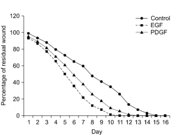
Fig. 6. Percentage of residual wound in nontreated, EGF treated and PDGF treated wound (split thickness wound group).
Fig. 7. Percentage of residual wound in nontreated, EGF treated and PDGF treated wound (full thickness wound group).
용량으로 치료했다.17) 완전히 상피화 되는 시간을 비교한 결과, EGF의 용량이 1 ug/g와 5 ug/g일 때에 완전히 100%
상피화가 되는 시간이 현저히 줄었다(Fig. 5).
은 등은 30마리의 쥐를 대상으로 부분층 두께의 표재성 상처와 전층 두께의 상처를 만들어서 EGF와 PDGF를 부분 도포하여 치료하였다20). 그들은 표재성 상처에서는 EGF로 치료하는 것이 PDGF로 치료한 것 보다 상피화가 더 빨리 된다고 보고하였다(p<0.005). 전층 두께 상처군에서는, 통 계적 의미는 없지만, EGF로 치료하는 것 보다는 PDGF로 치료한 것이 더 빠르게 상처 구축(wound contracture)과 상 피화가 진행되었다(p>0.005). 조직학적 관찰 소견에서는 EGF 치료군과 PDGF 치료군이 대조군보다 재상 피층의 두 께가 더 두꺼웠고 결체 조직(connective tissue)이 더 빨리 성숙되었다(Fig. 6, 7).
결 론
상처가 생기면, 우리 몸 자체에서 자율적으로 EGF를 손 상된 조직에 적절하게 공급하여 정상적으로 회복되도록 하 려고 한다. 그러나 EGF가 제대로 공급되지 않을 경우는 외 부에서 공급을 해주어 정상적으로 회복되도록 도와주어야 한다. EGF는 다양한 상처에서 그 효과가 입증되어졌다. 당
뇨성 족부 궤양, 창상, 각막 손상, 복부절개 수술, 미용 목적 의 박피수술, 노화된 피부의 개선, 위궤양의 치료, 화상 등 에서 이용 되고 있다. 물론 EGF가 상처 치료제로 독보적인 위치에 있다고는 할 수 없다. 이제까지의 여러 연구 보고를 살펴보면 다방면에서 상처 치료에 도움이 되는 치료 방법 임에는 틀림이 없다. 소아 화상 환자들은 부분층 화상을 입 는 경우가 많다. 특히 이 소아 화상환자에서 EGF로 치료하 여 완벽하게 화상을 치료 할 수 있다면 화상 치료의 방법 개선에 획기적인 역할을 할 수 있을 것으로 기대된다.
REFERENCES
1) Aikawa N. Cytokine storm in the pathogenesis of multiple organ dysfunction syndrome associated with surgical insults.
Nippon Geka Gakkai Zasshi. 1996;97(9):771-777.
2) Cohen S. Isolation of a mouse submaxillary gland protein
7) Varshney AC, Sharma DN, Singh M, Sharma SK, Nigam JM.
Therapeutic value of bovine saliva in wound healing. A histomorphological study. Indian J Exp Biol. 1997;35(5):5357.
8) Reim M, Kehrer T, Lund M. Clinical application of epidermal growth factor in patients of most severe eye burns.
Ophthalmologica. 1988;197:179-184.
9) Haber M, Cao Z, Panjwani N, Bedenice D, Li WW, Provost PJ. Effects of growth factors (EGF, PDGF-BB and TGF-1) on cultured equine epithelial cells and keratocytes: implications for wound healing. Veterinary Ophthalmology. 2003;6:211-217.
10) Jahovic N, Gzel E, Arbak S, Yegen B. The healing-promoting effect of saliva on skin burn is mediated by epidermal growth factor (EGF): role of the neutrophils. Burns. 2004;30:531-538.
11) Romano M, Kraus ER, Boland CR. Comparison between transforming growth factor alpha and epidermal growth factor in the protection of rat gastric mucosa against drug-induced injury. Ital J Gastroenterol. 1994;26:223-228.
12) Playford RJ, Marchbank T, Calnan DP. Epidermal growth factor is digested to smaller, less active forms in acidic gastric
16) Hong JP, Jung HD, Kim YW. Recombinant human epidermal growth factor (EGF) to enhance healing for diabetic foot ulcers. Ann Plast Surg. 2006;56:394-398.
17) Hong JP, Kim YW, Jung HD, Jung KI. The effect of various concentrations of human recombinant epidermal growth factor on split-thickness skin wounds. Int Wound J. 2006;3:
123-130.
18) Lee SW, Jung KI, Kim YW, Jung HD, Kim HS, Hong JP. Effect of epidermal growth factor against radiotherapy-induced oral mucositis in rats. Int J Radiation Oncology Biol Phys. 2007;
67(4):1172-1178.
19) Lee SW, Moon SY, Kim YH, Hong JP. The use of recombinant human epidermal growth factor to promote healing for chronic radiation ulcer. Int Wound J. 2007;4(3):216-220.
20) Eun SC, Heo CY, Baek RM, Minn KW. Cutaneous wound healing enhanced by epidermal growth factor and platelet derived growth factor. J Korean Wound Care Soc. 2007;3(1):
1-8.