28
서 론
마취 유도제의 투여에 의해서 수술환자가 의식이 소실되었을 때 용수환가나 기관튜브 삽관이 용이하 지 않을 때, 대부분의 마취과의사는 심리적으로 위 책임저자:염광원, 서울특별시 종로구 연건동 28번지
서울대학교 치과대학 치과마취과학교실 우편번호: 110-744
Tel: 02-760-3866, Fax: 02-766-9427 E-mail: [email protected]
하악전돌증 환자에서 기관내 삽관을 위한 기도평가에 관한 연구
서울대학교 치과대학 치과마취과학교실
이 승 주․김 현 정․염 광 원
Abstract
Airway Evaluation for Endotracheal Intubation of Mandibular Prognathic Patient
Sung-Ju Lee, Hyun-Jeong Kim, and Kwang-Won Yum
Department of Dental Anesthesiology, Seoul National University, College of Dentistry
Background: The fundamental responsibility of an anesthesiologist is to maintain adequate gas exchange. Failure to maintain a patent airway can result in brain damage or death. Generally, in patients with mandibular prognathism, who have the protruded mandible, the mask ventilation was thought to be not easy. The purpose of this study was to observe the degree of the difficulty of airway management in mandibular prognathism using some anatomic criteria for defining and grading difficulty of airway and difficulty of endotracheal intubation with direct laryngospoce.
Methods: The observations and measurements are done to the 54 patients with mandibular prognathism, who were scheduled for corrective esthetic surgery. The case study is done to the 30 patients with normal mandible for control group. In all patients, mouth opening distance (MOD), mouse opening angle (MOA), mandibular length (ML), mandibular depth (MD), thyromental distance (TMD), thyromental area (TMA), Mallampati grades, and Cormack and Lehane grades are measured. T-test and Chi-square test are done (P < 0.05).
Results: In the mandibular prognathism cases, the measurements of MD, TMD and TMA are more greater than those of controls (P < 0.05). Mallampati grades with tongue thrust are higher in the female mandibular prognathism cases than those of female controls. Most of the grades of the mandibular prognathism cases with Cormack and Lehane grading system are I or II being easy intubation cases (P<
0.05)
Conclusions: In the patients of mandibular prognathism, the intubation with laryngoscope will be easer than that of normal mandible in general. It is for that their laryngeal aperture can be easily visible when the laryngoscope are used. (JKDSA 2003; 3: 28∼33)
ꠚꠚꠚꠚꠚꠚꠚꠚꠚꠚꠚꠚꠚꠚꠚꠚꠚꠚꠚꠚꠚꠚꠚꠚꠚꠚꠚꠚꠚꠚꠚꠚꠚꠚꠚꠚꠚꠚꠚꠚꠚꠚꠚꠚꠚꠚꠚꠚꠚꠚꠚꠚꠚꠚꠚꠚꠚꠚꠚꠚꠚꠚꠚꠚꠚꠚꠚꠚꠚꠚꠚꠚꠚꠚꠚꠚꠚꠚꠚꠚꠚꠚꠚꠚꠚꠚ Key Words: Airway evaluation, General anesthesia, Intubation, Prognathism
축된다. 더 나아가 환자의 환기상태가 악화되어 호 흡정지 상태가 되거나 심폐정지가 오면 기도폐쇄가 일어나고, 수 분 후에 환자는 저산소증으로 인해 중 요 장기인 뇌나 심장의 손상을 경험하게 된다. 기도 와 관련된 의료분쟁의 85%가 뇌손상 또는 사망 사 례라는 것은 놀랄만한 사실은 아니다(Caplan 등, 1990).
기도유지의 실패는 마취와 관련된 위중한 합병증 및 사망 사례의 가장 중요한 요인으로 알려져 있다 (Biebuyck, 1991). 때문에 전신마취와 관련하여 가장 중요한 것은 기도유지이다. 즉 마취와 관련하여 수 술환자의 기도는 항상 안전하게 유지되어야 한다.
또한, 구강악안면외과 환자들은 기도평가에 관련 된 여러 구조물들이 정상인들에 비하여 변형을 보이 는 경우가 많고 기도유지가 어려운 경우가 많다 (Bavitz과 Collicott, 1995; 김현정 등, 1998; 김현정 등, 1999; 임현경 등, 2002). 그러나 이에 대한 체계 적인 연구들은 미미한 실정이다. 하악전돌증은 하악 이 상악에 비하여 과도하게 돌출 되어 있으므로 마 스크를 이용한 기도유지가 쉽지 않고, 기도유지평가 에 사용되는 여러 요소들이 정상인에 비하여 다를 수 있다. 그러나 하악전돌증과 관련된 임상연구들은 오히려 하악전돌증 교정수술 후 변화되는 기도에 대 한 해부학적 연구들이 대부분이다(Katakura 등, 1993;
Enacar 등, 1994; Tselnik과 Pogrel, 2000; Kawamata 등, 2000).
이에 본 연구에서는 하악의 변형이 동반되는 하악 전돌증 환자들을 대상으로 기도평가에 많이 사용되 는 여러 특징이 되는 요소들을 관찰하여 기관튜브의 기관내 삽관 난이도를 평가하고자 하였다.
대상 및 방법 실험대상
2001년 7월부터 2002년 8월까지 하악전돌증 교정 술을 위하여 서울대학교 치과병원 치과마취과에서 전신마취를 받았던 54명의 성인환자를 실험군으로 하고, 앵글 분류 I급의 구치관계를 가지며 구치부 반대교합이 없는 정상적인 상⋅하악 치열관계를 보 이는 30명의 환자를 대조군으로 하였다.
상기 84명의 환자는 미국마취과학회(ASA) 전신상 태 평가 기준에 의거하여 전신상태가 Class I 또는 Class II에 속하는 비교적 건강한 성인 수술환자들로 서 안과, 신경외과 질환, 천식, 비만, 열공 및 탈장 등의 질환을 동반하는 경우에는 본 연구에서 제외하 였다.
본 연구에서는 하악전돌증 수술환자와 대조군의 표본 크기를 동일하게 하지 못하였다. 이것은 정상 적인 하악을 가진 환자의 범주를 정하고 이에 맞는 수술환자를 선정하였으나, 조건을 만족하는 대상의 수가 제한적이었던 것이 원인이다.
실험방법
수술받기 전날 구강악안면 외과 병동에 입원한 수 술환자를 방문하여 이들 수술환자들의 기도 평가를 위하여 다음과 같이 개구거리(mouth opening length, MOD), 개구각도(mouth opening angle, MOA), 하악깊 이(mandibular depth, MD), 하악길이(mandibular length, ML), 갑상연골-턱 길이(thyromental distance, TMD), 양 측 하악하연 절흔(antegonial notch)간 거리 등을 측정
Fig. 1. The items of preoperative measurements (A; Mouth opening length, B; Mouth opening angle, C; Mandibular depth, D; Mandibular length, E; Thyro-mental distance).
하였다. 이들 측정치들 중 개구거리(mouth opening length, MOD), 개구각도(mouth opening angle, MOA), 하악깊이(mandibular depth, MD), 하악길이(mandibular length, ML) 및 갑상연골-턱 길이(thyromental distance, TMD) 등의 측정치들은 대조군 수술환자와 하악전돌 증 환자의 기관내 삽관과 관련하여 해부학적 구조물 을 비교분석하는 지표들로 이용되었다.
양측 하악하연 절흔간 거리의 측정치는 갑상연골- 턱 길이의 측정치와 곱하여 나온 측정치로서 갑상연 골-턱 면적(thyromental area, TMA)의 값을 구하여 지 표로 사용하였다.
수술받기 전날 이루어진 수술환자의 병실 방문에 서는 Mallampati 등에 의해 제안된 상기도 분류법에 의한 분류등급도 평가되었다. 이것은 개구 시 보여 지는 혀의 크기와 인두의 구조물들을 기준으로 등급
을 정하는 것으로, 등급이 증가할 수록 인두 구조물 들이 혀에 의해 가려져서 보이지 않게 되며, 따라서 기관 내 삽관의 난이도도 증가하게 된다.
따라서 수술전날에 이루어진 환자방문에서 측정한 것은 다음과 같다(Fig. 1 A-E).
∙개구거리(mouth opening length, MOD): 최대 개 구 시 전치부 사이의 길이
∙개구각도(mouth opening angle, MOA): 최대 개구 시 상⋅하악 치열이 이루는 각도
∙하악깊이(mandibular depth, MD): 전치의 절단면 부터 하악하연까지의 길이
∙하악길이(mandibular length, ML): 하악연합에서 하악각까지의 길이
∙갑상연골-턱 길이(thyromental distance, TMD): 갑 상연골에서 하악하연까지 거리
∙갑상연골-턱 면적(thyromental area, TMA): TMD × 하악의 internotch distance
∙Mallampati grading system (Fig. 2)
수술 당일에는 마취유도 직전에 심전도, 맥박산소 포화도 계측기, 자동혈압계, 체온계를 환자에게 거치 한 후 100% 산소를 3분 정도 안면 마스크를 통해 공 급하여 탈질소화 시켰다. 마취유도는 thiopental sodium (펜토탈소디움, 중외, Korea) 5 mg/kg를 정맥 내로 투 여하고, 근이완제로서 vecuronium (노큐론, 한화, Korea) 0.1 mg/kg을 정맥 내로 투여하였다. 이 후 안 면마스크로 산소-enflurane을 흡입시켜 마취를 유지하
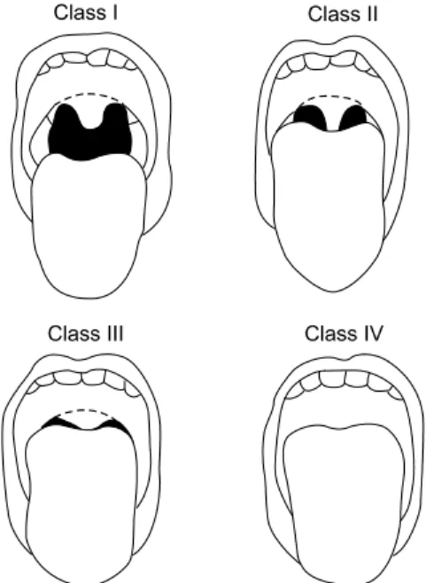
Fig. 2. Classification of the upper airway in terms of the size of the tongue and pharyngeal structures visible upon mouth opening. In class I patients, the soft palate, fauces, uvula, and anterior and posterior tonsillar pillars can be seen; in class II patients, all of the above can be seen except the tonsillar pillars, which are hidden by the tongue; in class III patients, just the base of the uvula can be seen; in class IV patients not even the uvula can be visualized. Reproduced with permission from Mallampati et al. (1985)
Fig. 3. The four grades of laryngeal view, as defined by Cormack and Lehane (1984) The four grades of laryngoscopic view, as defined by Cormack and Lehane.
Grade I is visualization of the entire laryngeal aperture;
grade II is visualization of just the posterior portion of the laryngeal aperture; grade III is visualization of only the epiglottis; and grade IV is the visualization of just the soft palate.
였다. 근이완제 주사 후 3-5분 후에 신경자극기로 train-of-four (TOF) 자극을 주어 완전한 신경근 차단을 확인한 후, 직접 후두경하에 관찰된 성문이 후두개에 의해 가려진 정도에 의해 등급을 분류한 Cormack과 Lehane (1984) grading system을 사용하여 기관내삽관 난이도를 평가하였다(Fig. 3).
통계분석은 SPSS 10.0을 이용하여 T-test 및 Chi- square test를 하였다(P < 0.05).
결 과
두 군간에 나이, 성별, 신장의 차이는 없었다. 그 러나 체중에서 남녀 사이에 차이가 있어 모든 통계 분석은 남녀로 나누어 시행하였다(Table 1).
수술전날 환자의 병실을 방문하여 측정한 개구거 리, 개구각도, 하악길이 등은 대조군과 실험군의 남․여에서 모두 유의할 만한 차이를 보이지 않았 다. 하지만 하악깊이, 갑상선-턱 길이, 갑상선-턱 면 적에서는 하악전돌증을 가진 수술환자군의 남․여 모두에서 유의할 만 한 차이를 발견할 수 있었다 (Table 2).
하악 깊이의 경우에는 남자 대조군 수술환자에서 5.1 ± 0.8 cm, 여자 대조군 수술환자에서 5.3 ± 0.9 cm이었으나 남자 하악전돌증 환자에서는 3.8 ± 0.9 cm, 여자 하악전돌증 환자에서는 3.4 ± 0.6 cm으로 하악전돌증을 가진 환자군에서 유의할 만 한 감소를 보였다(P < 0.05).
갑상선-턱 길이를 살펴보면 남자 대조군 수술환자
에서 6.1 ± 0.9 cm, 여자 대조군 수술환자에서 5.5
± 0.9 cm이었으나 남자 하악전돌증 환자에서는 8.1
± 1.2 cm, 여자 하악전돌증 환자에서는 6.7 ± 0.8 cm으로, 하악전돌증을 가진 환자군의 갑상선-턱 길 이가 유의할 만한 증가를 보였다(P < 0.05).
갑상선-턱 면적의 경우에는 남자 대조군 수술환자 에서 72.5 ± 4.5 cm2, 여자 대조군 수술환자에서 61.1 ± 11.8 cm2이었으나 남자 하악전돌증 환자에서 는 93.0 ± 17.5 cm2, 여자 하악전돌증 환자에서는 69.8 ± 11.1 cm2으로, 갑상선-턱 길이와 마찬가지로 하악전돌증을 가진 환자군에서 유의할 만 한 증가를 보였다(P < 0.05).
두부의 정상위치에서 Mallampati grade는 대조군 여자들에서 유의하게 증가되었으나, 두부 후굴 상태 에서는 두 군 사이에 유의한 차이를 보이지 않았다 (Table 3).
실제 기관내삽관할 때 관찰되는 성대의 관찰정도 에 따른 Cormack-Lehane grading system에 의한 기도
Table 2. Measurements of the Airway Evaluation Parameters
ꠧꠧꠧꠧꠧꠧꠧꠧꠧꠧꠧꠧꠧꠧꠧꠧꠧꠧꠧꠧꠧꠧꠧꠧꠧꠧꠧꠧꠧꠧꠧꠧꠧꠧꠧꠧꠧꠧꠧꠧꠧꠧꠧꠧꠧꠧꠧꠧꠧꠧꠧꠧꠧꠧꠧꠧꠧꠧꠧꠧꠧꠧꠧꠧꠧꠧꠧꠧꠧꠧꠧꠧꠧꠧꠧꠧꠧꠧꠧꠧꠧꠧꠧꠧꠧꠧꠧꠧꠧꠧꠧꠧꠧꠧꠧꠧꠧꠧꠧꠧ
Male Female Total
ꠏꠏꠏꠏꠏꠏꠏꠏꠏꠏꠏꠏꠏꠏꠏꠏꠏꠏꠏꠏꠏꠏꠏꠏꠏꠏ ꠏꠏꠏꠏꠏꠏꠏꠏꠏꠏꠏꠏꠏꠏꠏꠏꠏꠏꠏꠏꠏꠏꠏꠏꠏꠏꠏ ꠏꠏꠏꠏꠏꠏꠏꠏꠏꠏꠏꠏꠏꠏꠏꠏꠏꠏꠏꠏꠏꠏꠏꠏꠏꠏꠏꠏ Control Prognathism Control Prognathism Control Prognathism ꠏꠏꠏꠏꠏꠏꠏꠏꠏꠏꠏꠏꠏꠏꠏꠏꠏꠏꠏꠏꠏꠏꠏꠏꠏꠏꠏꠏꠏꠏꠏꠏꠏꠏꠏꠏꠏꠏꠏꠏꠏꠏꠏꠏꠏꠏꠏꠏꠏꠏꠏꠏꠏꠏꠏꠏꠏꠏꠏꠏꠏꠏꠏꠏꠏꠏꠏꠏꠏꠏꠏꠏꠏꠏꠏꠏꠏꠏꠏꠏꠏꠏꠏꠏꠏꠏꠏꠏꠏꠏꠏꠏꠏꠏꠏꠏꠏꠏꠏꠏ
MOD (cm) 4.7 ± 0.6 4.9 ± 0.5 4.7 ± 0.5 4.4 ± 0.4 4.7 ± 0.5 4.7 ± 0.5 MOA (cm) 3.6 ± 0.5 3.8 ± 0.5 3.7 ± 0.6 3.5 ± 0.4 3.6 ± 0.5 3.7 ± 0.5 ML (cm) 10.5 ± 1.1 11.1 ± 0.8* 10.4 ± 1.1 10.2 ± 0.5 10.4 ± 1.1 10.7 ± 0.8 MD (cm) 5.1 ± 0.8 3.8 ± 0.9 5.3 ± 0.9 3.4 ± 0.6 5.2 ± 0.8 3.6 ± 0.8*
TMD (cm) 6.1 ± 0.9 8.1 ± 1.2 5.5 ± 0.9 6.7 ± 0.8 5.8 ± 0.9 7.4 ± 1.3*
TMA (cm2) 72.5 ± 4.5 93.0 ± 17.5 61.1 ± 11.8 69.8 ± 11.1 66.4 ± 14.1 82.3 ± 18.8*
ꠏꠏꠏꠏꠏꠏꠏꠏꠏꠏꠏꠏꠏꠏꠏꠏꠏꠏꠏꠏꠏꠏꠏꠏꠏꠏꠏꠏꠏꠏꠏꠏꠏꠏꠏꠏꠏꠏꠏꠏꠏꠏꠏꠏꠏꠏꠏꠏꠏꠏꠏꠏꠏꠏꠏꠏꠏꠏꠏꠏꠏꠏꠏꠏꠏꠏꠏꠏꠏꠏꠏꠏꠏꠏꠏꠏꠏꠏꠏꠏꠏꠏꠏꠏꠏꠏꠏꠏꠏꠏꠏꠏꠏꠏꠏꠏꠏꠏꠏꠏ MOD: mouth opening distance, MOA: mouth opening angle, ML: mandibular length, MD: mandibular depth, TMD:
thyromental distance, TMA: thyromental area. All values are mean ± SD, *P < 0.05.
Table 1. Patient Characteristics
ꠧꠧꠧꠧꠧꠧꠧꠧꠧꠧꠧꠧꠧꠧꠧꠧꠧꠧꠧꠧꠧꠧꠧꠧꠧꠧꠧꠧꠧꠧꠧꠧꠧꠧꠧꠧꠧꠧꠧꠧꠧꠧꠧꠧꠧꠧꠧꠧ
Control Prognathism ꠏꠏꠏꠏꠏꠏꠏꠏꠏꠏꠏꠏꠏꠏꠏꠏꠏꠏꠏꠏꠏꠏꠏꠏꠏꠏꠏꠏꠏꠏꠏꠏꠏꠏꠏꠏꠏꠏꠏꠏꠏꠏꠏꠏꠏꠏꠏꠏ
Age (yr) 36.4 ± 15.6 22.4 ± 3.8
Sex (M:F) 14:16 29:25
Body weight (kg) 62.6 ± 11.0 59.0 ± 11.8*
Height (cm) 169.1 ± 6.5 168.8 ± 7.0 ꠏꠏꠏꠏꠏꠏꠏꠏꠏꠏꠏꠏꠏꠏꠏꠏꠏꠏꠏꠏꠏꠏꠏꠏꠏꠏꠏꠏꠏꠏꠏꠏꠏꠏꠏꠏꠏꠏꠏꠏꠏꠏꠏꠏꠏꠏꠏꠏ All values are mean ± SD, *P < 0.05
평가 등급도 하악전돌증을 가진 수술환자들에서는 기관내삽관이 쉬운 class I 또는 class II인 경우가 많 았고(Table 4), 이는 대조군 수술환자들과 비교해서 도 유의한 차이를 보였다(P < 0.05).
고 찰
1978년부터 1982년 5년 동안 프랑스의 마취 환자 들을 대상으로 보고된 바에 의하면 198,103명의 환 자 중 162명의 환자가 기도유지 실패로 인한 허혈성 뇌손상으로 사망한 것으로 보고되고 있다(Tiret 등, 1986). 기도유지 실패의 주요 3가지 원인으로는 식 도내 삽관, 환기 부전 및 기관 내 삽관이 어려운 경 우였다. 기관 내 삽관이 어려울 것으로 예상되는 경 우는 기관 내 삽간 시도 전 충분한 이학적 검사로 예측이 가능하다. 그러나 많은 마취과 의사들은 기 도평가에 많은 주의를 기울이지 않는 실정이다.
기관 내 삽관이라는 술기는 주로 경추, 환추-후두 관절(atalantooccipital joint), 하악, 구강내 연조직들, 목 및 설골 구조물들의 해부학적 구조 및 술자의 기 관내 삽관 기술에 의존한다고 알려져 있다. 따라서 이들 구조물들의 변형은 직접 후두경을 이용한 기관 내 삽관 시 및 안면 마스크를 이용한 기도유지 시 기도유지의 어려움을 야기할 수 있다. 하악전돌증 환자들에서도 하악의 구조변화로 인해 다른 연조직 들의 변화 역시 예상되며, 이러한 변화들이 기도평 가에 영향을 미칠 것으로 예상되나 하악전돌증 환자 에서는 이에 대하여 현재까지 보고된 바가 거의 없 었다. 하악전돌증과 연관 기도에 관한 연구는 오히 려 하악교정술 이후 변화되는 기도평가에 대한 것들
이 대부분이었다(Nakagawa 등, 1998; Achilleos 등, 2000; Kawamata 등, 2000).
하악과 관련되어 기도유지가 어려울 것으로 예상 되는 이학적 소견은 다음과 같다.
개구거리가 손가락 3개 폭 이하이거나 Treacher Collins 증후군과 Pierre Robin 증후군 등의 선천성 기형인 경우처럼 하악이 작고 뒤로 처진 경우, TMD 가 6 cm 이하인 경우, ML이 9 cm 이하이거나 하악 깊이가 증가된 경우들이다. 또한 정상적인 기도라면 개구거리는 3-4 cm 이상, TMD는 6.5 cm 이상, ML 는 9 cm 이상, Mallampati 등급은 1-2 정도이다.
본 연구 결과에 의하면 마취 전 기도평가 결과 하 악전돌증 환자들은 정상적인 기도를 가졌고, 후두경 을 이용한 기관내 삽관이 용이할 것으로 예상되었다.
후두경으로 관찰된 성대의 관찰 범위에 따른 등급 인 Cormack과 Lehane 등급에서도 대조군에 비하여 성대가 보다 온전하게 보이는 1급 또는 2급인 경우 가 대부분이어서 마취전 기도 평가에서 예상된 대로 기관내 삽관이 보다 용이하였다. 하지만 하악이 상 대적으로 발달하였고, 하악전돌증이 아주 심한 환자 들에서는 마취유도제 투여 후 안면 마스크를 이용한 기도유지가 쉽지 않았던 경우들도 관찰되었다.
결 론
하악전돌증 수술환자에서는 기도유지가 어려울 것 이라는 예상과는 달리 후두경을 이용하여 후두개곡 (vallecula)을 들었을 때 정상적인 하악을 가진 환자 들에 비하여 성대를 비롯한 후두 구조물이 보다 잘 Table 3. Distribution of the Mallampati Grade
ꠧꠧꠧꠧꠧꠧꠧꠧꠧꠧꠧꠧꠧꠧꠧꠧꠧꠧꠧꠧꠧꠧꠧꠧꠧꠧꠧꠧꠧꠧꠧꠧꠧꠧꠧꠧꠧꠧꠧꠧꠧꠧꠧꠧꠧꠧꠧꠧ
Mallampati Mallampati (tongue thrust) (neck extended) ꠏꠏꠏꠏꠏꠏꠏꠏꠏꠏꠏꠏꠏꠏꠏꠏꠏꠏ ꠏꠏꠏꠏꠏꠏꠏꠏꠏꠏꠏꠏꠏꠏꠏꠏꠏꠏ
Control Prognathism Control Prognathism ꠏꠏꠏꠏꠏꠏꠏꠏꠏꠏꠏꠏꠏꠏꠏꠏꠏꠏꠏꠏꠏꠏꠏꠏꠏꠏꠏꠏꠏꠏꠏꠏꠏꠏꠏꠏꠏꠏꠏꠏꠏꠏꠏꠏꠏꠏꠏꠏ
Grade I 13 25 21 32
Grade II 10 29 7 22
Grade III 6* 0 2 0
Grade IV 1 0 0 0
ꠏꠏꠏꠏꠏꠏꠏꠏꠏꠏꠏꠏꠏꠏꠏꠏꠏꠏꠏꠏꠏꠏꠏꠏꠏꠏꠏꠏꠏꠏꠏꠏꠏꠏꠏꠏꠏꠏꠏꠏꠏꠏꠏꠏꠏꠏꠏꠏ
*P < 0.05.
Table 4. Distribution of Laryngoscopic View of the Glottis by the Cormack-Lehane Grade
ꠧꠧꠧꠧꠧꠧꠧꠧꠧꠧꠧꠧꠧꠧꠧꠧꠧꠧꠧꠧꠧꠧꠧꠧꠧꠧꠧꠧꠧꠧꠧꠧꠧꠧꠧꠧꠧꠧꠧꠧꠧꠧꠧꠧꠧꠧꠧꠧ
Cormack & Lehan Cormack & Lehan (sniff position) (with pressure) ꠏꠏꠏꠏꠏꠏꠏꠏꠏꠏꠏꠏꠏꠏꠏꠏꠏꠏꠏ ꠏꠏꠏꠏꠏꠏꠏꠏꠏꠏꠏꠏꠏꠏꠏꠏꠏꠏ
Control Prognathism Control Prognathism ꠏꠏꠏꠏꠏꠏꠏꠏꠏꠏꠏꠏꠏꠏꠏꠏꠏꠏꠏꠏꠏꠏꠏꠏꠏꠏꠏꠏꠏꠏꠏꠏꠏꠏꠏꠏꠏꠏꠏꠏꠏꠏꠏꠏꠏꠏꠏꠏ
Class I 10 43* 15 50*
Class II 14 10* 13 4*
Class III 4 1* 0 0
Class IV 2 0 2 0
ꠏꠏꠏꠏꠏꠏꠏꠏꠏꠏꠏꠏꠏꠏꠏꠏꠏꠏꠏꠏꠏꠏꠏꠏꠏꠏꠏꠏꠏꠏꠏꠏꠏꠏꠏꠏꠏꠏꠏꠏꠏꠏꠏꠏꠏꠏꠏꠏ
*P < 0.05.
보여 일반적으로 기관내삽관이 쉬울 것으로 생각되 었다.
하악이 상대적으로 발달한 하악전돌증 수술환자들 에서는 의식이 소실된 후 기관튜브를 삽입하기 위해 수술환자를 산소화시키는 과정에서 마스크를 이용한 기도유지가 어려웠던 경우가 있었다. 하지만 이런 경우에는 환자의 의식이 소실되지 않은 상태에서 기 관튜브의 삽입을 시도하는 등의 술식을 적용한다면 역시 하악전돌증 환자들에서는 일반적으로 기도확보 가 보다 용이했다.
본 연구에 사용된 기도평가를 위한 지표들은 일반 적으로 하악과 관련된 해부학적 구조물들을 이용하 여 기관내삽관의 난이도를 예측하기 위해 사용되었 으며, 하악전돌증 뿐만이 아니라 일반적인 전신마취 와 관련하여 기관 내 삽관을 필요로 하는 수술환자 들에서 미리 그 난이도를 예측하는데 많은 도움이 될 것으로 생각되며, 매우 유용할 것이다.
하악전돌증을 가진 환자들의 기도평가에 사용된 지표들이 기관내삽관에 보다 용이한 수치들을 보이 는 것과 관련하여, 하악전돌증의 정도에 따른 변화 를 보기 위해 각 환자의 SNA, SNB, ANB 등을 측정 하여 비교를 해 보았으나 유의할 만 한 차이를 발견 하지는 못했다. 이것은 연구에 사용된 수술환자의 표본 크기가 작은데 원인이 있을 것이라 생각된다.
앞으로 하악전돌증만이 아니라 상악전돌증 혹은 하 악왜소증 환자들을 포함하는 연구가 이루어진다면 하악전돌의 양 혹은 하악의 크기와 기관 내 삽관 난 이도의 관계를 설정하는 데에 보다 도움이 될 것이 라고 생각된다.
참 고 문 헌
김현정, 김유영, 염광원, 이종호: Syngnathia 환자의 기도 관리. 대한구강악안면외과학회지 1998; 24: 323-5.
김현정, 이가영, 염광원: 소아에서 하악골절 전신마취시 굴곡성 내시경과 유도선을 이용한 경비 기관 내 삽관.
증례보고. 대한마취과학회지 1999; 36: 162-64.
임현경, 김태정, 이춘수, 이홍식, 박혜진, 정종권: 악안면 수술을 위한 악하 경구기관 내 삽관. 증례 보고. 대한마 취과학회지 2002; 43: 375-78.
Achilleos S, Krogstad O, Lyberg T: Surgical mandibular
setback and changes in uvuloglossopharyngeal mor- phology and head posture: a short- and long-term cephalometric study in males. Eur J Orthod 2000;
22(4): 383-94.
Bavitz JB, Collicott PE: Bilateral mandibular sub- condylar fractures contributing to airway obstruction.
Int J Oral Maxillofac Surg 1995; 24(4): 273-5.
Biebuyck JF: Management of the difficult adult airway.
Anesthesiology 1991; 75: 1087-110.
Caplan RA, Posner KL, Ward RJ, Cheney FW: Adverse respiratory events in anesthesia: a closed claims analysis. Anesthesiology 1990; 72(5): 828-33.
Cormack RS, Lehane J: Difficult tracheal intubation in obstetrics. Anaesthesia 1984; 39(11): 1105-11.
Davies JM, Eagle CJ: M.O.U.T.H.S Can J Anesth 1991;
38: 687-88.
Enacar A, Aksoy AU, Sencift Y, Haydar B, Aras K:
Changes in hypopharyngeal airway space and in tongue and hyoid bone positions following the surgical correction of mandibular prognathism. Int J Adult Orthodon Orthognath Surg 1994; 9(4): 285-90.
Katakura N, Umino M, Kubota Y: Morphologic airway changes after mandibular setback osteotomy for prognathism with and without cleft palate. Anesth Pain Control Dent 1993; 2(1): 22-6.
Kawamata A, Fujishita M, Ariji Y, Ariji E: Three- dimensional computed tomographic evaluation of morphologic airway changes after mandibular setback osteotomy for prognathism. Oral Surg Oral Med Oral Pathol Oral Radiol Endod 2000; 89(3): 278-87.
Mallampati SR, Gatt SP, Gugino LD, Desai SP, Waraksa B, Freiberger D, Liu PL: A clinical sign to predict difficult tracheal intubation: a prospective study. Can Anaesth Soc J 1985; 32(4): 429-34.
Nakagawa F, Ono T, Ishiwata Y, Kuroda T: Morphologic changes in the upper airway structure following surgical correction of mandibular prognathism. Int J Adult Orthodon Orthognath Surg 1998; 13(4): 299-306.
Tiret L, Desmonts JM, Hatton F, Vourc'h G. Com- plications associated with anaesthesia--a prospective survey in France. Can Anaesth Soc J 1986; 33(3 Pt 1):
336-44
Tselnik M, Pogrel MA: Assessment of the pharyngeal airway space after mandibular setback surgery. J Oral Maxillofac Surg 2000; 58(3): 282-5.