ABSTRACT
Ⅰ. Introduction
Direct resin composite restoration is considered as most conventional restoration in anterior and poste- rior restoration. However, there are several problems of posterior direct resin composite restoration such as
수종의 상아질 접착시스템이 즉시 및 지연 상아질 봉쇄의 미세인장결합강도에 미치는 영향
하진희∙김현철∙허복∙박정길*
부산대학교 치의학전문대학원 치과보존학교실
THE EFFECT OF VARIOUS BONDING SYSTEMS ON THE MICROTENSILE BOND STRENGTH OF IMMEDIATE AND DELAYED DENTIN SEALING
Jin-hee Ha, Hyeon-Cheol Kim, Bock Hur, Jeong-Kil Park*
Department of Conservative Dentistry, School of dentistry, Pusan National University
The purpose of this study was to compare the effect of various dentin bonding systems on microtensile bond strength of immediate dentin sealing (IDS) and delayed dentin sealing (DDS). Eighteen extracted permanent molars were used in this study. The teeth for DDS group were restored with a provisional restorations, and immersed in saline solution for 1 week, and divided into 3 subgroups according to various dentin bonding adhesives; SB subgroup (3 step total-etch adhesive), SE subgroup (2 step self-etch adhe- sive), XE subgroup (1 step self-etch adhesive). In IDS group, the teeth were divided into 3 subgroups, and applied with bonding adhesives as in DDS group. The teeth were restored with provisional restorations, and immersed in saline solution for 1 week. Indirect composite disc was cemented with resin cement, and all specimens were subjected to microtensile bond strength. The data were statistically analyzed with one- way ANOVA and Student t-test.
The results were as follows:
1. The IDS group showed significantly higher μTBS than DDS group in 3 step total-etch and 2 step self- etch adhesive (p < 0.05).
2. In IDS and DDS group, 3 step total-etch adhesive showed the highest μTBS value, followed by 2 step self-etch, and 1 step self-etch adhesive. In IDS group, the μTBS value for 1 step self-etch adhesive was significantly different from those of the other subgroups (p < 0.05), and in DDS group, there were sta- tistical differences in all subgroup (p < 0.05).
3. Failure modes of tested dentin bonding adhesives were mostly mixed failure and only 1 step self-etch adhesive showed adhesive failure. [J Kor Acad Cons Dent 33(6):526-536, 2008]
Key words: Immediate dentin sealing, Delayed dentin sealing, Indirect resin composite restoration, Dentin bonding adhesives, Microtensile bond strength
- Received 2008.8.27., revised 2008.9.3., accepted 2008.9.3-
*Corresponding Author : Jeong-Kil Park Department of Conservative Dentistry
School of Dentistry, Pusan National University, 1-10, Ami-dong, Seo-gu, 602-739, Busan, Korea Tel: 82-51-240-7454
E-mail: [email protected]
wear, polymerization stress, and inadequate proximal contact. Especially due to the residual polymerization stress, direct resin composite restorations are often contraindicated in large cavities1). Indirect resin com- posite and ceramic inlay / onlay restorations are advocated as alternatives in that situation2). Indirect resin composite restorations have improved clinical conditions with respect to proximal contact, occlusal anatomy and marginal adaptation3).
Successful dentin bonding is particularly important in the case of indirect resin composite restorations such as inlays, onlays, and veneers, because the final strength of the tooth-restoration complex is highly dependant on adhesive procedures4). Paul et al.5)and Bertschinger et al.6) described freshly cut dentin is the ideal substrate for dentin bonding, while dentin contamination due to provisional cements could reduce the potential for dentin bonding. “Immediate dentin sealing (IDS)”method was introduced. This method defined immediate application and polymer- ization of the dentin bonding adhesive to the freshly cut dentin, prior to impression taking4). This proce- dure appears to achieve increased bond strength, improved restoration adaptation, fewer gap forma- tions, decreased bacterial leakage, and reduced dentin sensitivity during provisional period. This approach to adhesion also has a positive influence on the structure preservation, patient comfort, and long-term survival of indirect bonded restoration. On the contrary, the conventional technique for indirect restorations, traditionally, consists in impression taking the cavity immediately after preparation.
Then, after the fabrication of the indirect restoration, provisional material and remnants of luting cement were removed and the adhesive or luting procedures are performed. This procedure was called “Delayed dentin sealing (DDS)”method1).
The success of “IDS”method might be attributed to the dentin bond strength. Although there is a ten- dency to simplify dentin bonding procedures, some authors7,8) described 3 step total-etch adhesive is indicated for IDS because of its ability to form a con- sistent and uniform layer. But, recently self-etch systems have been produced as an alternative to total-etch system9). Recent studies10,11) of self-etch systems have reported high bond strengths to dentin.
De Munck et al.12)reported that the 2 step self-etch adhesive resulted in bond strength close to that obtained with the 3 step total-etch adhesive.
Recently, 1 step self-etch adhesives have been intro- duced that combined the etchant, primer, and adhe- sive into one bottle13). 1 step self-etch adhesives are more hydrophilic due to a higher concentration of acidic monomers to properly etch the dentin surface14). These adhesives are clinically available and now widely used throughout the world.
However, the difference in bond strength between IDS and DDS for cementation of indirect restorations with various dentin adhesives has not yet to be eval- uated. The purpose of this study was to compare the effect of various dentin bonding adhesives on microtensile bonding strength (μTBS) of IDS and DDS for indirect resin composite restoration.
Ⅱ. Materials and methods
1. Tooth preparationEighteen extracted permanent molars without caries were stored in distilled water. The teeth were ground flat to expose occlusal dentin surface. The flat dentin surface was polished with wet 600-grit silicon carbide paper. The exposed dentin surface was eval- uated for the presence of remaining enamel under operating microscope (OPMI pico; Carl zeiss, Obercohen, Germany), which was removed by addi- tional trimming. The teeth were then randomly assigned to two groups of 9 teeth each, according to application of the dentin adhesive such as IDS and DDS (Figure 1).
2. Experimental group
D
Deellaayyeedd ddeennttiinn sseeaalliinngg ((DDDDSS))
The teeth for DDS group were restored with a pro- visional restoration material (Clip; VOCO, Cuxhaven, Germany), and immersed in saline solu- tion for 1 week. Following that delay, the provisional restoration was removed and dentin was cleaned by an intraoral air abrasion device (Danville Engineering Inc., Danville, CA, USA) filled with 50
㎛ aluminum oxide particles. And this group was
divided into 3 subgroups according to dentin bonding adhesive. All dentin bonding adhesives (Table 1) were manipulated and applied to the dentin surfaces according to the manufacturers’instructions (Ta ble 2). The specimen was light polymerized with LED curing unit (Bluephase; Ivoclar Vivadent, Shann, Liechtenstein) at 600 mW/㎠.
IImmmmeeddiiaattee ddeennttiinn sseeaalliinngg ((IIDDSS))
The teeth for IDS group were also divided into 3 subgroups. Like DDS group, all dentin bonding adhe- sives were manipulated and applied to the fresh dentin surfaces according to the manufacturers’
instructions. The specimen was light polymerized with LED curing unit at 600 mW/㎠.
Polymerization of the adhesive was followed by the application of an air-blocking barrier (glycerin jelly) and 10 seconds of additional light exposure with the same light unit to polymerize the oxygen-inhibition
layer as proposed by Magne4). The bonded surfaces were then isolated with petroleum gel not to attach between bonding surface and resin-based provisional restoration. Teeth were restored with a provisional restoration material, and immersed in saline solution for 1 week. Following that delay, the provisional restoration was removed and the sealed dentin was cleaned by an intraoral air abrasion device filled with 50 ㎛ aluminum oxide particles. An adhesive resin was applied once and polymerized.
3. Cementation
Eighteen indirect composite discs of 7mm in height and 10mm in diameter (Sinfony; 3M ESPE, St.
Paul, MN, USA) were prepared to simulate overlying laboratory-processed resin composite restorations.
The surface of each composite disc was sandblasted with 50㎛ aluminum oxide particles for 10 seconds Figure 1. Experimental groups and sequences
Tooth preparation
Dentin bonding procedure + Light polymerized
Provisional restoration (1 week) Provisional restoration (1 week)
Provisional restoration removed
Microabrasion of dentin
Dentin bonding procedure + Light polymerized
Cementation of indirect resin +Light polymerized
Microabrasion of sealed dentin surface
Additional adhesive application + Light polymerized
Cementation of indirect resin + Light polymerized Provisional restoration removed
DDS IDS
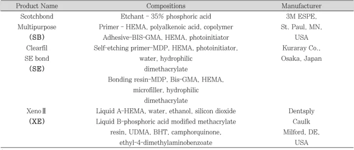
Figure 2. Schematic diagram of the specimen preparation for the microtensile bond test Table 1.Adhesives Investigated in This Study
Product Name Compositions Manufacturer
Scotchbond Etchant - 35% phosphoric acid 3M ESPE,
Multipurpose Primer - HEMA, polyalkenoic acid, copolymer St. Paul, MN,
((SSBB)) Adhesive-BIS-GMA, HEMA, photoinitiator USA
Clearfil Self-etching primer-MDP, HEMA, photoinitiator, Kuraray Co.,
SE bond water, hydrophilic Osaka, Japan
((SSEE)) dimethacrylate
Bonding resin-MDP, Bis-GMA, HEMA, microfiller, hydrophilic
dimethacrylate
XenoⅢ Liquid A-HEMA, water, ethanol, silicon dioxide Dentsply ((XXEE)) Liquid B-phosphoric acid modified methacrylate Caulk
resin, UDMA, BHT, camphorquinone, Milford, DE,
ethyl-4-dimethylaminobenzoate USA
* Abbreviations: HEMA = 2-hydroxyethyl methacrylate; BIS-GMA = bisphenol-A-glycidyl ether dimethacrylate;MDP = 10-methacryloyloxydecal
dihydrogen phosphate; UDMA = urethane dimethacrylate; BHT = butylhydroxy-toluene.
Table 2. Bonding Procedures
Bonding adhesive Procedures
S
SMM Etchant-Applied for 15 seconds, then rinsed
(3 step total-etch adhesive) thoroughly and gently dried for 2 seconds.
Primer-Applied, then dried gently for 5 seconds.
Adhesive- Applied, then light cured for 10 seconds.
S
SEE Self-etching primer -Primer is applied for 20
(2 step self-etch adhesive) seconds, air blown gently.
Bonding resin-Applied, then light cured for 10 seconds.
X
XEE Equal amounts of liquid A and B mixed for 5 seconds.
(1 step self-etch adhesive) Applied for 20 seconds, then light cured for 10 seconds.
from a distance of approximately 5mm. The compos- ite disc surface to be cemented was silanized with a Monobond-S (Ivoclar vivadent AG, Schaan, Liechtenstein) for one minute, and then air dried.
Rely X ARC resin cement (3M ESPE, St. Paul, MN, USA) was mixed according to the manufacturers’
instructions, and applied to the tooth specimen. A5
㎏ mass was applied to the composite disc during cementation. The excess resin cement was removed with a probe. The resin cement was photopolymer- ized for 40 seconds at 600 mW/㎠. The restored spec- imens were subsequently stored in distilled water at room temperature for 24 hours before testing.
4. Microtensile bond strength (μTBS) testing
The teeth were then cut longitudinally into six or seven sections perpendicular to the tooth / adhesive interface, with each slab being 1mm thick and 10mm long using the hard tissue cutter (Accutom-50;
Struers, Rфdovre, Denmark) under water cooling.
The sections were left attached to the remainder of the tooth for further sectioning to obtain sticks approximately 1×1mm thick and 10mm long (Figure 2). Each group was consisted of 36 rods. The speci- mens were glued to the jig of microtensile testing machine (BISCO Inc, Schaumburg, IL, USA) using cyanoacrylate cement (Zapit; Dental Ventures of America, Corona, CA, USA). Tensile load was applied until specimen was failed. Failure load was recorded for each specimen and then the μTBS was calculated.
5. Fracture mode investigations
1
1)) OOppttiiccaall mmiiccrroossccooppyy oobbsseerrvvaattiioonn
After testing, the failure mode of each beam was determined under operating microscope. Fractured test specimens were examined to record the type of bond failure (adhesive, cohesive, or mixed). Bond failure was characterized according to the area of resin remaining on the dentin surface. Adhesive fail- ures were characterized as having less than 25%
resin remaining at the interfacial bond area.
Cohesive failures had greater than or equal to 75%
resin remaining at the interfacial bond area, and
mixed failures had 25% to 75% resin remaining at the interfacial bond area.
2
2)) SSccaannnniinngg eelleeccttrroonn mmiiccrroossccooppyy ((SSEEMM)) oobbsseerrvvaattiioonn
The dentin sides of 6 fractured beams (mixed fail- ure or adhesive failure) from each group were air dried, sputter coated with gold / palladium (E1010 Ion Sputter, Hitachi Co., Mito City, Japan), and examined using SEM (S-3500N SEM, Hitachi Co., Mito City, Japan).
6. Statistical analysis
In each dentin bonding adhesive, the difference of μ TBS between IDS and DDS were analyzed statisti- cally by Student t-test. In IDS and DDS group, one- way ANOVA and Tukey’s test were used to deter- mined statistical difference of μTBS between the dentin bonding adhesives using SPSS 12.0 software (SPSS, Chicago, IL, USA). The level of significance was set at p < 0.05.
Ⅲ. Results
1. Microtensile bond strength (μTBS)
1
1)) CCoommppaarriissoonn bbeettwweeeenn IIDDSS aanndd DDDDSS iinn eeaacchh ddeennttiinn bboonnddiinngg aaddhheessiivvee
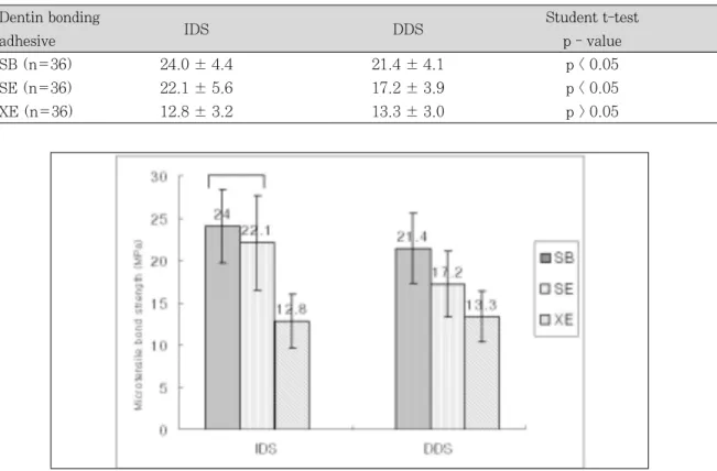
Table 3 lists the μTBS values of three dentin bond- ing adhesives to dentin in IDS and DDS group. The mean μTBS values from 12 to 25 MPa.
In 3 step total-etch SB and 2 step self-etch SE subgroup, IDS group showed higher bond strength than DDS group (p < 0.05). However, in 1 step self- etch XE subgroup, there was no significant difference between IDS and DDS group (p > 0.05).
2
2)) CCoommppaarriissoonn wwiitthh eeaacchh ddeennttiinn bboonnddiinngg a
addhheessiivvee iinn IIDDSS aanndd DDDDSS ggrroouupp
The μTBS values of three dentin bonding adhesives in IDS and DDS group were showed in Figure 3.
In IDS group, 3 step total-etch SB subgroup showed the highest μTBS, followed by 2 step self-etch SE, and 1 step self-etch XE subgroup. But, there
was no significant difference between 3 step total- etch SB and 2 step self-etch SE subgroup (p > 0.05).
1 step self-etch XE subgroup showed the lowest μ TBS value and was significantly different from the other subgroups (p < 0.05).
In DDS group, 3 step total-etch SB subgroup also exhibited the highest bond strength, followed by 2 step self-etch SE, and 1 step self-etch XE subgroup.
And, there was significant difference among all groups (p < 0.05).
2. Fracture mode
1
1)) OOppttiiccaall mmiiccrroossccooppyy oobbsseerrvvaattiioonn
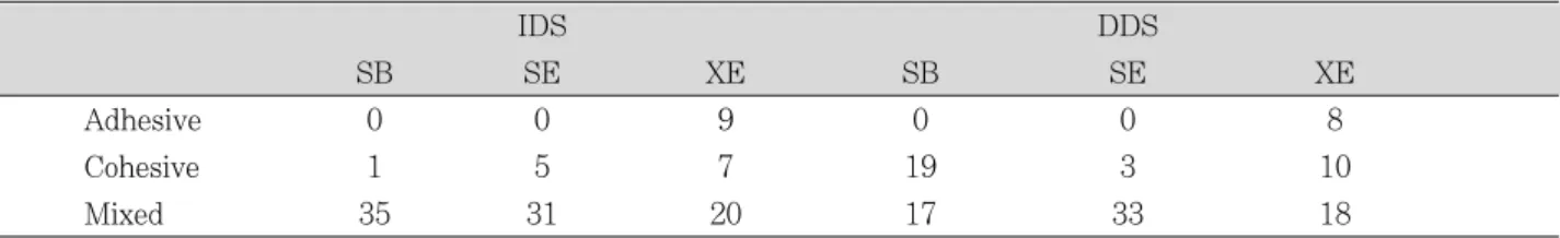
The fracture mode was different among the dentin bonding adhesives. These results were summarized in Table 4. In IDS and DDS group, the fractured beams for 3 step total-etch SB and 2 step self-etch SE subgroup demonstrated cohesive or mixed failure, and there was no specimen with adhesive failure.
However, 1 step self-etch XE subgroup showed adhe- sive failure both IDS and DDS group (25% and 22%
respectively).
2
2)) SSEEMM oobbsseerrvvaattiioonn
The fractured beam of 3 step total-etch SB and 2 step self-etch SE subgroup in both IDS and DDS groups showed a case of mixed failure. But, fractured beam of 1 step self-etch XE subgroup in IDS and DDS group showed a case of adhesive failure (Figure 4 and 5).
Ⅳ. Discussion
This study compared the μTBS of three dentin bonding adhesives with IDS and DDS. The results of this study demonstrated that the IDS method used when cementing indirect resin composite restoration may affect the μTBS of indirect restoration, depend- ing on the dentin bonding adhesive used.
For 3 step total-etch and 2 step self-etch subgroup,
Table 3.Microtensile bond strength of IDS and DDS according to dentin bonding adhesive (Mean ± SD, MPa)
Dentin bonding Student t-test
adhesive IDS DDS
p - value
SB (n=36) 24.0 ± 4.4 21.4 ± 4.1 p < 0.05
SE (n=36) 22.1 ± 5.6 17.2 ± 3.9 p < 0.05
XE (n=36) 12.8 ± 3.2 13.3 ± 3.0 p > 0.05
Figure 3. Microtensile bond strength of three dentin bonding adhesives according to IDS and DDS. Subgroups under the horizontal line were not significantly different (p > 0.05).
IDS group had a statistically higher mean value of bond strength than DDS group. Dietschi et al.15)sug- gested that using IDS and indirect bonded restora- tions, because of the delayed placement of the restoration and postponed occlusal loading, the dentin bond can increase over time and residual stress can dissipate, resulting in significantly improved restoration adaptation. And several studies
5,6) incorporating various dentin bonding adhesives
and application methods have shown that the IDS method increased bond strength values compared to the DDS method. This was related to formation of longer resin tags and a thicker hybrid zone.
Moreover, IDS method can protect the tooth from the consequences of microleakage by sealing the dentin tubules that are vulnerable to bacteria invasion, immediately after completion of the preparation16). Sealing of the dentin tubules also reduces sensitivity Table 4.Number of beams showing fracture modes
IDS DDS
SB SE XE SB SE XE
Adhesive 0 0 9 0 0 8
Cohesive 1 5 7 19 3 10
Mixed 35 31 20 17 33 18
Figure 4. Fractured beam in IDS exhibiting failure pattern (X 100)
(Left) Mixed failure in a beam of 3 step total-etch SB subgroup was shown (Middle) Mixed failure in a beam of 2 step self-etch SE subgroup was shown.
(Right) Adhesive failure in a beam of 1 step self-etch XE subgroup was shown.
(D: dentin,AL : adhesive layer)
Figure 5. Fractured beam in DDS exhibiting failure pattern (X 100)
(Left) Mixed failure in a beam of 3 step total-etch SB subgroup was shown . (Middle) Mixed failure in a beam of 2 step self-etch SE subgroup was shown.
(Right) Adhesive failure in a beam of 1 step self-etch XE subgroup was shown.
(D : dentin,AL : adhesive layer )
by preventing hydraulic fluid flow within the dentin tubules, which is associated with postoperative sensi- tivity17).
In this study, 3 step total-etch subgroup in IDS group exhibited the highest bond strength. According to recent study18), total-etch systems have shown high bond strength to dentin. Bouillaguet et al.19) demonstrated 3 step total-etch adhesive exhibited significantly higher bond strength values than some self-etch systems. In total-etch system, an acid etch- ing of dentin is necessary to efficiently dissolve the smear layer and the smear plugs and to promote a strong and impervious bond between dentin and adhesive mediated by a hybrid layer20). This system could be considered as more reliable in spite of their more time-consuming procedures and technique sen- sitivity.
The 2 step self-etch adhesive in this study present- ed good bond strength to dentin. The advantage of this adhesive is that it combines conditioning and priming into one step, avoiding a gap between inor- ganic component demineralization and primer infil- tration. Tanumiharja et al.21) reported that 2 step self-etch adhesive provides the simplest bonding technique and exhibited the highest bond strength to dentin. Laboratory studies22,23)have demonstrated the capability of self-etch systems to bond equally as well as phosphoric acid-etch based systems which dem- ineralize the tooth surface and require a wash and dry step prior to dentin application. In this study, SE Bond was used as 2 step self-etch adhesive, and then showed as high as μTBS of 3 step total-etch adhesive in IDS group. This is probably due to SE Bond’s mild acidic monomer content and high filler particle con- tent9). Another reason to show high μTBS of 2 step self-etch adhesive in IDS group described twice application of adhesive layer. First adhesive layer to the freshly cut dentin was done before impression taking. Second adhesive layer was applied before cementation. Several studies24,25) have reported that bond strength to dentin may be improved by applica- tion of a second adhesive layer. And due to the sec- ond adhesive layer, thicker adhesive layer contribute to greater reduction in polymerization shrinkage stress and the extent of microleakage in cavities24). IDS method is somewhat similar to a twice applica-
tions of adhesive on dentin bonding. Another advan- tage of 2 step self-etch adhesive has been associated with less postoperative sensitivity than 3 step total- etch adhesive26). It seems that 2 step self-etch adhe- sive in IDS showed as an alternative 3 step total- etch adhesive in IDS. But, the results of this study show that the bond strength of 2 step self-etch sub- group resulted in lower bond strength values than 3 step total-etch subgroup in DDS group.
In this study, XE subgroup bond strength is the lowest value in IDS and DDS group. 1 step self-etch adhesives have been shown to contain a higher con- centration of acid derivatives, methacrylated phos- phoric acid esters, water, and organic solvents than conventional bonding agents to simultaneously etch and infiltrate the dentin surface in 1 step. The low pH (1.5-2.5) of these 1 step self-etch adhesives makes them hydrolytically unstable as a result of the methacrylate-based components27). Meerbeek et al.8) described that μTBS of 1 step self-etch adhesive was significantly least favorable. Low bond strengths recorded with 1 step self-etch adhesive may indicate the single step material cannot yet fulfill all require- ments for the production of effective adhesive layers.
As reported in several studies28,29), most simplified 1 step self-etch adhesives are the least durable, while 2 step self-etch adhesives continue to show the best performance in terms of bond strength, aging, and stability of the bonded interface for IDS. Inoue et al.30) found that 1 step self-etch adhesive tended to have lower bond strengths than 2 step self-etch adhesive. In this study, in both IDS and DDS group, μTBS value of 1 step self-etch subgroup was lower than those of 2 step self-etch subgroup.
In this study, failure modes of tested dentin boning adhesive were mostly mixed or cohesive failure.
Cohesive failures represent integrity in the adhesive layer, protecting the dentin. In contrast, adhesive failures denote a rupture at the dentin / resin inter- face, characterized by open dentinal tubule and intertubular dentin. In this study, 1 step self-etch subgroup showed adhesive failure. This result is somewhat supported by the SEM microscopes show- ing poorly infiltrated smear layer. The low μTBS and the relatively high number of adhesive failures of the 1 step self-etch adhesive strongly suggest that the
incorporation of the smear layer might have decreased the adhesive properties or that they failed to optimally hybridize the smear layer covered dentin31).
For many years, some authors7,8) claimed the IDS method needed 3 step total-etch adhesive. But, the problem of 3 step total-etch adhesive showed tech- nique sensitivity and postoperative sensitivity. To overcome this problem, 2 step self-etch adhesives were produced. In this study, the μTBS of 2 step self- etch subgroup was as high as that of 3 step total- etch subgroup and failure mode of 2 step self-etch subgroup are similar to that of 3 step total-etch sub- group. As the result of this study, when clinician use the IDS method, 2 step self-etch adhesive may be advocated as alternatives of 3 step total-etch adhe- sive. But, 1 step self-etch adhesive is not recom- mended.
Ⅴ. Conclusions
With the limitations of the study, the following con- clusions were drawn,
1. The IDS group showed significantly higher μTBS than DDS group in 3 step total-etch and 2 step self-etch adhesive (p < 0.05).
2. In IDS and DDS group, 3 step total-etch adhe- sive showed the highest μTBS value, followed by 2 step self-etch, and 1 step self-etch adhesive. In IDS group, the μTBS value for 1 step self-etch adhesive was significantly different from those of the other subgroups (p < 0.05), and in DDS group, there were statistical differences in all subgroups (p < 0.05).
3. Failure modes of tested dentin bonding adhe- sives were mostly mixed failure and only 1 step self-etch adhesive showed adhesive failure.
References
1. Stavridakis MM, Krejci I, Magne P. Immediate dentin sealing of onlay preparations: thickness of pre-cured Dentin Bonding Agent and effect of surface cleaning.
Oper Dent 30:747-757, 2005.
2. Dietschi D, Magne P, Holz J. Recent trends in esthetic restorations for posterior teeth. Quintessence Int 25:659-677, 1994.
3. Touati B, Aidan N. Second generation laboratory com- posite resins for indirect restorations. J Esthet Dent
9:108-118. 1997.
4. Magne P. Immediate dentin sealing: a fundamental procedure for indirect bonded restorations. J Esthet Restor Dent 17:144-154, 2005.
5. Paul SJ, Scharer P. The dual bonding technique: a modified method to improve adhesive luting proce- dures. Int J Periodontics Restorative Dent 17:536-545, 1997.
6. Bertschinger C, Paul SJ, Luthy H, Scharer P. Dual application of dentin bonding agents: effect on bond strength. Am J Dent 9:115-119, 1996.
7. Magne P, Douglas WH. Porcelain veneers: dentin bonding optimization and biomimetic recovery of the crown. Int J Prosthodont 12:111-121, 1999.
8. Van Meerbeek B, De Munck J, Yoshida Y, Inoue S, Vargas M, Vijay P, Van Landuyt K, Lambrechts P, Vanherle G. Buonocore memorial lecture. Adhesion to enamel and dentin: current status and future chal- lenges. Oper Dent 28:215-235, 2003.
9. Ye¸silyurt C, Bulucu B. Bond strength of total-etch and self-etch dentin adhesive systems on peripheral and central dentinal tissue: a microtensile bond strength test. J Contemp Dent Pract 7:26-36, 2006.
10. Prati C, Chersoni S, Mongiorgi R, Pashley DH. Resin- infiltrated dentin layer formation of new bonding sys- tems. Oper Dent 23:185-194, 1998.
11. Van Meerbeek B, Van Landuyt K, De Munck J, Hashimoto M, Peumans M, Lambrechts P, Yoshida Y, Inoue S, Suzuki K. Technique-sensitivity of contempo- rary adhesives. Dent Mater J 24:1-13, 2005.
12. De Munck J, Van Meerbeek B, Satoshi I, Vargas M, Yoshida Y, Armstrong S, Lambrechts P, Vanherle G.
Microtensile bond strengths of one- and two-step self- etch adhesives to bur-cut enamel and dentin. Am J Dent 16:414-420, 2003.
13. Park JS, Kim JS, Kim MS, Son HH, Kwon HC, Cho BH. Aging effect on the microtensile bond strength of self-etching adhesives. J Kor Acad Cons Dent 31:415- 426, 2006.
14. Tay FR, Pashley DH, King NM, Carvalho RM, Tsai J, Lai SC, Marquezini L Jr. Aggressiveness of self-etch adhesives on unground enamel. Oper Dent 29:309- 316, 2004.
15. Dietschi D, Monasevic M, Krejci I, Davidson C.
Marginal and internal adaptation of class II restora- tions after immediate or delayed composite placement.
J Dent 30:259-269, 2002.
16. Nagaoka S, Miyazaki Y, Liu HJ, Iwamoto Y, Kitano M, Kawagoe M. Bacterial invasion into dentinal tubules of human vital and nonvital teeth. J Endod 21:70-73, 1995.
17. Suzuki S, Cox CF, White KC. Pulpal response after complete crown preparation, dentinal sealing, and pro- visional restoration. Quintessence Int 25:477-485, 1994.
18. Courson F, Bouter D, Ruse ND, Degrange M. Bond strengths of nine current dentine adhesive systems to primary and permanent teeth. J Oral Rehabil 32:296- 303, 2005.
19. Bouillaguet S, Gysi P, Wataha JC, Ciucchi B, Cattani M, Godin C, Meyer JM. Bond strength of composite to dentin using conventional, one-step, and self-etching adhesive systems. J Dent 29:55-61, 2001.
20. Rontani RM, Ducatti CH, Garcia-Godoy F, De Goes MF. Effect of etching agent on dentinal adhesive inter-
face in primary teeth. J Clin Pediatr Dent 24:205-209, 2000.
21. Tanumiharja M, Burrow MF, Tyas MJ. Microtensile bond strengths of seven dentin adhesive systems. Dent Mater 16:180-187, 2000.
22. Randall RC, Wilson NH. Glass-ionomer restoratives: a systematic review of a secondary caries treatment effect. J Dent Res 78:628-637, 1999.
23. Armstrong SR, Vargas MA, Fang Q, Laffoon JE.
Microtensile bond strength of a total-etch 3-step, total-etch 2-step, self-etch 2-step, and a self-etch 1- step dentin bonding system through 15-month water storage. J Adhes Dent 5:47-56, 2003.
24. Pashley EL, Agee KA, Pashley DH, Tay FR. Effects of one versus two applications of an unfilled, all-in-one adhesive on dentine bonding. J Dent 30:83-90, 2002.
25. Park JG, Cho KH, Cho YG. Effects of application methods of a self-etching primer adhesive system on enamel bond strength. J Kor Acad Cons Dent 33:90- 97, 2008.
26. Opdam NJ, Roeters FJ, Feilzer AJ, Verdonschot EH.
Marginal integrity and postoperative sensitivity in
Class 2 resin composite restorations in vivo. J Dent 26:555-562, 1998.
27. Nishiyama N, Suzuki K, Yoshida H, Teshima H, Nemoto K. Hydrolytic stability of methacrylamide in acidic aqueous solution. Biomaterials 25:965-969, 2004.
28. Tay FR, Pashley DH, Yoshiyama M. Two modes of nanoleakage expression in single-step adhesives. J Dent Res 81:472-476, 2002.
29. Son CY, Kim HC, Hur B, Park JK. Effects of one or two applications of all-in-one adhesive on microtensile bond strength to unground enamel. J Kor Acad Cons Dent 31:445-451, 2006.
30. Inoue S, Vargas MA, Abe Y, Yoshida Y, Lambrechts P, Vanherle G, Sano H, Van Meerbeek B. Microtensile bond strength of eleven contemporary adhesives to dentin. J Adhes Dent 3:237-245, 2001.
31. Tay FR, Carvalho R, Sano H, Pashley DH. Effect of smear layers on the bonding of a self-etching primer to dentin. J Adhes Dent 2:99-116, 2000.
수종의 상아질 접착시스템이 즉시 및
지연 상아질 봉쇄의 미세인장결합강도에 미치는 영향
하진희∙김현철∙허복∙박정길*
부산대학교 치의학전문대학원 치과보존학교실
이 연구의 목적은 수종의 상아질 접착시스템이 즉시 및 지연 상아질 봉쇄에서 미세인장결합강도에 미치는 영향을 평 가하는 것이었다. 18개의 발거된 대구치를 사용하여 지연 상아질 봉쇄그룹은 노출된 상아질면을 임시수복하였으며, 1 주간 보관 후, 접착제에 따라 3개의 소그룹으로 나누어 도포하였다; SB 그룹 (3 단계 산 부식 접착제), SE 그룹 (2단계 자가 부식 접착제), XE 그룹 (1단계 자가 부식 접착제). 즉시 상아질 봉쇄그룹은 3개의 소그룹으로 나누어 접착제를 도 포하고 임시수복 후 1주간 보관하였다. 모든 시편은 간접 복합레진과 레진 시멘트로 합착하고 미세인장결합강도를 측정 하여, 다음과 같은 결과를 얻었다.
1. 즉시 상아질 봉쇄그룹은 지연 상아질 봉쇄그룹에 비해 3단계 산 부식과 2단계 자가 부식 접착제에서 높은 미세인 장결합강도를 보였다 (p < 0.05).
2. 즉시 및 지연 상아질 봉쇄그룹 모두 미세인장결합강도는 3단계 산 부식, 2단계 자가 부식, 1단계 자가 부식 접착제 순으로 감소하였고, 즉시 상아질 봉쇄그룹에서는 1단계 자가 부식 접착제와 다른 소그룹간 유의한 차이가 있었으며 (p
< 0.05), 지연 상아질 봉쇄 그룹에서는 모든 소그룹간 유의한 차이가 있었다 (p < 0.05).
3. 파절 양상은 대부분 혼합성 파절을 보였으며, 1단계 자가 부식 접착제에서만 접착성 파절을 보였다.
주요단어 : 즉시 상아질 봉쇄, 지연 상아질 봉쇄, 간접 복합레진 수복물, 상아질 접착제, 미세인장결합강도 국문초록