http://dx.doi.org/10.3988/jcn.2012.8.1.43 J Clin Neurol 2012;8:43-50
Introduction
It is being increasingly recognized that the prevalence of vas- cular cognitive impairment (VCI) is higher than originally thought. Since VCI is considered to develop under the influence of various cardiovascular risk factors, it is emerging as a poten- tially treatable and preventable form of dementing disorders.1
However, currently available therapeutic options for treatment of dementia, such as cholinesterase inhibitors, were originally devised for Alzheimer’s disease (AD), and their application in vascular dementia (VaD) has produced limited and uncertain clinical benefit.2
It is widely recognized that at the incipient stage, AD pa- tients generally complain about memory problems, whereas VaD patients usually present with cognitive deficits involving nonmemory domains.3 However, a substantial proportion of stroke survivors suffer from poststroke memory dysfunction, and several factors, including medial temporal atrophy (MTA)
Medial Temporal Atrophy and Memory Dysfunction in Poststroke Cognitive Impairment-No Dementia
Beom Joon Kim,a,b Mi-Young Oh,a Myung Suk Jang,b Moon-Ku Han,b Jisung Lee,c Juneyoung Lee,c Yeonwook Kang,d Kyung-Ho Yu,e Byung-Chul Lee,e Sangyun Kim,b Byung-Woo Yoon,a Hee-Joon Baeb
aDepartment of Neurology, Seoul National University Hospital, Seoul National University College of Medicine, Seoul, Korea
bDepartment of Neurology, Seoul National University Bundang Hospital, Seoul National University College of Medicine, Seongnam, Korea
cDepartment of Biostatistics, Korea University College of Medicine, Seoul, Korea
dDepartment of Psychology, Hallym University, Chuncheon, Korea
eDepartment of Neurology, Hallym University College of Medicine, Chuncheon, Korea
Received April 29, 2011 Revised July 18, 2011 Accepted July 18, 2011 Correspondence Hee-Joon Bae, MD, PhD Department of Neurology, Seoul National University Bundang Hospital, 82 Gumi-ro 173beon-gil, Bundang-gu, Seongnam 463-707, Korea Tel +82-31-787-7464 Fax +82-31-787-4059 E-mail [email protected]
Background and PurposezzIt was recently reported that the prevalence of poststroke memory dysfunction might be higher than previously thought. Stroke may exist concomitantly with under- lying Alzheimer’s disease (AD), and so we determined whether post-stroke memory dysfunction indicates manifestation of underlying subclinical AD.
MethodszzOf 1201 patients in a prospective cognitive assessment database, we enrolled subjects with poststroke amnestic vascular cognitive impairment-no dementia (aVCIND; n=48), poststroke nonamnestic vascular cognitive impairment-no dementia (naVCIND; n=50), and nonstroke amnestic mild cognitive impairment (aMCI; n=65). All subjects had cognitive deficits, but did not meet the criteria for dementia. A standardized neuropsychological test battery and magnetic resonance im- aging were performed at least 90 days after the index stroke (mean, 473 days). Visual assessment of medial temporal atrophy (MTA) was used as a measure of underlying AD pathology.
ResultszzThe MTA score was significantly lower in the naVCIND group (0.64±0.85, mean±SD) than in the aVCIND (1.10±1.08) and aMCI (1.45±1.13; p<0.01) groups. Multivariable ordinal lo- gistic regression analysis revealed that compared with naVCIND, aVCIND [odds ratio (OR)=2.69;
95% confidence interval (CI)=1.21-5.99] and aMCI (OR=5.20; 95% CI=2.41-11.23) were signif- icantly associated with increasing severity of MTA.
ConclusionszzOur findings show that compared with poststroke naVCIND, the odds of having more-severe MTA were increased for poststroke aVCIND and nonstroke aMCI.
J Clin Neurol 2012;8:43-50 Key Wordszz vascular cognitive impairment, memory dysfunction, stroke, poststroke dementia.
Open Access
cc This is an Open Access article distributed under the terms of the Cre- ative Commons Attribution Non-Commercial License (http://creative- commons.org/licenses/by-nc/3.0) which permits unrestricted non-com- mercial use, distribution, and reproduction in any medium, provided the ori- ginal work is properly cited.
or white matter hyperintensities (WMHs), are associated with poststroke memory dysfunction.4 Furthermore, there is increas- ing evidence that coexisting cerebrovascular disease precipi- tates and unmasks underlying preclinical AD.5 Stroke and AD are highly prevalent and the two conditions share common vas- cular risk factors, and so it can be hypothesized that patients with poststroke memory dysfunction may have had subclini- cal AD before their stroke, and their memory deficits may be the divulgence of underlying AD triggered by stroke. This is- sue has not been studied in populations with cognitive impair- ment-no dementia (CIND), which is clearly the foremost tar- get of therapeutic interventions for preventing dementia.6 We sought to test this hypothesis by measuring the severity of MTA, which is an acknowledged neuroimaging index of AD.7 For this purpose, we compared patients with poststroke amnes- tic vascular cognitive impairment-no dementia (aVCIND) and patients with nonstroke amnestic mild cognitive impairment (aMCI) to patients with poststroke nonamnestic vascular cog- nitive impairment-no dementia (naVCIND).
Methods
Subject recruitment
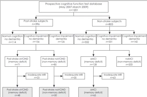
This was a retrospective analysis based on a prospective cog- nitive test database. The patient selection process is summa- rized in Fig. 1. A battery of standard neuropsychological tests was administered to 1201 patients at Seoul National Universi- ty Bundang Hospital between May 2007 and March 2009. Of these 1201 patients, 396 had a documented history of clinical stroke with which neuroimaging findings were compatible; the remaining 805 patients were designated as nonstroke subjects.
Stroke was defined using the World Health Organization def-
inition of “rapidly developed clinical signs of focal or global disturbance of cerebral function, lasting more than 24 hours or leading to death, with no apparent cause other than a vascu- lar origin”.8 CIND was defined as having cognitive impairment but not meeting the Diagnostic and Statistical Manual of Men- tal Disorders, Fourth Edition, Text Revision (DSM-IV-TR) cri- teria for dementia.6,9 We also applied an Instrumental Activity of Daily Living score of <0.43 as indicating “no dementia”.10
For poststroke patients, a standardized neuropsychological battery was administered at least 90 days after stroke onset.
The interval from stroke onset to neuropsychological evalua- tion was 473±311 days (mean±SD). We selected poststroke VCIND patients (n=156) who had an impairment in one or more cognitive domains but did not meet the DSM-IV-TR criteria for dementia. Cognitive impairment in language, visuospatial function, or memory domains was defined as a score in the each of the domain-specific tests of less than the 7th percen- tile (mean-1.5 SD). Cognitive impairment in the frontal do- main was determined by a score of less than the 7th percentile in two or more of the five frontal-domain-specific tests (details of the neuropsychological tests used are given below). Post- stroke VCIND was further classified into poststroke amnestic VCIND (aVCIND; n=71) and poststroke nonamnestic VCIND (naVCIND; n=85), according to the presence of memory im- pairment. Among 805 nonstroke patients, 331 were identified as having CIND; 128 patients who had cognitive impairment in the memory domain were further categorized into the aMCI group. Patients without coronal magnetic resonance imaging (MRI) results were excluded (23 from the aVCIND group, 35 from the naVCIND group, and 63 from the aMCI group). These criteria resulted in 163 patients (48 aVCIND, 50 naVCIND, and 65 aMCI) being included in this study. All of the included
Prospective cognitive function test database (May 2007-March 2009)
n=1201
Post-stroke subjects n=396
Normal cognition no dementia
n=114
Normal cognition no dementia
n=332 Cognitive impairment
no dementia n=156
Post-stroke aVCIND (memory deficit)
n=71
Inadequate MRI n=23
Post-stroke aVCIND (memory deficit)
n=48
Post-stroke naVCIND (non-memory deficit)
n=50
(memory deficit)aMCI n=65 Inadequate MRI
n=35 Inadequate MRI
n=63 Post-stroke naVCIND
(non-memory deficit) n=85
(memory deficit)aMCI n=128
naMCI (non-memory deficit)
n=203 Cognitive impairment
no dementia n=331 Cognitive impairment
dementia n=126
Cognitive impairment dementia
n=142 Non-stroke subjects
n=805
Fig. 1. Recruitment of the study popula- tion. Gray boxes denote the enrolled sub- jects who were included in the final anal- yses. aVCIND: amnestic vascular co- gnitive impairment-no dementia, na- VCIND: nonamnestic vascular cognitive impairment-no dementia, aMCI: am- nestic mild cognitive impairment.
patients were free of any other organic medical or neurologi- cal conditions that may adversely affect cognitive functions.
The study protocols were approved by the local institutional review board.
Cognitive assessment
A battery of standard neuropsychological tests was adapted from the 60-minute neuropsychology protocol of the Vascu- lar Cognitive Impairment Harmonization Standards (VCI- HS).11 The Korean-VCIHS Neuropsychologic Battery was standardized for the Korean population, and its validity and feasibility were examined and confirmed in a multicenter epi- demiological study involving 15 hospitals with nationwide coverage.12 The Korean-VCIHS Neuropsychologic Battery comprises the following cognitive tests of various cognitive domains: frontal executive/activation (animal naming test, phonemic fluency test, Digit Symbol Coding, Trail Making Test), language/lexical retrieval (Korean-Boston Naming Test - short form), visuospatial (Rey-Osterrieth Complex Figure Test: Copy), memory (Seoul Verbal Learning Test), depressive mood (Geriatric Depression Scale), and others (Informant Questionnaire for Cognitive Decline in the Elderly, Korean Mini Mental State Examination, and Instrumental Activity of Daily Living). Trained clinical psychometricians who were blinded to the clinical and radiological profile of each patient administered the battery. The score on each cognitive test was transformed into a standardized Z-score [Z-score=(individual score - population mean score)/(population SD)].
Vascular risk factors and MRI evaluation of the patients
The baseline demographic and clinical characteristics of pa- tients included in the study were collected, including age, gen- der, years of education, handedness, and vascular risk factors, such as presence of hypertension (previous use of antihyper- tensive medication, systolic blood pressure >140 mm Hg, or diastolic blood pressure >90 mm Hg), diabetes mellitus (pre- vious use of glucose-lowering medication, fasting blood glu- cose >7.0 mmol/L, or 2-hours-postprandial blood glucose
>11.1 mmol/L), dyslipidemia (previous use of lipid-lowering medication, total cholesterol >6.0 mmol/L, or low-density li- poprotein cholesterol >4.14 mmol/L), and smoking (current smoker or stopped smoking within the past 5 years). For post- stroke patients, the severity of stroke was assessed using the National Institutes of Health Stroke Scale score, which was measured at hospital admission for the most-recent stroke, and the modified Rankin Scale score, measured at discharge from that admission.
All participants underwent brain MRI, and the mean inter- val between MRI and test for the neuropsychological battery
was 153 days. The MRI studies were performed using a 1.5-tes- la superconducting magnet (Intera, Philips Healthcare, Eind- hoven, The Netherlands). The standardized MRI protocols consisted of an axial T2-weighted spin echo, coronal T2- weighted spin echo, fluid-attenuated inversion recovery im- age, gradient-echo image, and axial T1-weighted image. MTA was rated using a 5-point rating scale, as described previously.13 Both sides were rated simultaneously, and in the case of notice- able asymmetry, the score of the more affected side was chosen as being representative.14 WMHs were rated visually using a 10-point rating scale, based on the fluid-attenuated inversion recovery image.15 Information regarding the subtype, location, and laterality of the stroke and the involvement of the medial temporal lobe was obtained from the prospective stroke regis- try and by reviewing the electronic medical records and MRI findings. Two reviewers (B.J. Kim and M.-Y. Oh) indepen- dently and blindly rated the MTA and WMHs (Spearman’s correlation coefficient: 0.79 for MTA and 0.73 for WMHs), and any disagreement was settled by consensus.
Statistical analysis
Differences in baseline characteristic variables among the poststroke aVCIND, poststroke naVCIND, and aMCI groups were examined by using Pearson’s χ2 test, the Kruskal-Wallis test with pairwise Mann-Whitney U test, or one-way analysis of variance with Bonferroni’s post-hoc adjustment for multiple comparisons, as appropriate. Z-scores were described as mean
±SE values. Bivariate analyses and multivariable ordinal lo- gistic regression analyses performed by taking the MTA score as a dependent variable were used to assess whether covariates were associated with an increasing severity of MTA. To build ordinal logistic regression models, three patients with the most-severe MTA (MTA score=4) were combined with the group of patients with an MTA score of 3. The distribution of patients by MTA score used in the model was as follows: 60 (36.8%), 52 (31.9%), 29 (18.0%), and 22 (13.5%) patients with MTA scores of 0, 1, 2, and 3 or 4, respectively. The propor- tional odds assumption for each ordinal logistic regression model was examined and found to be satisfactory. Variables with a bivariate p<0.10 for their association with MTA were selected for adjustment in multivariate models, including age, WMHs, hypertension, and diabetes. Statistical significance was defined as a probability value of p≤0.05. All statistical analy- ses were performed using SPSS 15.0 (SPSS, Chicago, IL, USA).
Results
The distributions of gender and MTA differed significantly among the three CIND groups; however, age, dominant hand, education, WMHs, and cardiovascular risk factors did not dif-
fer (Table 1). Stroke-related features, the location and laterali- ty of stroke lesions, the stroke subtype, and the severity of the stroke did not differ significantly between poststroke aVCIND and naVCIND patients. The MTA score varied significantly among the three types of CIND (p<0.01, Kruskal-Wallis test).
Pairwise comparisons revealed that the MTA score was signif- icantly lower in the poststroke naVCIND group (median, in- terquartile range; 1, 0-2) than in the poststroke aVCIND group (1, 0-1; p=0.022) and the aMCI group (1, 0-2; p<0.01; the sig- nificance level was set to p≤0.017 after adjusting for pairwise comparison). However, the MTA score did not differ between the poststroke aVCIND and aMCI groups (p=0.10).
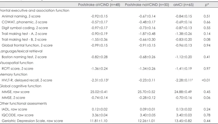
Analysis of variance showed that with the exception of the memory score, the domain-specific test scores did not differ among the three CIND groups (Table 2) (Fig. 2). The memory domain was used to differentiate poststroke aVCIND from poststroke naVCIND. Global Frontal Function score, obtained by averaging the Z-scores of the five frontal domain-specific tests, did not differ between the groups (p=0.94).
Ordinal logistic regression analyses showed that there was a significant association between the severity of MTA and the type of CIND (Table 3). In multivariable analysis, the odds ra- tio for poststroke aVCIND was 2.69 (95% confidence inter- val=1.21-5.99) and the odds ratio for aMCI was 5.20 (95% con- Table 1. Comparison of baseline characteristics among the three CIND groups
Poststroke aVCIND (n=48) Poststroke naVCIND (n=50) aMCI (n=65) p*
Demographic factors
Male gender 39 (81.3%) 27 (54.0%) 45 (69.2%) 0.02
Age (years) 66.31±9.99 67.64±9.70 66.63±7.94 0.75
Right-handedness 44 (91.7%) 48 (96.0%) 61 (93.8%) 0.87
Years of education 0.07
0-6 years 11 (22.9%) 18 (36.0%) 12 (18.5%)
7-12 years 25 (52.1%) 16 (32.0%) 38 (58.5%)
>13 years 12 (25.0%) 16 (32.0%) 15 (23.1%)
Cardiovascular risk factors
Hypertension 32 (66.7%) 32 (64.0%) 47 (72.3%) 0.62
Diabetes 19 (39.6%) 14 (28.0%) 19 (29.2%) 0.39
Hyperlipidemia 24 (50.0%) 20 (40.0%) 22 (33.8%) 0.22
Smoking 24 (50.0%) 22 (44.0%) 37 (56.9%) 0.39
Neuroimaging factors
WMHs, mean±SD 2.52±1.44 2.52±2.17 3.06±2.10 0.23
MTA score
0 17 (35.4%) 27 (54.0%) 16 (24.6%)
1 16 (33.3%) 17 (34.0%) 19 (29.2%)
2 9 (18.8%) 3 (6.0%) 17 (26.2%)
3 5 (10.4%) 3 (6.0%) 11 (16.9%)
4 1 (2.1%) 0 (0%) 2 (3.1%)
Median (IQR) 1 (0, 2) 0 (0, 1) 1 (0, 2) <0.01
Stroke-related factors
Ischemic stroke 42 (87.5%) 48 (96.0%) - 0.16
Supratentorial stroke 43 (89.6%) 47 (94.0%) - 0.48
Laterality of stroke 0.36
Left 29 (60.4%) 28 (56.0%) -
Right 13 (27.1%) 19 (38.0%) -
Bilateral 6 (12.5%) 3 (6.0%) -
Involvement of medial temporal lobe 3 (6.3%) 2 (4.0%) - 0.67
Admission NIHSS score, median (IQR) 2 (1, 4) 2 (1, 6) - 0.91
Discharge mRS score, median (IQR) 1 (1, 2) 1 (0, 3) - 0.82
Values are number of patients (percentages) except where indicated otherwise.
*Pearson’s Chi-squared test, analysis of variance (ANOVA), Kruskal-Wallis test, or Mann-Whitney’s U test, as appropriate.
WMHs: white-matter hyperintensities, CIND: cognitive impairment-no dementia, aVCIND: amnestic vascular cognitive impairment and no dementia, naVCIND: nonamnestic vascular cognitive impairment-no dementia, aMCI: amnestic mild cognitive impairment, MTA: medial temporal atrophy, NIHSS: National Institutes of Health Stroke Scale, IQR: interquartile range, mRS: modified Rankin Scale.
fidence interval=2.41-11.23) relative to poststroke naVCIND.
When compared with aMCI, poststroke aVCIND was not significantly associated with the severity of MTA (p=0.11). In
addition, age and diabetes mellitus were independently associ- ated with increasing severity of MTA (Table 3).
Table 2. Comparison of cognitive function and other related tests among the three CIND groups
Poststroke aVCIND (n=48) Poststroke naVCIND (n=50) aMCI (n=65) p*
Frontal executive and association function
Animal naming, Z-score -0.92±0.15 -0.67±0.14 -0.84±0.15 0.51
COWAT, phonemic, Z-score -0.57±0.17 -0.48±0.17 -0.69±0.16 0.66
Digit symbol coding, Z-score -0.97±0.17 -0.73±0.16 -0.87±0.13 0.55
Trail making test - A, Z-score -0.90±0.19 -1.87±0.48 -1.38±0.26 0.14
Trail making test - B, Z-score -1.55±0.36 -0.66±0.30 -0.83±0.20 0.08
Global frontal function, Z-score -0.99±0.15 -0.91±0.15 -0.96±0.13 0.94
Language/lexical retrieval
Boston naming test, Z-score -0.82±0.28 -0.68±0.26 -1.12±0.20 0.41
Visuospatial function
RCFT score, Z-score -1.36±0.24 -1.34±0.26 -1.41±0.19 0.97
Memory function
HVLT-R, delayed recall, Z-score -2.31±0.13† -0.23±0.11 -2.28±0.11† <0.01
Global cognitive function
MMSE, raw score 25.02±0.41 25.70±0.52 24.88±0.49 0.45
MMSE, Z-score -0.74±0.14 -0.28±0.12 -0.70±0.16 0.06
Other functional assessments
IADL, raw score 0.12±0.02 0.09±0.01 0.13±0.02 0.24
IQCODE, raw score 3.36±0.04 3.40±0.05 3.40±0.03 0.78
Geriatric Depression Scale, raw score 11.81±1.10 12.26±1.01 13.45±0.82 0.44
Each score is the mean±SE value.
*ANOVA, †Significant difference from poststroke naVCIND in multiple comparisons with Bonferroni’s post-hoc adjustment.
aMCI: amnestic mild cognitive impairment, aVCIND: amnestic vascular cognitive impairment and no dementia, naVCIND: nonam- nestic vascular cognitive impairment-no dementia, CIND: cognitive impairment-no dementia, COWAT: Controlled Oral Word Associ- ation Test, HVLT-R: Hopkins Verbal Learning Test-Revised, IADL: Instrumental Activity of Daily Living, IQCODE: Informant Questionnaire for Cognitive Decline in the Elderly, MMSE: Mini-Mental Status Examination, RCFT: Rey-Osterrieth Complex Figure Test.
Post-stroke aVCIND
Post-stroke
aVCIND Post-stroke
aVCIND Post-stroke
aVCIND Post-stroke
naVCIND
Post-stroke
naVCIND Post-stroke
naVCIND Post-stroke
naVCIND aMCI
aMCI aMCI
aMCI p value=0.937
p value=0.973 p value<0.001
p value=0.407 0.0
-0.2 -0.4 -0.6 -0.8 -1.0 -1.2
0.0 -0.2 -0.4 -0.6 -0.8 -1.0 -1.2 -1.4 -1.6
0.0 -0.5 -1.0 -1.5 -2.0 -2.5 0.0 -0.2 -0.4 -0.6 -0.8 -1.0 -1.2 -1.4
Z-score global frontal cognitive functionZ-score visuospatial function Z-score memory functionZ-score language function
Fig. 2. Comparison of Z-scores among the three vascular cognitive impairment and no dementia (CIND) groups. aMCI:
amnestic mild cognitive impairment, aVCIND: amnestic vascular cognitive impairment-no dementia, naVCIND: non- amnestic vascular cognitive impairment- no dementia.
Discussion
The patients in this study were divided into two groups accord- ing to the presence of memory dysfunction so as to make it possible to evaluate the association between MTA and memo- ry dysfunction in patients with poststroke CIND. The effect of stroke was evaluated by comparing nonstroke aMCI patients with poststroke CIND patients. With the exception of memory, patients in the three CIND groups defined in our study had similar cognition and functional ability profiles. We found that the odds of having more-severe MTA were increased for poststroke aVCIND and nonstroke aMCI relative to poststroke naVCIND. Older age and diabetes mellitus were also found to be independently associated with MTA.
The concept of poststroke memory dysfunction seems coun- terintuitive in that stroke does not usually affect the memory- processing structures.16 However, one-third to one-half of stroke survivors were reported to have memory dysfunction around 3 months after the stroke,4 and the risk of dementia doubles with a history of stroke.17 Both MTA and imaging markers of cerebrovascular disease are useful in predicting the development of clinical dementia.18 Experimental studies have also suggested that certain mechanisms are responsible for pro- gressive memory deterioration. Decreased cerebral blood flow, which occurs with ischemic stroke, may modulate traf- ficking of β-amyloid between the blood and the brain, result- ing in the attenuation of β-amyloid clearance from the brain.19,20 Moreover, small ischemic injuries that develop in the paraven- Table 3. Results from univariate and multivariate ordinal logistic regression analyses for the increasing severity of MTA
Univariate OR (95% CI) Multivariable OR† (95% CI) Demographic factors
Male gender 0.99 (0.55-1.80) -
Age (per 1-year increase) 1.09 (1.05-1.13) 1.10 (1.06-1.14)
Right-handedness 0.77 (0.24-2.43) -
Years of education
0-6 years 01.00 (reference) -
7-12 years 1.25 (0.57-2.73)
>13 years 1.22 (0.61-2.42)
Neuroimaging factors Types of CIND
Poststroke aVCIND 2.35 (1.11-4.96) 2.69 (1.21-5.99)
aMCI 4.23 (2.08-8.63) 05.20 (2.41-11.23)
Poststroke naVCIND 01.00 (reference) 01.00 (reference)
WMHs (0-9 scale) (per 1-grade increase) 1.33 (1.15-1.55) 1.14 (0.96-1.34)
Cardiovascular risk factors
Hypertension 2.02 (1.09-3.74) 1.69 (0.85-3.34)
Diabetes 2.48 (1.35-4.56) 2.01 (1.05-3.86)
Hyperlipidemia 0.93 (0.53-1.64) -
Smoking 1.15 (0.66-2.02) -
Stroke-related factors*
Ischemic stroke 1.67 (0.41-6.80) -
Supratentorial stroke 0.61 (0.16-2.29) -
Location of stroke
Left 0.77 (0.34-1.71) -
Bilateral 1.21 (0.31-4.70) -
Right 01.00 (reference)
Stroke involving medial temporal lobe 02.44 (0.48-12.45) -
Admission NIHSS score (per 1-point increase) 1.03 (0.92-1.15) -
Discharge mRS score (per 1-point increase) 1.16 (0.87-1.56) -
Univariate or multivariable ordinal logistic regression analyses were performed by taking the MTA score as a dependent variable.
*Univariate ORs of stroke-related factors were calculated based on patients with poststroke aVCIND and naVCIND, excluding the nonstroke aMCI group, †A multivariable model was constructed using variables with p<0.10 in univariate analyses.
aMCI: amnestic mild cognitive impairment, aVCIND: amnestic vascular cognitive impairment-no dementia, CI: confidence interval, CIND: cognitive impairment-no dementia, MTA: medial temporal atrophy, naVCIND: nonamnestic vascular cognitive impairment-no dementia, OR: odds ratio, WMHs: white-matter hyperintensities.
tricular area and deep structures may cause widespread dis- connection of cholinergic innervations to the cortex,21,22 there- by inducing additive attrition of cognitive reserve.23
We deliberately recruited CIND patients from our large population of patients with neuropsychological assessments.
Cholinesterase inhibitors and memantine, which are used to treat AD patients, seem to provide little benefit to VaD patients,2 and there has been no clinical trial regarding their efficacy in the treatment of poststroke dementia. According to DSM-IV- TR criteria, the presence of stroke per se in a demented indi- vidual excludes a diagnosis of AD. However, our results, as well as those from other recent studies, show that VaD patients usually have “mixed” pathology.24,25 In this context, our find- ings suggests that poststroke CIND can be regarded as subclin- ical AD if there is MTA or memory dysfunction, and that cho- linesterase inhibitors or memantine can be beneficial for these individuals. In addition, the consideration of MTA when recruit- ing subjects for VCI trials or in secondary analysis of VCIND research would improve the homogeneity of study populations.
The association between MTA and poststroke dementia has been addressed by previous studies. However, the clinical im- plications of the results obtained in those studies were limited because they disregarded CIND or were case series of stroke at a specific location.26-29 In addition to the type of CIND, our study detected increases in the severity of MTA with the pres- ence of diabetes, which is consistent with a previous report.30 WMHs and hypertension were not associated with severity of MTA in multivariable analyses, in spite of their significance in univariate analyses (Table 3). A lack of power due to the small sample is a possible explanation for this finding. Furthermore, previous studies also found that the association between WMHs and MTA is controversial.31,32 Contrary to the concept of early executive dysfunction in VaD, frontal function did not differ among the three CIND groups in the present study. How- ever, the development of executive dysfunction has been re- ported to be as common in AD as in VaD.33
There are several reasons why our results should be inter- preted with caution. First, this study was conducted at a single center, had a cross-sectional design, and a relatively small sam- ple. Second, it should be noted that approximately 5% of MCI patients are reported to return to normal over time and that the natural course of progressive memory deterioration is thought to be dynamic.4,34 Third, single- and multidomain CINDs were not distinguished in our study due to the small sample. Fourth, the visual rating scale of MTA was used instead of a volumet- ric assessment. However, volumetry is cumbersome because it requires expensive software and a time-consuming render- ing process, which limits its usefulness for research purposes.
Moreover, it has been reported that visual and volumetric as- sessments of MTA are equally accurate.35,36 Fifth, prestrike
neuropsychological assessments were not available for our subjects.
Our results do not indicate that poststroke memory dysfunc- tion is essentially AD or that such patients should receive cho- linesterase inhibitors - only a prospective cohort study with an adequate duration of follow-up can clarify the association between AD and poststroke memory dysfunction. Consider- ing the increasing burden of stroke and dynamic interrelation- ships among vascular etiologies, degenerative changes in the brain, and host factors with respect to VCI,37,38 our findings support the notion that a stroke event may trigger subclinical AD-type neurodegeneration and result in poststroke memory dysfunction.
Conflicts of Interest
The authors have no financial conflicts of interest.
Acknowledgements
This study was supported by a grant from the Korea Healthcare Tech- nology R&D Project, Ministry of Health, Welfare & Family Affairs, Re- public of Korea (A102065). The funding organization did not participate in the design, conduct, or analysis of the study, or in the preparation of this report.
REFERENCES
1. Hachinski V. The 2005 Thomas Willis Lecture: stroke and vascular cognitive impairment: a transdisciplinary, translational and transaction- al approach. Stroke 2007;38:1396.
2. Kavirajan H, Schneider LS. Efficacy and adverse effects of cholines- terase inhibitors and memantine in vascular dementia: a meta-analysis of randomised controlled trials. Lancet Neurol 2007;6:782-792.
3. Traykov L, Baudic S, Thibaudet MC, Rigaud AS, Smagghe A, Boller F. Neuropsychological deficit in early subcortical vascular dementia:
comparison to Alzheimer’s disease. Dement Geriatr Cogn Disord 2002;
14:26-32.
4. Snaphaan L, de Leeuw FE. Poststroke memory function in nondement- ed patients: a systematic review on frequency and neuroimaging cor- relates. Stroke 2007;38:198-203.
5. Kalaria RN. The role of cerebral ischemia in Alzheimer’s disease.
Neurobiol Aging 2000;21:321-330.
6. Rockwood K, Wentzel C, Hachinski V, Hogan DB, MacKnight C, Mc- Dowell I. Prevalence and outcomes of vascular cognitive impairment.
Vascular Cognitive Impairment Investigators of the Canadian Study of Health and Aging. Neurology 2000;54:447-451.
7. Korf ES, Wahlund LO, Visser PJ, Scheltens P. Medial temporal lobe atrophy on MRI predicts dementia in patients with mild cognitive im- pairment. Neurology 2004;63:94-100.
8. The World Health Organization MONICA Project (monitoring trends and determinants in cardiovascular disease): a major international col- laboration. WHO MONICA Project Principal Investigators. J Clin Epi- demiol 1988;41:105-114.
9. American Psychiatric Association. Diagnostic and Statistical Manual of Mental Disorders: DSM-IV-TR. 4th ed, text revision. Washington, DC: American Psychiatric Association, 2000.
10. Kang SJ, Choi SH, Lee BH, Kwon JC, Na DL, Han SH; Korean De- mentia Research Group. The reliability and validity of the Korean in- strumental activities of daily living (K-IADL). J Korean Neurol Assoc 2002;20:8-14.
11. Hachinski V, Iadecola C, Petersen RC, Breteler MM, Nyenhuis DL,
Black SE, et al. National Institute of Neurological Disorders and Stroke- Canadian Stroke Network vascular cognitive impairment harmoniza- tion standards. Stroke 2006;37:2220-2241.
12. Kang Y. Recent developments in the Korean VCI neuropsychology har- monization efforts. The 4th Congress of the International Society for Vas- cular Behavioural and Cognitive Disorders (VAS-COG). Singapore:
2009.
13. Scheltens P, Leys D, Barkhof F, Huglo D, Weinstein HC, Vermersch P, et al. Atrophy of medial temporal lobes on MRI in “probable” Alzheim- er’s disease and normal ageing: diagnostic value and neuropsycho- logical correlates. J Neurol Neurosurg Psychiatry 1992;55:967-972.
14. Scheltens P, Launer LJ, Barkhof F, Weinstein HC, van Gool WA. Visu- al assessment of medial temporal lobe atrophy on magnetic resonance imaging: interobserver reliability. J Neurol 1995;242:557-560.
15. Wong TY, Klein R, Sharrett AR, Couper DJ, Klein BE, Liao DP, et al.
Cerebral white matter lesions, retinopathy, and incident clinical stroke.
JAMA 2002;288:67-74.
16. Wagner AD, Schacter DL, Rotte M, Koutstaal W, Maril A, Dale AM, et al. Building memories: remembering and forgetting of verbal experi- ences as predicted by brain activity. Science 1998;281:1188-1191.
17. Savva GM, Stephan BC; Alzheimer’s Society Vascular Dementia Sys- tematic Review Group. Epidemiological studies of the effect of stroke on incident dementia: a systematic review. Stroke 2010;41:e41-e46.
18. Staekenborg SS, Koedam EL, Henneman WJ, Stokman P, Barkhof F, Scheltens P, et al. Progression of mild cognitive impairment to de- mentia: contribution of cerebrovascular disease compared with medi- al temporal lobe atrophy. Stroke 2009;40:1269-1274.
19. Niwa K, Kazama K, Younkin L, Younkin SG, Carlson GA, Iadecola C. Cerebrovascular autoregulation is profoundly impaired in mice over- expressing amyloid precursor protein. Am J Physiol Heart Circ Physiol 2002;283:H315-H323.
20. Iadecola C. Neurovascular regulation in the normal brain and in Al- zheimer’s disease. Nat Rev Neurosci 2004;5:347-360.
21. Selden NR, Gitelman DR, Salamon-Murayama N, Parrish TB, Me- sulam MM. Trajectories of cholinergic pathways within the cerebral hemispheres of the human brain. Brain 1998;121:2249-2257.
22. Román GC, Kalaria RN. Vascular determinants of cholinergic deficits in Alzheimer disease and vascular dementia. Neurobiol Aging 2006;
27:1769-1785.
23. Fratiglioni L, Paillard-Borg S, Winblad B. An active and socially inte- grated lifestyle in late life might protect against dementia. Lancet Neu- rol 2004;3:343-353.
24. Viswanathan A, Rocca WA, Tzourio C. Vascular risk factors and de- mentia: how to move forward? Neurology 2009;72:368-374.
25. Zekry D. Is it possible to treat vascular dementia? Front Neurol Neuro- sci 2009;24:95-106.
26. Hénon H, Pasquier F, Durieu I, Pruvo JP, Leys D. Medial temporal lobe atrophy in stroke patients: relation to pre-existing dementia. J Neu- rol Neurosurg Psychiatry 1998;65:641-647.
27. Pohjasvaara T, Mäntylä R, Salonen O, Aronen HJ, Ylikoski R, Hi- etanen M, et al. MRI correlates of dementia after first clinical ischemic stroke. J Neurol Sci 2000;181:111-117.
28. Jokinen H, Kalska H, Ylikoski R, Hietanen M, Mäntylä R, Pohjas- vaara T, et al. Medial temporal lobe atrophy and memory deficits in el- derly stroke patients. Eur J Neurol 2004;11:825-832.
29. Van der Werf YD, Scheltens P, Lindeboom J, Witter MP, Uylings HB, Jolles J. Deficits of memory, executive functioning and attention fol- lowing infarction in the thalamus; a study of 22 cases with localised le- sions. Neuropsychologia 2003;41:1330-1344.
30. den Heijer T, Vermeer SE, van Dijk EJ, Prins ND, Koudstaal PJ, Hof- man A, et al. Type 2 diabetes and atrophy of medial temporal lobe structures on brain MRI. Diabetologia 2003;46:1604-1610.
31. van de Pol LA, Verhey F, Frisoni GB, Tsolaki M, Papapostolou P, No- bili F, et al. White matter hyperintensities and medial temporal lobe at- rophy in clinical subtypes of mild cognitive impairment: the DESCRI- PA study. J Neurol Neurosurg Psychiatry 2009;80:1069-1074.
32. Appel J, Potter E, Bhatia N, Shen Q, Zhao W, Greig MT, et al. Associ- ation of white matter hyperintensity measurements on brain MR imag- ing with cognitive status, medial temporal atrophy, and cardiovascular risk factors. AJNR Am J Neuroradiol 2009;30:1870-1876.
33. Moorhouse P, Song X, Rockwood K, Black S, Kertesz A, Gauthier S, et al. Executive dysfunction in vascular cognitive impairment in the con- sortium to investigate vascular impairment of cognition study. J Neu- rol Sci 2010;288:142-146.
34. Kelley BJ, Petersen RC. Mild cognitive impairment. In: Miller BL, Boeve BF. The behavioral neurology of dementia. New York: Cam- bridge University Press, 2009;172-187.
35. Wahlund LO, Julin P, Johansson SE, Scheltens P. Visual rating and vol- umetry of the medial temporal lobe on magnetic resonance imaging in dementia: a comparative study. J Neurol Neurosurg Psychiatry 2000;
69:630-635.
36. DeCarli C, Frisoni GB, Clark CM, Harvey D, Grundman M, Petersen RC, et al. Qualitative estimates of medial temporal atrophy as a predic- tor of progression from mild cognitive impairment to dementia. Arch Neurol 2007;64:108-115.
37. Erkinjuntti T, Inzitari D, Pantoni L, Wallin A, Scheltens P, Rockwood K, et al. Limitations of clinical criteria for the diagnosis of vascular de- mentia in clinical trials. Is a focus on subcortical vascular dementia a so- lution? Ann N Y Acad Sci 2000;903:262-272.
38. Hong KS, Kim J, Cho YJ, Seo SY, Hwang SI, Kim SC, et al. Burden of ischemic stroke in Korea: analysis of disability-adjusted life years lost. J Clin Neurol 2011;7:77-84.