© 2018 Korean Breast Cancer Society. All rights reserved. http://ejbc.kr | pISSN 1738-6756
INTRODUCTION
Axillary staging remains a primary prognostic discriminant in breast cancer and is important for tailoring treatment [1].
Recent studies have shown that sentinel lymph node (SLN) biopsy without axillary lymph node dissection (ALND) offers excellent regional control, and this regimen is likely to be a reasonable option for management [2,3].
SLN biopsy indicates that the first nodes in the lymphatic chain are at risk for metastasis, and post-SLN metastasis re- flects the involvement of subsequent nodes. However, the pat- tern of axillary metastasis has not yet been defined. While
SLN metastasis is well known, few studies have investigated post-SLN metastasis. The transition to non-SLNs through nonphysiological lymphatic flow is of interest in breast cancer.
Indocyanine green (ICG) is considered an accurate tool for studying patterns of metastasis [4,5]. Several studies have shown that ICG is a safe and practical compared blue dye and isotope [6-9]. Furthermore, ICG has been shown to identify SLN at rates greater than 90%. Injection of ICG allows real- time transcutaneous lymphography with direct visualization of the lymphatic pathways, which can be traced to the axilla [7,9-11]. Near-infrared (NIR) fluorescence imaging provides visualization of subcutaneous lymphatic flow and allows sur- geons to directly observe the axillary nodes [8]. ICG is more effective at traveling to the lymph nodes after the surveillance lymph node, compared with the conventional blue or radioac- tive colloid pigments [5].
The purposes of this study were to evaluate the lymphatic pattern by using ICG in breast cancer with cytologically prov- en axillary metastasis, and to investigate risk factors for non- physiological lymphatic metastasis.
Patterns of Axillary Lymph Node Metastasis in Breast Cancer: A Prospective Single-Center Study
Hee Jun Choi, Jae-Myung Kim, Jai Min Ryu, Isaac Kim, Seok Jin Nam, Jonghan Yu, Se Kyung Lee, Jeong Eon Lee, Seok Won Kim
Division of Breast Surgery, Department of Surgery, Samsung Medical Center, Sungkyunkwan University School of Medicine, Seoul, Korea ORIGINAL ARTICLE
Purpose: The recent trend in breast cancer treatment is to mini- mize axillary dissection. However, no pattern of axillary metasta- sis has been precisely established. The purpose of this study was to evaluate the metastatic lymphatic pattern using near-in- frared fluorescence imaging with indocyanine green (ICG) in breast cancer with cytologically proven axillary metastasis.
Methods: This was a prospective single-center study. We evalu- ated 147 patients with breast cancer involving cytologically proven axillary metastasis, and compared physiological and nonphysiological lymphatic metastasis. Results: We performed lymphatic mapping for 64 patients who exhibited level II lym- phatic flow on near-infrared fluorescence imaging with ICG, and
found that all had axillary metastasis: 51 patients who did not re- ceive neoadjuvant chemotherapy (NAC) and 13 patients post- NAC. Of patients who did not receive NAC, 32 had physiological lymphatic metastasis and 19 had nonphysiological lymphatic metastasis. The risk factors for nonphysiological lymphatic me- tastasis were age ≥55 years, high Ki-67 index (>20%), and perinodal extension in both univariate and multivariate analysis (p<0.05). Conclusion: Patients with identified risk factors in cyto- logically-proven axillary metastasis who did not receive NAC may have nonphysiological lymphatic metastasis.
Key Words: Axilla, Breast neoplasms, Lymphatic metastasis
Correspondence to: Seok Won Kim https://orcid.org/0000-0002-6130-7570
Division of Breast Surgery, Department of Surgery, Samsung Medical Center, Sungkyunkwan University School of Medicine, 81 Irwon-ro, Gangnam-gu, Seoul 06351, Korea
Tel: +82-2-3410-3726, Fax: +82-2-3410-6982 E-mail: [email protected]
This research was supported by Samsung Medical Center grant (SMC 1161651 & SMO1170021).
Received: June 5, 2018 Accepted: September 8, 2018
Cancer
METHODS
In this prospective single-arm study, we included 147 pa- tients diagnosed with invasive breast cancer with cytologically proven axillary lymph node metastasis, who then underwent curative surgery at Samsung Medical Center between May 2016 and December 2017 and three breast surgeons partici- pated. Sixty-eight patients had received neoadjuvant chemo- therapy (NAC) and 79 patients did not receive NAC. Of the 147 total patients, 31 experienced ICG failure because of
whole or inflammatory breast cancer, and 28 achieved axillary pathologic complete response after NAC. Among the 88 re- maining patients, 64 displayed axillary level II lymphatic flow on NIR fluorescence imaging with ICG and 24 patients with- out axillary level II lymphatic flow on ICG were excluded. We evaluated these patients and performed lymphatic mapping with ICG. For accurate analysis, we divided the patients into without NAC and post-NAC.
For the ICG staining, ICG was diluted 100 times, and then 5 cc of the diluted ICG was injected intradermally and subcuta-
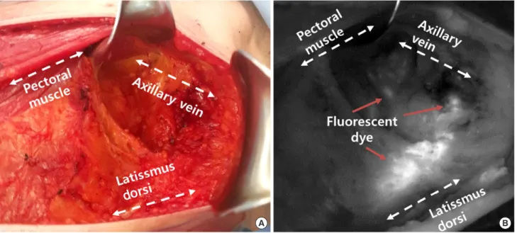
Figure 1. Axillary lymph node dissection with lymphatic mapping on indocyanine green. (A) Axillary lymph node dissection seen in surgical field. (B) Axillary lymph node dissection seen in near-infrared fluorescence.
A B
Figure 2. Classification of axillary lymph node dissection by indocyanine green. (A) Level I sentinel lymph node in near-infrared fluorescence. (B) Level I non-sentinel lymph node in near-infrared fluorescence. (C) Level II lymph node in near-infrared fluorescence.
A B C
neously into the breast using a 24-gauge needle. Each patient then underwent ALND, which was performed using a fluores- cence camera. First, we removed the fluorescent lymph node at axillary levels I and II. We then removed the remaining axil- lary lymph nodes at levels I and II. We obtained the results for the axillary lymph node by classification (Figures 1 and 2).
In this study, the non-SLN was defined as the lymph node that was not stained along with the ICG node but as an exist- ing lymph node at axillary level I. Physiologic lymphatic me- tastasis was defined as metastasis that was SLN-positive at ax- illary level I, non-SLN-negative and axillary level II lymph- node–negative or positive. Nonphysiological lymphatic me- tastasis was defined as the other physiology pattern of lym- phatic metastasis, with nonfluorescent lymph node metastasis by ICG (Figures 3 and 4).
We used the chi-square test and Spearman correlation coef- ficient to compare discrete variables. Differences were as- sumed to be statistically significant when the p-value was less than 0.05. We used SPSS version 23 (IBM Inc., Armonk, USA) for the chi-square tests and for Spearman correlation coefficient. This study was approved by the Institutional Re- view Board of Samsung Medical Center, Seoul, Korea (IRB file no. 2015-01-046). All patients provided written informed consent.
RESULTS
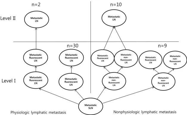
We performed lymphatic mapping for 64 patients with level II lymphatic flow using ICG. Among 64 patients, the without NAC group comprised 51 patients and the post-NAC group comprised 13 patients. Without NAC group, there were 32 patients with SLN metastasis and without non-SLN metasta- sis, and all of them had physiological lymphatic metastasis. Of these patients, two (6.3%) had lymph node metastasis in level II and 30 patients (93.1%) did not have lymph node metasta- sis in level II. The number of patients with non-SLN metasta- sis was 19, of which 10 (52.6%) had lymph node metastasis in level II and nine (47.3%) patients had no lymph node metas- tasis in level II (Figure 3). In the post-NAC group, there were five patients with SLN metastasis and without non-SLN me- tastasis, and all of these had physiologic lymphatic metastasis.
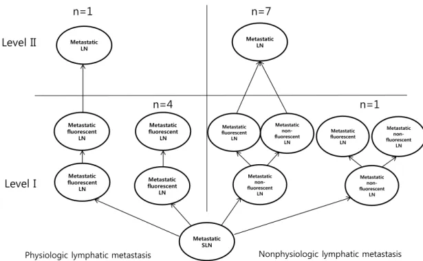
Among these patients, one (20.0%) had lymph node metasta- sis in level II and four patients (80.0%) did not have lymph node metastasis in level II. The number of patients with non- SLN metastasis was eight, of which seven (87.5%) had lymph node metastasis in level II and one (12.5%) patients had no lymph node metastasis in level II (Figure 4).
Of 51 patients who did not receive NAC, 32 had axillary metastasis with physiologic lymphatic metastasis and 19 pa-
Figure 3. Algorithm of axillary metastasis between physiologic lymphatic metastasis and nonphysiologic lymphatic metastasis without neoadjuvant chemotherapy.
LN=lymph node; SLN=sentinel lymph node.
Metastatic SLN Metastatic
fluorescent
Level I LN
Level II Metastatic LN
n=30 n=2
n=9
Metastatic non- fluorescent
LN
n=10
Physiologic lymphatic metastasis Nonphysiologic lymphatic metastasis
Metastatic fluorescent
LN
Metastatic fluorescent
LN Metastatic fluorescent
LN
Metastatic LN
Metastatic fluorescent
LN
Metastatic fluorescent
LN
Metastatic non- fluorescent
LN
Metastatic non- fluorescent
LN
Metastatic fluorescent non-
LN
tients had nonphysiological lymphatic metastasis. We com- pared the clinicopathological characteristics between these groups (Table 1). The risk factors for nonphysiologic lymphat-
ic metastasis were high Ki-67 index (>20%), perinodal exten- sion (PNE), and age ≥55 years in both univariate and multi- variate analyses (p<0.05) (Tables 2 and 3).
Figure 4. Algorithm of axillary metastasis between physiologic lymphatic metastasis and nonphysiologic lymphatic metastasis with neoadjuvant che- motherapy.
LN=lymph node; SLN=sentinel lymph node.
Metastatic SLN Metastatic
fluorescent
Level I LN
Level II Metastatic LN
n=4 n=1
n=1
Metastatic non- fluorescent
LN
n=7
Physiologic lymphatic metastasis Nonphysiologic lymphatic metastasis
Metastatic fluorescent
LN
Metastatic fluorescent
LN Metastatic fluorescent
LN
Metastatic LN
Metastatic fluorescent
LN
Metastatic fluorescent
LN
Metastatic non- fluorescent
LN
Metastatic non- fluorescent
LN
Metastatic non- fluorescent
LN
Characteristic
Physiologic lymphatic metastasis (n=32) No. (%)
Nonphysiologic lymphatic metastasis (n=19) No. (%)
p-value
Age (yr) 0.069
<55 26 (81.3) 11 (57.9)
≥55 6 (18.8) 8 (42.1)
Type of surgery 0.291
Total mastectomy 12 (37.5) 10 (52.6)
BCS 20 (62.5) 9 (47.4)
T stage 0.451
1 8 (25.0) 2 (10.5)
2 20 (62.5) 14 (73.7)
3 4 (12.5) 3 (15.8)
N stage <0.001
1 24 (75.0) 3 (15.8)
2 6 (18.8) 10 (52.6)
3 2 (6.3) 6 (31.6)
Perinodal extension 18 (56.3) 16 (84.2) 0.035
LVI 15 (46.9) 8 (42.1) 0.741
Characteristic
Physiologic lymphatic metastasis
(n=32) No. (%)
Nonphysiologic lymphatic metastasis
(n=19) No. (%)
p-value
Tumor location 0.381
UOQ 16 (50.0) 10 (52.6)
UIQ 3 (9.4) 3 (15.8)
LOQ 3 (9.4) 2 (10.5)
LIQ 2 (6.3) 3 (15.8)
Central/whole 8 (25.0) 1 (5.3)
BMI (kg/m2) 0.313
≤25 21 (65.6) 15 (78.9)
>25 11 (34.4) 4 (21.1)
ER positive 32 (100) 18 (94.7) 0.190
PR positive 30 (93.8) 19 (100) 0.266
HER2 positive 1 (3.1) 3 (15.8) 0.104
Ki-67 (%) 0.044
≤20 25 (78.1) 10 (52.6)
>20 7 (21.9) 9 (47.4)
BCS=breast-conserving surgery; LVI=lymphovascular invasion; UOQ=upper outer quadrant; UIQ=upper inner quadrant; LOQ=lower outer quadrant; LIQ=lower inner quadrant; BMI=body mass index; ER=estrogen receptor; PR=progesterone receptor; HER2=human epidermal growth factor receptor 2.
Table 1. Comparison of clinicopathological characteristics between patients with physiologic versus nonphysiologic lymphatic metastasis without neoadjuvant chemotherapy
DISCUSSION
In this prospective study, we performed lymphatic flow im- aging using ICG of cytologically proven axillary metastasis.
Imaging was used for intraoperative navigation to identify SLNs and non-SLNs. The results of previous studies regarding SLN detection by NIR fluorescence using ICG for breast can- cer showed that the identification rate of SLNs was 92%–100%
[4,9,12-15]. This indicates that the discrimination of SLNs us- ing ICG is very reliable.
The American College of Surgeons Oncology Group (ACOSOG) Z0011 trial investigated women with T1 or T2 invasive primary breast cancer and 1 or 2 SLNs containing metastases. In this trial, the 10-year overall survival for pa- tients treated with SLN dissection alone was noninferior to that for those treated with ALND [3]. In the After Mapping of the Axilla: Radiotherapy or Surgery (AMAROS) trial, SLN bi- opsy without ALND offered excellent regional control, indi- cating that this regimen may be a reasonable option for man- agement [2]. Although our study involved patients who were axillary lymph node-positive before surgery, 32 patients had positive SLNs and negative non-SLNs, including two patients (6.3%) with level II lymph node metastasis.
A recent study determined the percentage of patients with invasive breast cancer who had N2 or N3 stage disease, and developed a nomogram for the prediction of N2 or N3 stage disease in these patients. The nomogram revealed that lym- phovascular invasion, T2 stage, metastatic lymph node ratio, and PNE were independent predictors of N2 or N3 stage dis-
ease [16]. PNE, found in frozen sections from breast cancer, is a predictor [17]. Our study showed that 29 of 35 patients (82.9%) with N2 or N3 stage disease had PNE, and 31 of 35 patients (88.6%) with N2 or N3 stage disease had stage T2 or greater disease.
Selective ALND involves only the fluorescent lymph nodes at level I and II visualized using ICG. Selective dissection may lead to less arm morbidity and lymphedema, compared with routine ALND. A study showed that selective ALND after re- verse mapping prevented breast-cancer–related lymphedema [18]. However, patients aged ≥55 years with high Ki-67 indi- ces (>20%) and perinodal status, such as those in our study, might not be good candidates for selective axillary dissection, as these patients may have nonphysiological lymphatic metas- tasis.
Skip metastasis of axillary lymph nodes is an important phenomenon. The mechanism of skip metastasis of axillary lymph node is unclear. In some article, skip metastasis has been defined as level I absence but level II and/or III involve- ment [19]. However skip metastasis was not observed in our study. Chemotherapy can induce axillary lymph node fibrosis and lymphatic flow block. Nonphysiologic lymphatic metasta- sis may be affected by these factors. In our study, eight (61.5%) and 19 patients (37.3%) were found in the post-NAC and without NAC groups with nonphysiological lymphatic pat- terns, respectively. There was no statistical difference (p=
0.114) but patients in the post-NAC group showed a greater tendency of non-physiological lymphatic metastasis.
This study had several limitations. First, it was limited to a single comprehensive cancer institution. Second, our study was a prospective design study of lymphatic passage in pa- tients with cytologically proven axillary metastasis, and we analyzed only those patients in whom SLN metastasis was confirmed and all ICG levels were elevated to level II. There- fore, it is difficult to mention the identification rate of SLNs and false negative rate of SLNs using ICG in this study. Third, this study did not enroll a large population, limiting the statis- tical power. However, this study is meaningful even if the number of patients was relatively small, because it had a pro- spective design with clinically suspected node-positive pa- Table 2. Univariate analysis of factors associated with nonphysiologic
lymphatic metastasis without neoadjuvant chemotherapy Characteristic Univariate analysis
Exp (B) 95% CI p-value
LVI 0.399 0.065–2.422 0.320
Perinodal extension 5.177 0.952–28.161 0.048
Age ≥55 yr 15.779 2.009–123.937 0.009
BCS 0.314 0.054–1.824 0.197
BMI >25 kg/m2 0.098 0.008–1.206 0.070 Tumor location
UOQ 0.762
UIQ 3.678 0.191–70.781 0.388
LOQ 0.564 0.037–8.543 0.680
LIQ 0.871 0.024–31.836 0.940
Central/whole 0.296 0.020–4.307 0.373
HER2 positive 5.292 0.074–377.963 0.444
Ki-67 >20% 10.549 1.887–58.970 0.007
CI=confidence interval; LVI=lymphovascular invasion; BCS=breast-conserv- ing surgery; BMI=body mass index; UOQ=upper outer quadrant; UIQ=upper inner quadrant; LOQ =lower outer quadrant; LIQ =lower inner quadrant;
HER2=human epidermal growth factor receptor 2.
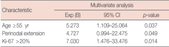
Table 3. Multivariate analysis of factors associated with nonphysiologic lymphatic metastasis
Characteristic Multivariate analysis
Exp (B) 95% CI p-value
Age ≥55 yr 5.273 1.109–25.064 0.037
Perinodal extension 4.727 0.994–22.475 0.049
Ki-67 >20% 7.030 1.476–33.476 0.014
CI=confidence interval.
tients confirmed by cytology. In addition, there has been little research conducted regarding post-SLN metastasis. Further- more, we obtained the results of axillary lymph nodes in axil- lary level I (SLNs and non-SLNs) and level II lymph nodes by classification.
In conclusion, lymphatic metastatic flow in cytologically proven axillary metastasis can be either physiological or non- physiological lymphatic metastasis. Axillary metastases in nonphysiological lymphatic flow are more frequently level II lymph node metastases. In without NAC group, patients with cytologically proven axillary metastasis aged ≥55 years, with high Ki-67 indices (>20%), and with PNE may have non- physiological lymphatic metastasis. Patients with these risk factors might not be good candidates for selective axillary dis- section.
CONFLICT OF INTEREST
The authors declare that they have no competing interests.
REFERENCES
1. Dewis R, Gribbin J. National Institute for Health and Clinical Excel- lence: guidance. In: Dewis R, Gribbin J. Breast Cancer: Diagnosis and Treatment: An Assessment of Need. Cardiff: National Collaborating Centre for Cancer; 2009. p.13-35.
2. Donker M, van Tienhoven G, Straver ME, Meijnen P, van de Velde CJ, Mansel RE, et al. Radiotherapy or surgery of the axilla after a positive sentinel node in breast cancer (EORTC 10981-22023 AMAROS): a randomised, multicentre, open-label, phase 3 non-inferiority trial.
Lancet Oncol 2014;15:1303-10.
3. Giuliano AE, Ballman KV, McCall L, Beitsch PD, Brennan MB, Kelemen PR, et al. Effect of axillary dissection vs no axillary dissection on 10-year overall survival among women with invasive breast cancer and sentinel node metastasis: the ACOSOG Z0011 (Alliance) randomized clinical trial. JAMA 2017;318:918-26.
4. Guo J, Yang H, Wang S, Cao Y, Liu M, Xie F, et al. Comparison of sentinel lymph node biopsy guided by indocyanine green, blue dye, and their combination in breast cancer patients: a prospective cohort study.
World J Surg Oncol 2017;15:196.
5. Schaafsma BE, Mieog JS, Hutteman M, van der Vorst JR, Kuppen PJ, Löwik CW, et al. The clinical use of indocyanine green as a near-infrared fluorescent contrast agent for image-guided oncologic surgery. J Surg Oncol 2011;104:323-32.
6. Jung SY, Kim SK, Kim SW, Kwon Y, Lee ES, Kang HS, et al. Comparison of sentinel lymph node biopsy guided by the multimodal method of
indocyanine green fluorescence, radioisotope, and blue dye versus the radioisotope method in breast cancer: a randomized controlled trial.
Ann Surg Oncol 2014;21:1254-9.
7. Hirche C, Murawa D, Mohr Z, Kneif S, Hünerbein M. ICG fluores- cence-guided sentinel node biopsy for axillary nodal staging in breast cancer. Breast Cancer Res Treat 2010;121:373-8.
8. Sugie T, Kassim KA, Takeuchi M, Hashimoto T, Yamagami K, Masai Y, et al. A novel method for sentinel lymph node biopsy by indocyanine green fluorescence technique in breast cancer. Cancers (Basel) 2010;
2:713-20.
9. Wishart GC, Loh SW, Jones L, Benson JR. A feasibility study (ICG-10) of indocyanine green (ICG) fluorescence mapping for sentinel lymph node detection in early breast cancer. Eur J Surg Oncol 2012;38:651-6.
10. Alford R, Simpson HM, Duberman J, Hill GC, Ogawa M, Regino C, et al. Toxicity of organic fluorophores used in molecular imaging: litera- ture review. Mol Imaging 2009;8:341-54.
11. Gilmore DM, Khullar OV, Gioux S, Stockdale A, Frangioni JV, Colson YL, et al. Effective low-dose escalation of indocyanine green for near- infrared fluorescent sentinel lymph node mapping in melanoma. Ann Surg Oncol 2013;20:2357-63.
12. Hokimoto N, Sugimoto T, Namikawa T, Funakoshi T, Oki T, Ogawa M, et al. A novel color fluorescence navigation system for intraoperative transcutaneous lymphatic mapping and resection of sentinel lymph nodes in breast cancer: comparison with the combination of gamma probe scanning and visible dye methods. Oncology 2018;94:99-106.
13. Zhang X, Li Y, Zhou Y, Mao F, Lin Y, Guan J, et al. Diagnostic perfor- mance of indocyanine green-guided sentinel lymph node biopsy in breast cancer: a meta-analysis. PLoS One 2016;11:e0155597.
14. Chi C, Ye J, Ding H, He D, Huang W, Zhang GJ, et al. Use of indocya- nine green for detecting the sentinel lymph node in breast cancer pa- tients: from preclinical evaluation to clinical validation. PLoS One 2013;
8:e83927.
15. Pitsinis V, Provenzano E, Kaklamanis L, Wishart GC, Benson JR. Indo- cyanine green fluorescence mapping for sentinel lymph node biopsy in early breast cancer. Surg Oncol 2015;24:375-9.
16. Kim I, Ryu JM, Kim JM, Choi HJ, Lee SK, Yu JH, et al. Development of a nomogram to predict N2 or N3 stage in T1-2 invasive breast cancer patients with no palpable lymphadenopathy. J Breast Cancer 2017;
20:270-8.
17. van der Loo EM, Sastrowijoto SH, Bril H, van Krimpen C, de Graaf PW, Eulderink F. Less operations required due to perioperative frozen section examination of sentinel nodes in 275 breast cancer patients.
Ned Tijdschr Geneeskd 2001;145:1986-91.
18. Gennaro M, Maccauro M, Sigari C, Casalini P, Bedodi L, Conti AR, et al. Selective axillary dissection after axillary reverse mapping to prevent breast-cancer-related lymphoedema. Eur J Surg Oncol 2013;39:1341-5.
19. Wang H, Mao XY, Zhao TT, Zheng XY, Jin F, Li JG. Study on the skip metastasis of axillary lymph nodes in breast cancer and their relation with Gli1 expression. Tumour Biol 2012;33:1943-50.